莫迪司核磁共振增强扫描联合AFP、CA125、CEA在早期原发性肝癌诊断中的效果及诊断效能研究
2021-03-28赖婳妤
赖婳妤

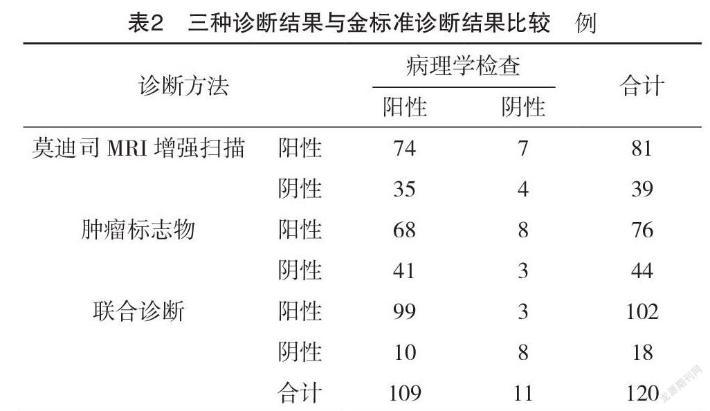

【摘要】 目的:研究探討莫迪司核磁共振(MRI)增强扫描联合甲胎蛋白(AFP)、糖类抗原125(CA125)、血清癌胚抗原(CEA)在早期原发性肝癌诊断中的效果及诊断效能。方法:选取2020年1-12月本院收治的120例肝脏占位疑似肝癌患者为研究对象,患者入院后均进行莫迪司MRI增强扫描,并检测血清肿瘤标志物(AFP、CA125、CEA)。比较组织病理学、莫迪司MRI增强扫描、肿瘤标志物及联合诊断的诊断阳性率;以术后病理学检测结果为金标准,比较莫迪司MRI增强扫描、肿瘤标志物及联合诊断与金标准诊断结果的一致性;比较各检测方法对早期原发性肝癌的诊断效能。结果:组织病理学诊断结果显示,120例患者中109例确诊为肝癌。莫迪司MRI增强扫描、肿瘤标志物的诊断阳性率与组织病理学比较,差异均有统计学意义(P<0.05);联合诊断的诊断阳性率与组织病理学比较,差异无统计学意义(P>0.05)。经Kappa一致性检验,联合诊断结果与金标准有高度一致性(Kappa=0.878,P<0.05),莫迪司MRI增强扫描检查、血清肿瘤标志物检查与金标准结果一致性一般(Kappa=0.529、0.503,P<0.05)。三种诊断方法灵敏度、特异度、阳性预测值、阴性预测值、准确度比较,差异均有统计学意义(P<0.05),且联合诊断的各诊断效能指标最高。结论:莫迪司核磁共振增强扫描联合AFP、CA125、CEA血清肿瘤标志物检查在早期原发性肝癌诊断中有较高的应用价值,能够提高原发性肝癌的诊断准确率,提高灵敏度、特异度,可作为组织病理学检查的替代手段广泛应用于临床,值得进一步推广应用。
【关键词】 原发性肝癌 莫迪司核磁共振增强扫描 甲胎蛋白 糖类抗原125 血清癌胚抗原
[Abstract] Objective: To investigate the effect and diagnostic efficacy of MODIS enhanced MRI combined with alpha fetoprotein (AFP), carbohydrate antigen 125 (CA125) and serum carcinoembryonic antigen (CEA) in the early diagnosis of primary liver cancer. Method: A total of 120 cases of suspected liver cancer patients with liver occupying space admitted to our hospital from January to December 2020 were selected as the research objects. After admission, all patients underwent MODIS enhanced MRI, and serum tumor markers (AFP, CA125, CEA) were detected. The positive rates of histopathology, MODIS enhanced MRI, tumor markers and combined diagnosis were compared. The results of postoperative pathology were used as the gold standard to compare the consistency of MODIS enhanced MRI, tumor markers and combined diagnosis with the gold standard diagnosis. The diagnostic efficacy of each detection method for early primary liver cancer was compared. Result: Histopathological findings showed that 109 of the 120 patients were diagnosed with liver cancer. Compared with histopathology, the positive rates of MODIS enhanced MRI and tumor markers were significantly different (P<0.05); there was no significant difference between the positive rate of combined diagnosis and histopathology (P>0.05). The results of the combined diagnosis were highly consistent with the gold standard (Kappa=0.878, P<0.05) by Kappa consistency test. The MODIS enhanced MRI and serum tumor marker examination were generally consistent with the gold standard results (Kappa=0.529, 0.503, P<0.05). The differences in sensitivity, specificity, positive predictive value, negative predictive value and accuracy of the three diagnostic methods were statistically significant (P<0.05), and the combined diagnosis had the highest diagnostic efficiency indexes. Conclusion: MODIS enhanced MRI scan combined with AFP, CA125, CEA serum tumor markers has high application value in the diagnosis of early primary liver cancer, can improve the diagnostic accuracy of primary liver cancer, improve the sensitivity and specificity of diagnosis, and can be widely used in clinic as an alternative means of histopathological examination, which is worthy of further promotion and application.
[Key words] Primary liver cancer MODIS enhanced MRI AFP CA125 CEA
First-authors address: Yuedong Hospital, The Third Affiliated Hospital of Sun Yat-sen University, Meizhou 514000, China
doi:10.3969/j.issn.1674-4985.2021.19.001
原发性肝癌是临床最为常见的恶性肿瘤疾病之一,患者发病后表现为明显的肝区疼痛,出现腹痛腹胀等症状,并随着患者病情的进展,会出现肝脏组织的坏死、破裂,可引起腹腔內出血,出现腹膜刺激征,进而严重影响患者的生命健康和生活质量,并最终危及患者的生命[1-2]。随着社会工作压力的增加和生活习惯的改变,原发性肝癌的发病率逐年升高,已成为威胁人类生命安全的最重要的癌症肿瘤疾病,对其临床诊断治疗研究也在不断地深入。在对于原发性肝癌的诊断中,多数患者在确诊时多已处于中晚期阶段,不利于患者诊疗活动的开展导致患者病情迅速恶化进展至终末期[3-4]。……
