CT扫描联合磁共振成像在肋骨骨折中的诊断价值及临床意义
2021-10-22陈钦龙李建伟
陈钦龙 李建伟
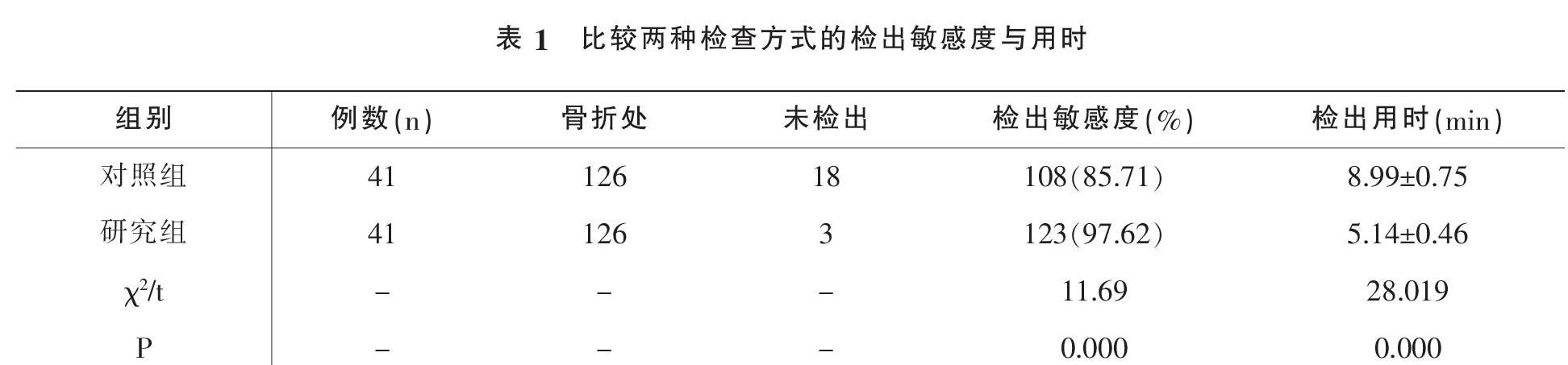
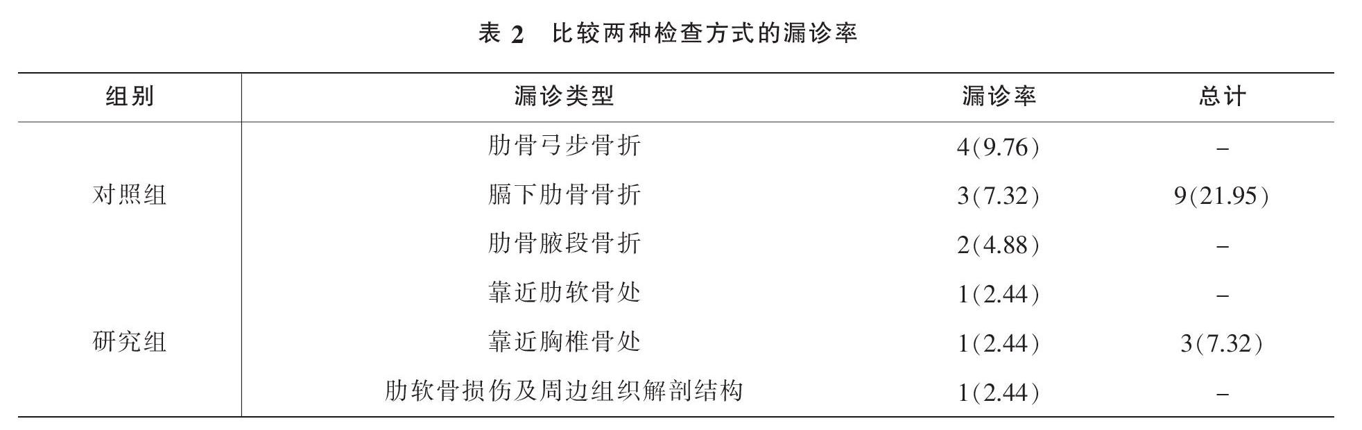
摘要:目的:探讨CT扫描联合磁共振成像用于诊断肋骨骨折,阐述其诊断价值及临床意义。方法: 选择2019年8月-2021年2月期间本院和东莞市人民医院收治的82例胸部外伤患者为研究对象,随机分为2组,各41例,对照组给予CT扫描检查,研究组采用CT扫描联合磁共振成像检查,观察肋骨及肋软骨损伤情况及周边组织解剖结构关系,以及肋软骨损伤的诊断准确性。结果:对照组的漏诊率为 26.21%(27 处骨折),研究组的漏诊率为 4.85%(5 处骨折)。与对照组相比,用CT扫描联合磁共振成像诊断的漏诊率更低(P<0.05)。结论:CT扫描联合磁共振成像在诊断肋骨骨折中的应用价值较高,可明显降低漏诊率,准确性显著提高。
关键词:CT扫描;磁共振成像;肋骨骨折;诊断价值
中图分类号:R445.2;R683文献标识码:BDOI:10?郾3969/j.issn.1001-0270.2021.05.09
Diagnostic Value and Clinical Significance of CT Scanning Combined with Magnetic Resonance Imaging in Rib Fracture
Chen Qin-long1, Li Jian-wei2
(1. Department of Radiology, Peoples Hospital of Xinhui District, Jiangmen City, Guangdong 529100,China; 2. Department of Radiology, Peoples Hospital of Dongguan City, Guangdong 523000, China)
Abstract: Objective: The diagnostic value and clinical significance of CT scan combined with magnetic resonance imaging(MRI) were investigated in the diagnosis of rib fracture. Methods: Eighty-two patients with chest trauma admitted to our hospital and Dongguan Peoples Hospital from August 2019 to February 2021 were selected as the study subjects and randomly divided into two groups, 41 cases for each group. The control group was given CT scan examination, and the study group was examined by CT scan combined with magnetic resonance imaging, i.e. To observe the injury of ribs and costal cartilage and the anatomical structure of surrounding tissues, as well as the diagnostic accuracy of costal cartilage injury. Results: 26.21% (27 fractures) of the patients in the control group and 4.85% (5 fractures) of the patients in the study group were missed. Compared with the control group, the missed diagnosis rate of rib fractures in this group of patients diagnosed with CT scan combined with magnetic resonance imaging was lower(P<0.05).Conclusion: CT scan combined with magnetic resonance imaging is of high value in the diagnosis of rib fracture. It can significantly reduce the missed diagnosis rate of rib fracture and significantly improve the accuracy.
Key Words: CT scan; magnetic resonance imaging; rib fracture; diagnostic value
臨床上肋骨骨折是常见的胸部损伤之一,最常出现在胸部钝挫伤后,但也可由重度咳嗽、体育活动以及非意外性创伤引起[1]。肋骨骨折的伴发损伤及并发症从轻微不适至危及生命的疾病(例如气胸、脾裂伤及肺炎)不等。目前,影像学检查是诊断此病的主要手段。既往临床上常采用DR检查诊断肋骨骨折,但诊断效果并不理想。CT成为胸部外伤诊断的主要检测手段,尤其针对事故性外伤引起的肋骨骨折,也是后续责任鉴定的重要判定依据[2-3]。但肋骨形态和走行特殊,较多重叠处,而常规CT横断面观察敏感度较低[4]。有研究报道,DR检查无法准确定位患者骨折的具体位置,发生漏诊或误诊概率较大。近年来,CT检查、磁共振成像在诊断肋骨骨折方面得到了广泛的应用[5]。目前,临床术前分期诊断主要采用CT和磁共振成像(MRI),这两种诊断方式均具有较高的诊断价值,但临床上对于术前分期选择哪种诊断方式能确保分期诊断的准确率仍存在极大的争议。……