应用MR T2 mapping序列对肾衰竭患者膝关节软骨损伤 的研究
2019-10-21徐震陈其春王龙胜郑穗生
徐震 陈其春 王龙胜 郑穗生

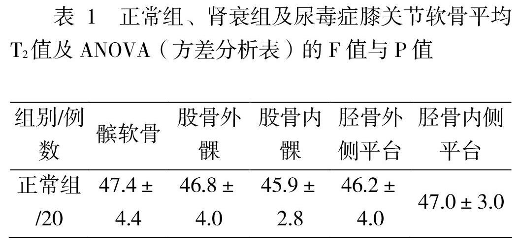

摘要:目的 测量慢性肾功能不全衰竭期及尿毒症期患者膝关节和正常对照组关节软骨T2弛豫时间并比较差异性,评价磁共振T2 mapping成像在慢性肾功能不全衰竭期患者及尿毒症期患者膝关节软骨损伤中的应用价值。材料与方法 采用西门子3.0T 磁共振,选取正常组20个膝关节,慢性肾功能不全衰竭期21个膝关节,尿毒症期20个膝关节;应用T2 mapping序列观察和测量正常组(20例)和慢性肾功能不全衰竭期(21例)及尿毒症期(20例)髌软骨、股骨内侧髁、外侧髁、胫骨内侧平台、外侧平台5处关节软骨的T2值,并进行统计学分析。结果 慢性肾功能不全衰竭期及尿毒症期T2值均高于正常组;正常组对比慢性肾功能不全衰竭期以及尿毒症期软骨T2值差异具有统计学意义(P<0.05);慢性肾功能不全衰竭期与尿毒症期关节软骨T2值差异不具有统计学意义(P>0.05);结论 T2 mapping成像可以早期发现膝关节软骨内组织成分的变化,进而评价软骨的损伤,对肾功能不全患者膝关节软骨损伤的诊断和监测具有一定的临床应用价值;肾功能不全患者存在膝关节软骨的损伤。
关键词:软骨;肾功能不全;尿毒症;磁共振成像;T2值
【中图分类号】R692 【文献标识码】A 【文章编号】2107-2306(2019)03-019-03
[Abstract] Objective To measure the relaxation time of articular cartilage T2 in knee joints of patients with chronic renal failure and uremia, and to evaluate the value of magnetic resonance T2 mapping imaging in knee joint cartilage injury of patients with chronic renal failure and uremia. Materials and Methods Twenty knees in normal group, 21 knees in chronic renal failure stage and 20 knees in uremic stage were selected by using Siemens 3.0T magnetic resonance imaging. Patellar cartilage, medial femoral condyle, lateral condyle, medial tibial plateau and lateral tibial plateau in normal group (20 cases) and chronic renal failure (21 cases) and uremic stage (20 cases) were observed and measured by T2 mapping sequence. T 2 values of articular cartilage at 5 lateral plateaus were analyzed statistically. Results The T2 values of articular cartilage in chronic renal failure and uremia were higher than those in normal group; there were significant differences between normal group and chronic renal failure and uremia (P < 0.05); there was no significant difference between chronic renal failure and uremia (P > 0.05); Conclusion T2 mapping imaging can detect knee joint early (P > 0.05). The changes of tissue composition in articular cartilage and the evaluation of cartilage injury have certain clinical value in the diagnosis and monitoring of knee cartilage injury in patients with renal insufficiency, and there is knee cartilage injury in patients with renal insufficiency.
[keywords] cartilage; renal insufficiency; uremia; magnetic resonance imaging; T2 value
膝關节是人体最大的关节,膝关节损伤在日常生活中很常见,损伤的同时往往最早发生关节软骨的损伤;关节软骨损伤是影响关节稳定性的重要因素[1]。目前关于关节软骨T2 mapping方面的研究主要集中在骨性关节炎和软骨修复等方面,但是关于肾功能不全病人关节软骨损伤的探索还比较少,本文旨在探索肾功能不全患者膝关节软骨损伤的临床应用价值。