乳腺导管内癌的X线表现与分子分型的对照研究
2017-08-22黄社磊马捷蒋华景
黄社磊+马捷+蒋华景
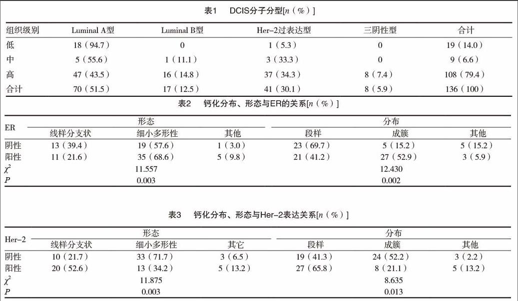
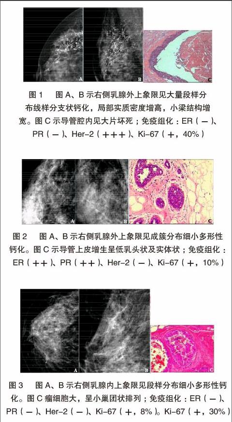
[摘要] 目的 探討乳腺导管内癌的X线表现与分子分型的关系。 方法 回顾性分析133例136侧DCIS的X线表现及组织病理学、免疫组织化学结果。 结果 (1)136侧乳腺导管内癌X线表现为单纯钙化(32.4%)、钙化伴局灶性不对称/肿块(29.4%),钙化出现率61.8%。钙化形态以细小多形性(58.3%)、段样分布多见(54.8%);形态分布组合以细小多形性钙化成簇分布多见。肿块的形态多为不规则形(62.5%)、边缘模糊(43.8%)、高密度(75%)。(2)DCIS以Luminal A型多见,Her-2过表达型次之。(3)ER/PR(+)、Her-2(-)X线表现单纯钙化多见35.6%、34.6%,形态为细小多形性68.6%、71.7%、成簇分布52.9%、52.2%;ER/PR(-)、Her-2过表达多表现为钙化伴局灶性不对称/肿块38.8%、36.2%,钙化形态前者为细小多形性57.6%、段样分布69.7%,后者为线样分支状52.6%、段样分布65.8%。(4)Ki-67增殖指数≤10%时,83.3%ER(+)、80.6%Her-2(-),钙化形态多为细小多形性(88.2%);当Ki-67增殖指数>31%时,56.4%ER(+)、48.7%Her-2(-),钙化形态为线样分支状48.3%。 结论 DCIS分子分型以Luminal A型多见。X线以钙化为主要表现,形态多为细小多形性、线样分支状;分布以段样及成簇多见。钙化的分布、形态及形态分布组合与ER、PR、Her-2及Ki-67增殖指数有关。
[关键词] 数字化乳腺X线摄影;癌;导管;激素受体;人表皮生长因子受体;分子分型
[中图分类号] R737.9 [文献标识码] A [文章编号] 2095-0616(2017)13-13-04
[Abstract] Objective To explore the relationship the X-ray appearance and molecule subtype of the Breast DCIS (Ductal Carcinoma In Situ,DCIS),in order to know more about them. Methods The main X-ray appearance,histopathology and immunhistochemistry of the 133 cases DCIS (all 136 latero-breast) were retrospectively analyzed. Results (1)The main X-ray appearance of the 136 laterals DCIS were calcification 32.4%,calcification with focal dissymmetry/mass 29.4%,the frequency of calcification of all cases was 61.8%.The main morphous and distribution of calcifications was fine pleomorphic calcifications 58.3% and segmental 54.8% respectively,the main combination of morphous and distribution of calcifications was clustering fine pleomorphic calcifications.The appearance of the mass were irregular 62.5%,ambigutiy boundary 43.8%,high density 75%.(2)The molecule subtype of the Luminal A was the most frequent found in the different grade DCIS.(3)The main X-ray appearance of the ER/PR(+) and Her-2(-) DCIS were purely calcifications (35.6% vs 34.6%),the appearance of calcifications of them were fine pleomorphic calcifications (68.6% vs 71.7%),cluster(52.9% vs 52.2%).However,the main X-ray appearance of the ER/PR(-) and Her-2(+) DCIS were calcification with focal dissymmetry/mass (38.8% vs 36.2%),the appearance of calcifications the ER/PR(-)DCIS were fine pleomorphic (57.6%),segment (69.7%).While the Her-2(+) DCIS were linear branching (52.6%),segment (65.8%).(4)The proliferation index of the Ki-67≤10%,the proportion of ER(+) and Her-2(-) DCIS were 83.3% vs 80.6%,and the morphous of calcification was fine pleomorphic calcifications (88.2%).While the proliferation index of the Ki-67>31%,the proportion of ER (+) and Her-2(-) DCIS were 56.4% vs 48.7%,and the morphous of calcification was linear branching (48.3%). Conclusion The Luminal A is the most frequent found in the molecule subtypes of the DCIS.The main X-ray appearance of the DCIS is calcifications,the main morphous of the calcifications are fine pleomorphic and linear branching,the main distrutions of them are segment and clustered.The morphous,distrutions,combination of the morphous and distrutions of the calcifications are relevanted with ER, PR, Her-2 and the proliferation index of the Ki-67.
[Key words] Digital mammographic screening;Carcinoma;Duct;Hormone receptors;Her-2;Molecular typing
乳腺导管内癌(ductal carcinoma in situ,DCIS)是局限于导管内基底膜内而不侵犯间质的一类乳腺导管上皮细胞恶性增生,约占全部乳腺癌的20%,影像学(X线等)的发展和广泛应用显著提高了DCIS的检出率[1]可根据雌激素受体(ER)、孕激素受体(PR)、人类上皮生长因子(HER-2)的表达水平以及细胞增殖指数(Ki-67)将乳腺癌划分为4类分子亚型:包括Luminal A型、Luminal B型、HER-2阳性和三阴性乳腺癌,分子分型与乳腺癌治疗和预后具有密切关系[2]。本……
