侧脑室中枢神经细胞瘤的MRI分析
2017-05-30李吉华李艳丽陈国云钟颖洁朱昱衡
李吉华 李艳丽 陈国云 钟颖洁 朱昱衡
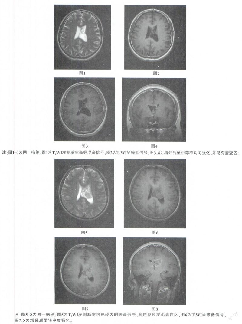
摘要:目的:分析侧脑室中枢神经细胞瘤的MRI表现。方法:回顾性分析经手术证实的CNC6例。男4例,女2例,年龄15-48岁,平均年龄38.5岁。结果:本组6例病例肿瘤均位于侧脑室透明隔近孟氏孔处。在MRI T1WI像呈不均匀等或低信号,T2WI呈不均匀高信号,肿瘤内可有囊变区,增强扫描呈不均匀中等强化,边界清楚。结论:中青年位于侧脑室腔明隔的肿瘤,应考虑致CNC的可能。结合临床及MRI表现有助于此病的诊断。
关键词:中枢神经细胞瘤;磁共振成像;透明隔
中图分类号:R445.2;R322.81 文獻标识码:B DOI:10.3969/j.issn.1001-0270.2017.02.29
Abstract: Objective: To analyze the MRI findings of the lateral ventricle. Methods: The clinical data of CNC 6 cases confirmed by operation were analyzed retrospectively. There were 4 males and 2 females with an average age of years (range, 15-48 years). Results: 6 cases of this group of tumors were located in the lateral ventricle near the foramen of Monro at septum pellucidum. In MRI T1WI, such as uneven or low signal, T2WI showed uneven high signal, the tumor can have cystic change area, enhanced scan showed uneven moderate enhancement, clear boundary. Conclusion: CNC should be considered in young and middle-aged patients with tumors located in the septum of the lateral ventricle. Combined with clinical and MRI findings, it is helpful for the diagnosis of the disease.
Key Words: Central Neurocytoma; Magnetic Resonance Imaging (MRI); Septum Pellucidum
中枢神经细胞瘤(central neurocytoma,CNC),临床较少见。1982年Hassoun等[1]首次报告。WHO中枢神经系统肿瘤组织学分类将中枢神经细胞瘤归类于神经元和混合神经元—神经胶质肿瘤。以往国内文献多为零散报道,故确切的发病率尚不清楚,不到中枢神经系统肿瘤的1%。笔者搜集6例经手术病理证实的CNC的MRI影像表现进行回顾性分析,以提高对CNC的诊断,以帮助临床选择正确的治疗方案。
1 材料与方法
1.1 临床资料
6例经手术和病理证实的中枢神经细胞瘤中,男4例,女2例,年龄15-48岁,平均年龄38.5岁。6例均出现颅内压增高症状,出现头痛(6例),呕吐(4例),视物不清(2例),病史1周至2年。
1.2 检查方法
使用Simens1.5和3.0T超导磁共振扫描仪,头颅表面线圈,常规行SE T1WI,FSE T2WI横轴面及矢状面MR扫描,辅以冠状面,扫描层厚5mm,层间距0。6例均按0.1mmol/kg体重静脉注射钆喷酸葡胺(Gd-DTPA)行SE T1WI横轴面、矢状面及冠状面增强扫描。
2 结果
2.1 MRI表现
5例均行MRI检查(常规平扫及增强扫描),病灶均位于侧脑室透明隔区域,邻近脑实质未受累及。……