双源CT增强扫描及碘含量对肾脏占位的评估及鉴别
2021-11-22王慎峰
王慎峰

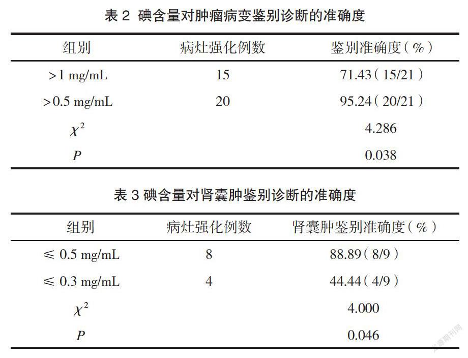
摘 要:目的 探討双源CT增强扫描及碘含量对肾脏占位评估及鉴别的应用价值。方法 回顾性分析枣庄市肿瘤医院2019年2月~2021年6月收治的57例肾脏占位患者的CT影像资料, 对照组29例采取常规双源CT平扫,并以3D重建获取图像,研究组28例采取双源CT增强扫描,注射碘帕醇后进行扫描,并以虚拟VNC分析所得碘图像,由2名经验技术相当的医师分析比较两组平均CT值、碘含量,并对图像质量进行评分。同时以病理诊断为金标准,分析碘含量对肾脏占位病变的鉴别准确度。结果 研究组肿瘤病变的平均CT值、碘含量及图像质量评分均高于对照组(P<0.05);两组肾囊肿的平均CT值差异无统计学意义(P>0.05),研究组肾囊肿的碘含量、图像质量评分均高于对照组(P<0.05);肿瘤病变碘含量>0.5 mg/mL的鉴别准确度显著高于碘含量>1 mg/mL(P<0.05),肾囊肿碘含量≤0.5 mg/mL的鉴别准确度显著高于碘含量≤0.3 mg/mL(P<0.05)。结论 双源CT增强扫描对临床肾脏占位诊断有显著效果,不同碘含量可区分肾脏占位不同病变,0.5 mg/mL碘含量可以作为临床肿瘤病变及肾囊肿的鉴别依据,但需后续临床扩充样本量进一步研究。
关键词:双源CT;碘剂量;肾脏占位;诊断
中图分类号:R575文献标识码:A文章编号:1009-8011(2021)-14-000-03
Evaluation and differentiation of Renal mass by Dual Source CT Enhanced Scanning and Iodine Content
WANG Shen-feng
(Department of Imaging, Zaozhuang Tumor Hospital, Shandong Zaozhuang, 277500, China)
Abstract: Objective To explore the value of dual source CT enhanced scanning and iodine content in the evaluation and differentiation of renal space occupying lesions.Methods The CT image data of 57 patients with renal space occupying lesions in Zaozhuang tumor hospital from February 2019 to June 2021 were analyzed retrospectively. They were digital randomly divided into two groups.The control group(29 cases) was scanned with conventional dual source CT and obtained with 3D reconstruction, and the study group(28 cases) was scanned with dual source CT enhancement, and scanned after injection of iodopanol, and analyzed the iodine images with virtual VNC. Two doctors with equivalent experience and technology analyzed the average CT value and iodine content of the two groups, and scored the image quality. At the same time, taking pathological diagnosis as the gold standard, the differential accuracy of iodine content in renal space occupying lesions was analyzed.Results The average CT value, iodine content and image quality score of tumor lesions in the study group were higher than those in the control group(P<0.05); There was no significant difference in the average CT value of renal cyst between the two group (P>0.05). The iodine content and image quality score of renal cyst in the study group were higher than those in the control group (P<0.05); The discrimination accuracy of tumor lesions with iodine content >0.5 mg/mL was significantly higher than that with iodine content >1 mg/mL(P<0.05). The discrimination accuracy of renal cyst with iodine content ≤0.5 mg/mL was significantly higher than that with iodine content ≤0.3 mg/mL (P<0.05).Conclusion Dual source CT enhanced scanning has a significant effect on the clinical diagnosis of renal space occupying lesions Different iodine content can distinguish different renal space occupying lesions. 0.5 mg/mL iodine content can be used as the basis for the differentiation of clinical tumor lesions and renal cysts, but it needs to be further studied by expanding the sample size in follow-up clinical practice.
Keywords: Dual source CT; Iodine; Renal space occupying; Diagnosis
肾脏占位鉴别诊断对患者后续选择不同治疗方式有重要的指导作用,临床早期诊断一般建议通过超声、CT扫描等手段进行鉴别[1]。但由于病变类型不同、病变强化程度不同,常规的CT平扫的CT值评估准确性有待提高,影响CT值测量因素较多,会出现一定比例的误诊[2]。双源CT增强扫描的碘含量分析对各病变病灶的诊断提出了更高可行性。碘含量分析通过直接获取病灶器官的碘含量,更真实地得到病变强化情况,进而鉴别病变类型[3-4]。本研究对57例肾脏占位患者进行双源CT增强扫描,对比其与常规CT平扫对肾脏占位的鉴别差异,并通过对肾脏占位不同病变类型的碘含量分析,根据Chandarana H等[5]研究的0.5 mg/mL阈值进一步分析,研究碘含量对肾脏占位不同病变类型的鉴别准确度。