共聚焦激光显微内镜与普通结肠镜对溃疡性结肠炎患者炎症活动度及黏膜屏障功能改变的判定价值比较
2021-07-01王贵龙王爱瑶
王贵龙 王爱瑶
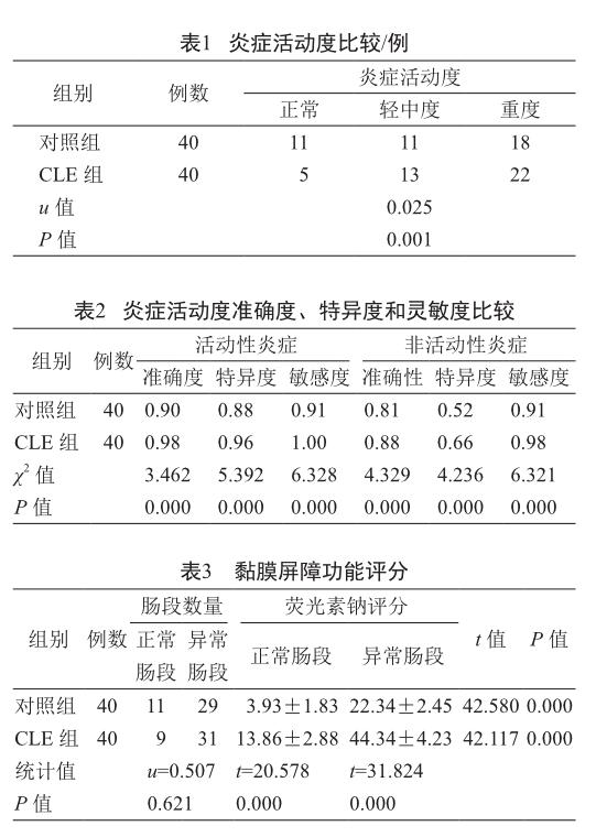
摘 要 目的:比較共聚焦激光显微内镜(CLE)与普通结肠镜对溃疡性结肠炎(UC)患者炎症活动度及黏膜屏障功能改变的判定效果。方法:采用CLE的白光模式和显微模式分别对CLE组(n=40)和对照组(n=40)UC患者进行实时观察,记录两组患者的炎症活动度,评价2种方法的准确度、灵敏度和特异度;在进行荧光素渗透试验后,记录两组患者的正常肠段和异常肠段数量,并完成荧光素渗透评分。结果:两组患者正常、轻中度、重度检测结果差异具有统计学意义(P<0.05),且CLE组炎症活动度的灵敏度、特异度、准确度度均显著高于对照组(P<0.05);CLE组和对照组的功能正常肠段和异常肠段数量差异无统计学意义(P>0.05),CEL组荧光素评分则差异有统计学意义(P<0.05),2项评分均显著高于对照组。结论:采用CLE技术诊断UC患者的炎性活动度具有较高的灵敏度、准确度和特异度,同时该技术也能特异性评估患者结肠黏膜屏障功能改变情况,对于指导临床诊断具有较高的应用价值。
关键词 溃疡性结肠炎 共聚焦激光显微内镜 炎症活动度 黏膜屏障功能
中图分类号:R574.62 文献标志码:B 文章编号:1006-1533(2021)07-0060-04
The comparison of confocal laser microscopy and common colonoscopy to determine changes in inflammatory activity and mucosal barrier function in ulcerative colitis patients
WANG Guilong1*, WANG Aiyao2
(1. Department of Gastroenterology, Peoples Hospital of Hengfeng County, Jiangxi Hengfeng 334300, China; 2. Department of Gastroenterology, Jiangxi Provincial Peoples Hospital, Nangchang 330000, China)
ABSTRACT Objective: To compare the evaluating value of confocal laser microscopy (CLE) with common colonoscopy on the changes of inflammatory activity and mucosal barrier function in patients with ulcerative colitis (UC). Methods: The white light mode and microscopic mode of CLE were used to observe UC patients in CLE group (n=40) and control group (n=40) in real time, and the inflammatory activity of the two groups was recorded. The accuracy, sensitivity and specificity of the two methods were evaluated. After the luciferin permeation test, the number of normal and abnormal intestinal segments in the two groups of patients was recorded, and the luciferin permeation score was completed. Results: There were significant differences in normal, mild, moderate and severe test results between the two groups (P<0.05), and the sensitivity, specificity and sensitivity of inflammatory activity in CLE group were significantly higher than those in the control group (P<0.05). There was no significant difference in the number of normal and abnormal intestinal segments between CLE group and control group (P>0.05), while there was a significant difference in luciferin score (P<0.05). Both scores in CEL group were significantly higher than those in control group. Conclusion: CLE technique has high sensitivity, accuracy and sensitivity in the diagnosis of inflammatory activity in UC patients, and it can also specifically evaluate the changes of colonic mucosal barrier in patients, which has a high application value in guiding clinical diagnosis.
KEy WORDS ulcerative colitis; confocal laser microendoscopy; inflammatory activity; mucosal barrier function
结肠炎(colitis)是由细菌、真菌、病毒等多种病原体引起的结肠炎性病变,因病因不同可分为特异性和非特异性炎性病变,前者包括感染性、缺血性和伪膜性结肠炎,后者主要包括溃疡性结肠炎(ulcerative colitis, UC)和結肠Crohn病[1]。UC作为结肠炎中的重要发病类型之一,也是我国消化系统中的常见病症,具有较高的发病率和复发率[2]。UC的早期诊断对于准确评估炎症的活动度具有重要意义。目前,UC的诊断方式主要依赖于临床表现、普通内镜、病理学、影像学以及实验室检查结果的综合评估,其中组织病理学检查是公认的“金标准”[3-4]。但黏膜活检耗时长、耗材多,而使用普通肠镜,内镜医师缺乏对病情分期、病变范围以及严重程度的判定依据,因此该类方法仍存在一定局限性。随着内镜技术的发展,临床疾病的诊疗手段也呈现日新月异的变化。……