下颌磨牙近中根管解剖形态分析
2021-05-10李少茜荣丽
李少茜 荣丽

【摘要】 根管治疗是目前临床上治疗牙髓根尖周疾病的首选方法,而对根管系统解剖形态的充分掌握是根管治疗成功的基础。下颌磨牙是临床上牙髓病的多发牙位,但其根管解剖尤其是近中根管形态复杂多样,给临床根管治疗带来很大困难,增加了根管治疗的失败率。本文对下颌第一恒磨牙及下颌第二恒磨牙近中根管解剖形态的研究做一综述,为临床根管治疗提供依据。
【关键词】 下颌恒磨牙 近中根管 解剖形态 弯曲度
[Abstract] The root canal therapy is the first choice in the clinical treatment of endodontic and apical disease at present, and the thorough understanding of the anatomical morphology of the root canal system is the basis of successful root canal therapy. Mandibular molar is one of the most common dental sites in clinical pulp disease, but its root canal anatomy is complex and varied, especially in the mesial root canal, make great difficulty to clinical root canal therapy, increase the failure rate of root canal therapy. In this paper, the anatomical configuration of the mandibular first permanent molars and mandibular second permanent molars mesial root canal were reviewed, to provide evidence for clinical root canal therapy.
[Key words] Mandibular permanent molars Mesial root canal Anatomical morphology Curvature
First-authors address: Affiliated Hospital of Binzhou Medical University, Binzhou 256603, China
doi:10.3969/j.issn.1674-4985.2021.08.042
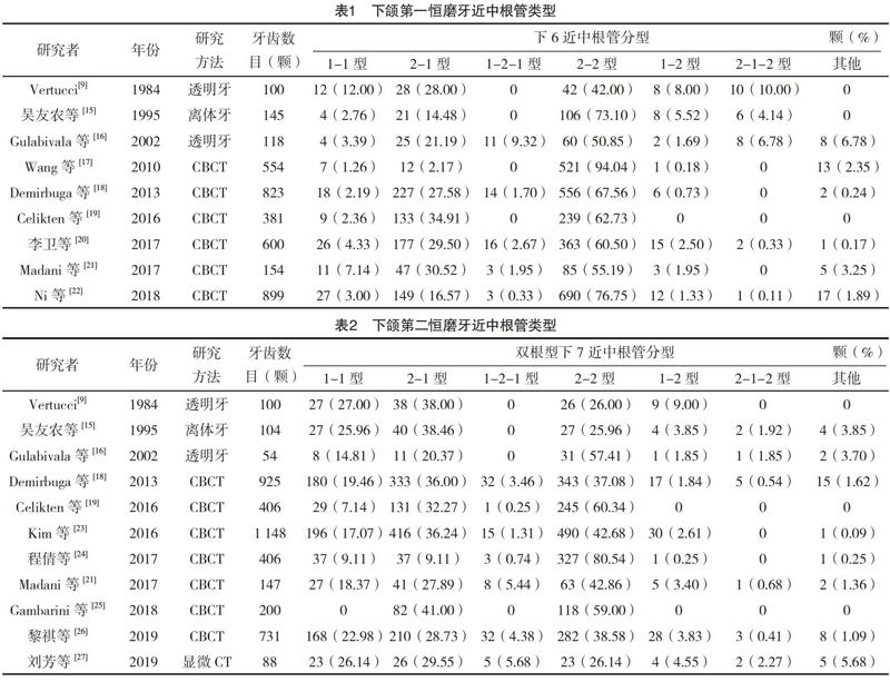
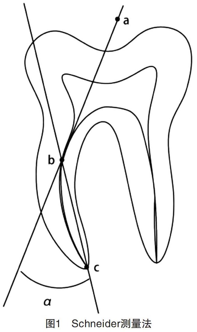
下頜恒磨牙是牙髓病的好发牙位,但其牙根及根管解剖结构复杂,变异较多,增加了临床治疗的难度。文献[1-2]研究发现,下颌第一恒磨牙牙根为两根(近中根、远中根)的发生率最高,其次为三根,分别为近中根、远中根及远中舌根。其根管形态变异以近中根管为甚,形状及数量都有较大差异。1981年美国人首次发现下颌第一恒磨牙近中第三根管[3],近年来有下颌第一恒磨牙7根管(近中3个,远中4个)的发现[4-5]。复杂变异的根管系统在临床根管治疗时易导致根管侧穿、器械折断及台阶等并发症,或因遗漏根管而引起根尖周炎,从而导致根管治疗的失败。髓腔形态是临床进行牙体牙髓疾病治疗的重要依据,充分掌握其形态特点包括根管的数目及类型、根管口的位置、根管弯曲程度及方向等可帮助更彻底地清理根管,减少并发症的发生,对提高根管治疗成功率尤为重要。
多年来国内外学者对下颌磨牙解剖结构的研究也越来越深入和全面。……