一种基于改进U形网络的眼底图像视网膜新生血管检测方法
2021-05-06邹北骥易博松刘晴
邹北骥 易博松 刘晴
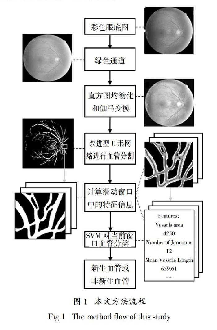
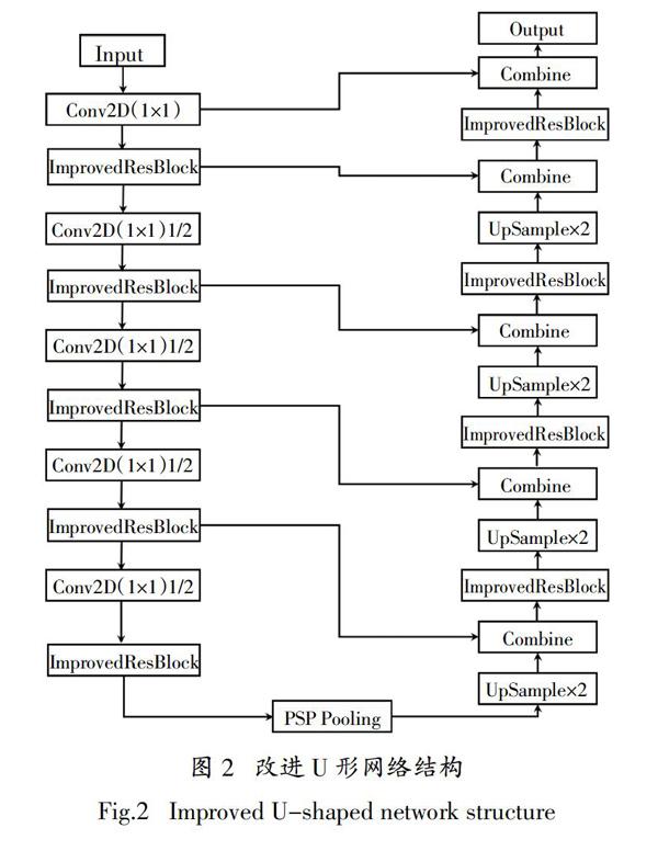
摘 要:糖尿病性视网膜病变(简称糖网病)是主要的致盲眼疾病之一,视网膜新生血管的出现是糖网病恶化的重要标志. 为了更准确地检测出视网膜新生血管,本文提出了一种基于彩色眼底图的视网膜新生血管检测方法. 首先通过一种改进的U形卷积神经网络对血管进行分割;然后利用滑动窗口提取特定区域内血管的形态特征,通过支持向量机将窗口内的血管分为普通血管和新生血管. 使用来自MESSIDOR数据集和Kaggle数据集的含有视网膜新生血管的彩色眼底图对实验进行训练和测试,结果表明该方法对视网膜新生血管检测的准确率为95.96%;该方法在糖网病计算机辅助诊断方面有潜在的应用前景.
关键词:视网膜新生血管检测;血管分割;U形网络;深度学习
中图分类号:TP391.41 文献标志码:A
A Method of Retinal Neovascularization Detection on
Retinal Image Based on Improved U-net
ZOU Beiji2,3,YI Bosong1,3,LIU Qing2,3
(1. School of Automation,Central South University,Changsha 410083,China;
2. School of Computing,Central South University,Changsha 410083,China;
3. Hunan Machine Vision and Intelligent Medical Research Center,Changsha 410083,China)
Abstract:Diabetic retinopathy (DR) is one of the major causes of blindness, and the appearance of retinal neovascularization (RN) is an important sign of DR deterioration. In order to detect RN more accurately, a method based on color fundus photograph for retinal neovascularization detection is proposed. First, an improved U-shaped convolutional neural network is used to segment the blood vessels. Then, a sliding window is used to extract the morphological characteristics of blood vessels in the specific area. A support vector machine (SVM) is used to classify the blood vessels into normal vessels and retinal neovascularization in the window. The experiments use color fundus photographs with retinal neovascularization from the MESSIDOR dataset and the Kaggle dataset for training and testing. The result shows that the accuracy of this method for the RN detection is 95.96%; This method has potential application prospects in the computer-aided diagnosis of diabetic retinopathy.
Key words:retinal neovascularization detection;segmentation of blood vessels;U-shaped neural network;deep learning
糖尿病性视网膜病变是糖尿病的微血管主要并发症之一,也是主要的致盲疾病之一[1].据估计,到2030年全世界将有约3.6 亿人罹患糖尿病. 我国目前的糖尿病患病率为 9.7%,约有 9 400 万人罹患糖尿病[1]. 临床上视网膜新生血管的出现是非增殖期糖尿病性视网膜病变恶化至增殖期糖尿病性视网膜病变的主要标志,也是医生是否需要对患者立刻进行积极治疗的关键判断依据[1-2].
目前已有的对于视网膜新生血管的检测方法主要是通过传统图像处理方法对彩色眼底图中的新生血管进行分割,去除背景和大部分图像噪声,只提取血管图像,再使……