内侧开放性胫骨高位截骨治疗内翻膝关节炎的临床疗效
2020-04-17董翔宇黄钡冬姚运峰荆珏华
董翔宇 黄钡冬 姚运峰 荆珏华
【摘要】 目的 探討内侧开放性胫骨高位截骨(HTO)治疗内翻膝关节炎的临床疗效。方法 34例内翻膝关节炎患者, 均采用内侧开放性胫骨高位截骨治疗, 并对其进行随访, 观察分析患者的手术效果及术后并发症发生情况, 同时比较患者术前及术后末次随访时的膝关节功能美国特种外科医院(HSS)评分。结果 34例患者均获得随访, 随访时间6~12个月, 平均随访8.9个月。术后末次随访时, 患者中优20例, 良13例, 可1例, 差0例。患者术后末次随访时的膝关节功能HSS评分(89.6±3.9)分高于术前的(63.2±6.4)分, 差异具有统计学意义(P<0.05)。术后所有患者的膝关节内侧疼痛症状均得到有效改善, 未出现血管神经损伤、内固定断裂及感染等并发症;其中 2例外侧铰链皮质骨折患者延迟下地, 定期复查后均愈合良好;2例患者因合并皮肤疾病及糖尿病出现伤口愈合不良, 经多次换药及伤口护理, 于术后1个月左右伤口愈合并给予拆线, 术后复查X线提示患者矫形效果满意、下肢力线良好。结论 内翻膝关节炎患者采用内侧开放性胫骨高位截骨治疗效果显著, 可有效改善患者膝关节疼痛症状, 且术后并发症少。
【关键词】 内翻膝关节炎;内侧开放性胫骨高位截骨;膝关节功能
【Abstract】 Objective To discuss the clinical efficacy of medial opening high tibial osteotomy (HTO) in treatment of knee osteoarthritis associated with varus tibial deformity. Methods A total of 34 patients with knee osteoarthritis associated with varus tibial deformity all treated by medial opening high tibial osteotomy, and they were followed up. The surgical effect and postoperative complications were observed and analyzed. Meanwhile, the hospital for special surgery (HSS) score of knee function before operation and at the last follow-up after operation was compared. Results 34 patients were followed up for 6-12 months, with an average of 8.9 months. At the last follow-up, there was 20 excellent cases, 13 good cases, 1 fair case and 0 poor case. The HSS score of knee function (89.6±3.9) points at the last follow-up after operation was higher than (63.2±6.4) points before operation, and the difference was statistically significant (P<0.05). The pain symptoms of the medial knee joint in all patients were effectively improved, and there were no complications such as vascular nerve injury, internal fixation fracture and infection. 2 patients with side hinge cortical fracture were delayed to go to the ground, and healed well after regular reexamination; 2 patients suffered from poor wound healing due to skin diseases and diabetes mellitus, and after multiple dressing changes and wound care, the wound healed and the suture was removed about 1 month after the operation. The reexamination of X-ray after the operation indicated that the patients had satisfactory orthopedic effect and good lower limb strength line. Conclusion Medial opening high tibial osteotomy shows remarkable therapeutic effect for patient with knee osteoarthritis associated with varus tibial deformity, and it can effectively improve the pain symptom of knee joint with less postoperative complications.
【Key words】 Knee osteoarthritis associated with varus tibial deformity; Medial opening high tibial osteotomy; Knee function
胫骨高位截骨对于内侧间室膝关节骨关节炎和内翻畸形的患者是非常有效的治疗手段。近年来开放楔形胫骨高位截骨被更多医生所接受并逐渐取代传统外侧闭合楔形截骨[1]。因为这样避免了腓骨截骨和损伤腓总神经的风险[2]。内侧截骨手术剥离更少、可以更方便、准确的矫正畸形[3, 4]。而且对于以后需行膝关节置换的患者既能方便手术入路更好的在于HTO已经矫正了胫骨近端的畸形从而使关节置换手术更简单方便[5]。本研究分析对34例内翻膝关节炎患者行HTO手术治疗的效果, 报告如下。
1 资料与方法
1. 1 一般资料 选取2017年5月~2019年1月在本院接受治疗的34例内翻膝关节炎患者作为研究对象, 其中男11例, 女23例; 年龄40~65岁, 平均年龄(52±8.9)岁;左侧内翻膝关节炎20例, 右侧内翻膝关节炎14例。纳入标准[6, 7]:对运动有适当要求的患者, 膝关节内翻畸形伴内侧间室变窄的症状性关节炎, 无屈曲挛缩畸形, 膝关节屈曲角度至少90°, 无膝关节不稳, 保守治疗无效。排除标准:股骨髁和胫骨平台严重骨缺损, 类风湿性关节炎, 感染性关节炎, 膝关节外侧间室明显关节炎, 膝关节屈伸活动明显受限[8]。
1. 2 方法 ①术前计划:负重位双下肢全长片, 其可明确下肢的力线和需要矫形的度数, 膝关节磁共振检查初步评估患者半月板交叉韧带病变、是否有骨内病变、骨与软骨缺损、骨坏死以及软骨下水肿等情况。②手术过程:患者仰卧位置于可透视手术床, 常规使用止血带, 在胫骨结节和胫骨后内侧缘之间作5 cm左右纵行切口, 将鹅足肌腱附着点从胫骨处剥离并暴露内侧副韧带浅层, 切断内侧副韧带浅层远端, 钝头骨拨至于胫骨及内侧副韧带后方保护后方的血管神经, 找到髌韧带内侧缘后从胫骨结节到胫骨后内缘进行骨膜下分离, 内侧关节间隙下3.5~4 cm斜向外上朝向腓骨头尖的方向置入两枚定位克氏针, 透视下见位置良好后使用摆锯于克氏针下进行截骨, 确保截骨线从胫骨结节和胫骨后内缘到距胫骨外侧皮质内侧1 cm處, 并且在矢状面平行于胫骨斜坡, 使用外翻力量将截骨处打开, 如果认为截骨处张开不理想, 可使用2~3个骨凿置于张开间歇处以避免关节内骨折的风险, 最后放置矫正好的楔形金属垫块使截骨处矫形达到理想位置, 在透视下确保股骨头中心和踝关节中心的连线位于胫骨平台的外侧, 大约62%的位置[9, 10], 透视见矫形效果满意后植入锁定Tomofix钢板, 对于撑开间隙>10 cm的病例使用患者自体髂骨进行撑开间隙填充。逐层关闭切口。③术后康复:患者术后即刻开始股四头肌等长收缩锻炼和被动膝关节活动, 开始的3周患者只能进行脚趾触地的部分负重, 然后进行渐进的负重, 术后6周可进行完全负重。
1. 3 观察指标 患者均进行了随访, 统计比较患者术前及术后末次随访时的膝关节功能HSS评分, 总分100分, 包括疼痛30分、功能32分、活动度18分、肌力10分、稳定性10分, 分值越高表明患者膝关节功能越好。采用美国膝关节功能HSS评分判定术后末次随访时疗效, 疗效判定标准:优:85~100分;良:70~84分;可:60~70分;差:<60分。同时统计分析患者的术后并发症发生情况。
1. 4 统计学方法 采用SPSS19.0统计学软件处理数据。计量资料以均数±标准差( x-±s)表示, 采用t检验;计数资料以率(%)表示, 采用χ2检验。P<0.05表示差异有统计学意义。
2 结果
2. 1 手术效果 34例患者均获得随访, 随访时间6~12个月, 平均随访8.9个月。术后末次随访时, 患者中优20例, 良13例, 可1例, 差0例。患者术后末次随访时的膝关节功能HSS评分(89.6±3.9)分高于术前的(63.2±6.4)分, 差异具有统计学意义(P<0.05)。
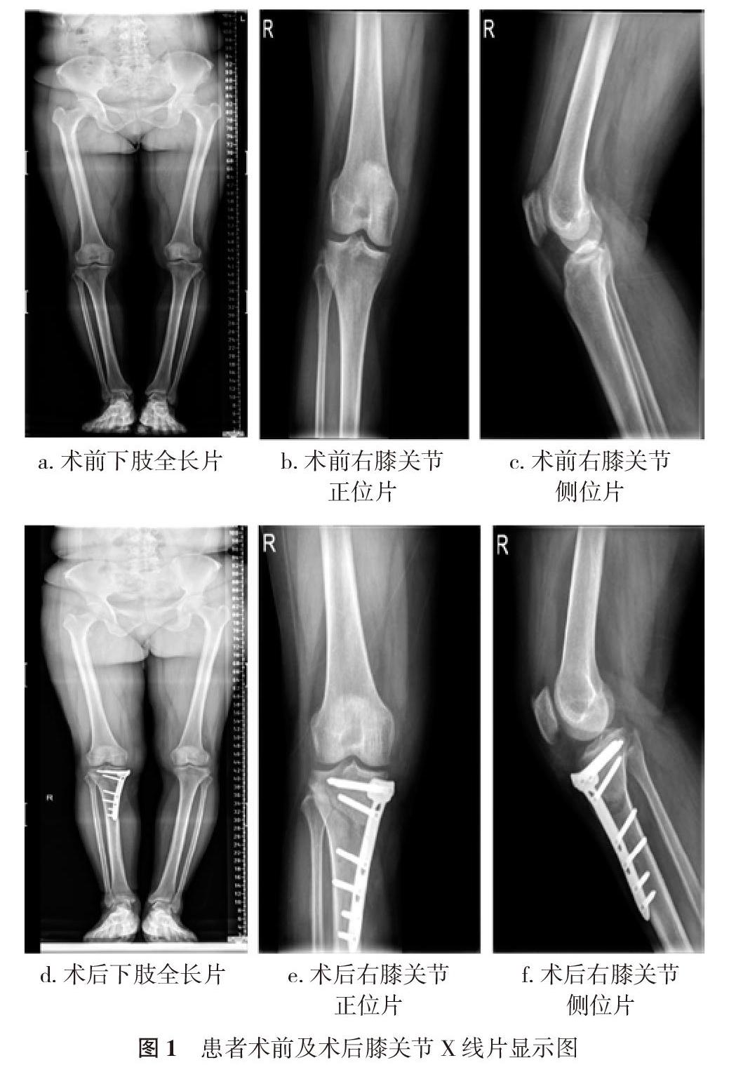
2. 2 术后并发症 术后所有患者的膝关节内侧疼痛症状均得到有效改善, 未出现血管神经损伤、内固定断裂及感染等并发症;其中 2例外侧铰链皮质骨折患者延迟下地, 定期复查后均愈合良好;2例患者因合并皮肤疾病及糖尿病出现伤口愈合不良, 经多次换药及伤口护理, 于术后1个月左右伤口愈合并给予拆线, 术后复查X线提示患者矫形效果满意、下肢力线良好。见图1。
3 讨论
内侧开放楔形截骨的目的是在内侧使用锁定钢板的基础上以外侧皮质作为生物力学的稳定结构。这种手术可以允许早期负重, 膝关节屈伸活动和为骨的愈合提供充分的时间[11, 12]。
HTO过程中进行轻度过度矫正能得到满意的结果已经成为业内共识。然而, 对于理想的外翻角度仍存在争议, Dugdale 建议3~5°的机械轴外翻角是合适的, 也有很多作者持有不同意见。Conventry等[13]报道了术后不同外翻角的10年有效率, 5°外翻角的10年有效率为63%, 6~7°外翻角的10年有效率为87%, >8°外翻角的10年有效率为94%。本文使用的是Dugdale推荐的方法:负重位力线(股骨头中心到踝关节中心的连线)通过胫骨平台自内向外的62%处, 即胫骨髁间棘外侧稍外处, 矫形不够会引起再次内翻, 过度矫正则会导致形态和功能较差。
HTO术内侧一般应用钢板进行稳定支撑, 钢板的种类也有很多。然而, Tomofix钢板被认为是金标准, 被认为有促进骨快速愈合的特性[14]。很多影像学结果表明Tomofix钢板对于大的矫形度数和小的矫形度数均有良好表现[15, 16]。
在临床工作中, 术前计划很重要, 尤其是矫正角度。如果内侧截骨后撑开间隙>10 mm, 作者使用患者自体髂骨做为填充植骨材料进行支撑, 术后患者部分负重6周以减少骨折风险。为了达到理想的角度稳定内固定作者推荐使用至少遠端4枚锁定钉近端4枚锁定钉的锁定钢板。同时也要注意关节倾斜角和畸形的过度矫正。医源性的胫骨近端角度>93°被认为是病理性的。一旦发生这种情况采用股骨远端和胫骨近端的双重截骨来实现术后的关节线在机械轴的范围内, 本次研究的病例均不在此范围。
据报道[17], 外侧皮质铰链的骨折发生率高达30%。这种截骨受很多手术操作的影响, 首先是内侧截骨的高度和长度以及外侧铰链位置的高度。截骨的止点在腓骨头水平相对是安全的, 因为这样截骨后胫骨前后的皮质基本相同也避免的骨折发生的可能性[18, 19]。并且有生物力学研究表明外侧皮质铰链上的定位克氏针孔不会减少应力及骨折的
风险[20, 21]。其他手术并发症包括感染、延迟愈合及不愈合、内固定失效等。随访病例有限是这次分析研究的不足之处。
综上所述, 在仔细的术前计划和细心的术中操作前提下, 内侧开放性胫骨高位截骨治疗内翻膝关节炎是标准的、安全的和可靠的, 能显著改善患者膝关节疼痛症状, 且术后并发症少。
参考文献
[1] Poignard A, Flouzat Lachaniette CH, Amzallag J, et al. Revisiting high tibial osteotomy: Fifty years of experience with the opening-wedge technique. J Bone Joint Surg Am, 2010(2):187-195.
[2] Gaasbeek RD, Nicolaas L, Rijnberg WJ, et al. Correction accuracy and collateral laxity in open versus closed wedge high tibial osteotomy. A one-year randomised controlled study. International orthopaedics, 2010, 34(2):201-207.
[3] Hankemeier S, Mommsen P, Krettek C, et al. Accuracy of high tibial osteotomy: Comparison between open- and closed-wedge technique. Knee surgery, sports traumatology, arthroscopy, 2010, 18(10):1328-1333.
[4] Bito H, Takeuchi R, Kumagai K, et al. A predictive factor for acquiring an ideal lower limb realignment after opening-wedge high tibial osteotomy. Knee surgery, sports traumatology, arthroscopy, 2009, 17(4):382-389.
[5] Hui C, Salmon LJ, Kok A, et al. Long-term survival of high tibial osteotomy for medial compartment osteoarthritis of the knee. The American journal of sports medicine, 2011, 39(1):64-70.
[6] Sabzevari S, Ebrahimpour A, Roudi MK, et al. High tibial osteotomy: A systematic review and current concept. The archives of bone and joint surgery, 2016, 4(3):204-212.
[7] King A, Wall O. Osteotomies around the knee. 2014, 28(6):388-395.
[8] Erquicia J, Gelber PE, Perelli S, et al. Biplane opening wedge high tibial osteotomy with a distal tuberosity osteotomy, radiological and clinical analysis with minimum follow-up of 2 years. Journal of experimental orthopaedics, 2019, 6(1):10.
[9] Huang Meng-Quan, Li Yu-Biao, Liao Chun-Lai, et al. Open-wedge high tibial osteotomy and unicomartmental knee arthroplasty in treating medial compartment osteoarthritis of the knee: a Meta analysis. Zhongguo Gu Shang, 2019, 32(5):428-433.
[10] Kuriyama S, Morimoto N, Shimoto T, et al. Clinical efficacy of preoperative 3D planning for reducing surgical errors during open-wedge high tibial osteotomy. Journal of orthopaedic research, 2019, 37(4):898-907.
[11] Imhoff FB, Imhoff AB. Editorial commentary: Lateral hinge fracture in high tibial osteotomy: Risk or annex?. Arthroscopy : the journal of arthroscopic & related surgery, 2018, 34(11):3080-3081.
[12] Thami B, Abderrazak H, Mohamed Lr, et al. High tibial osteotomy for medial osteoarthritis of the knee: 15 years follow-up. International Orthopaedics, 2010, 34 (2):209-215.
[13] Coventry MB, Ilstrup DM, Wallrichs SL. Proximal tibial osteotomy. A critical long-term study of eighty-seven cases. The Journal of bone and joint surgery American volume, 1993, 75(2):196-201.
[14] Diffo Kaze A, Maas S, Waldmann D, et al. Biomechanical properties of five different currently used implants for open-wedge high tibial osteotomy. Journal of experimental orthopaedics, 2015, 2(1):14.
[15] Staubli AE, De Simoni C, Babst R, et al. Tomofix: A new lcp-concept for open wedge osteotomy of the medial proximal tibia-early results in 92 cases. Injury, 2003(34):B55-62.
[16] Staubli AE, Jacob HA. Evolution of open-wedge high-tibial osteotomy: Experience with a special angular stable device for internal fixation without interposition material. International orthopaedics, 2010, 34(2):167-172.
[17] van Raaij TM, Brouwer RW, de Vlieger R, et al. Opposite cortical fracture in high tibial osteotomy: Lateral closing compared to the medial opening-wedge technique. Acta orthopaedica, 2008, 79(4):508-514.
[18] Ogawa H, Matsumoto K, Akiyama H. The prevention of a lateral hinge fracture as a complication of a medial opening wedge high tibial osteotomy: A case control study. The bone & joint journal, 2017, 99-B(7):887-893.
[19] Nakamura R, Komatsu N, Fujita K, et al. Appropriate hinge position for prevention of unstable lateral hinge fracture in open wedge high tibial osteotomy. The bone & joint journal, 2017, 99-B(10):1313-1318.
[20] Bujnowski K, Getgood A, Leitch K, et al. A pilot hole does not reduce the strains or risk of fracture to the lateral cortex during and following a medial opening wedge high tibial osteotomy in cadaveric specimens. Bone & joint research, 2018, 7(2):166-172.
[21] Cotic M, Vogt S, Feucht MJ, et al. Prospective evaluation of a new plate fixator for valgus-producing medial open-wedge high tibial osteotomy. Knee surgery, sports traumatology, arthroscopy, 2015, 23(12):3707-3716.
[收稿日期:2019-11-19]