多排螺旋CT增强扫描在肾盂癌诊断中的应用价值
2020-04-05苟军武庆勇于祥艳宋旭路漫
苟军 武庆勇 于祥艳 宋旭 路漫
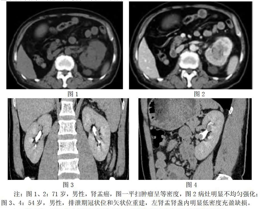
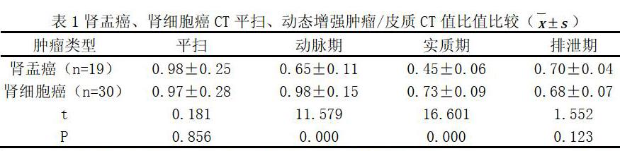
【摘 要】目的:評价多排螺旋CT增强扫描在肾盂癌诊断中的应用。方法:回顾性分析2017年6月至2019年6月在我院49例经病理证实为肾脏恶性肿瘤的CT检查资料,其中肾盂癌19例,肾细胞癌30例。结果:多层螺旋CT增强扫描中,肾盂癌轻度强化,排泄期呈低密度充盈缺损或管壁不规则增厚。平扫肿瘤/皮质CT比值与肾细胞癌比较显示P>0.05,差异不存在统计学意义;增强扫描皮质期、髓质期肿瘤/皮质CT比值均较肾细胞癌低,P<0.05,差异存在统计学意义。 结论:多排螺旋CT增强扫描结合三维后处理技术能发现肾盂癌的多种特征性影像学表现,有利于医师鉴别诊断,提高诊断准确率高,为治疗提供依据。
【关键词】多排螺旋CT;动态增强;排泄期;肾盂癌;充盈缺损;
【中图分类号】R737 【文献标识码】A 【文章编号】1672-3783(2020)02-0076-02
The value of multi-slice spiral CT enhanced scan in the diagnosis of renal pelvic carcinoma
Gou Jun1,Wu Qiongyong2,Yu Xiangyan2,Song Xu2,Lu Man2(Corresponding Author)
(1.Department of Radiology,the TCM Hospital of Mianyang City,621000, Sichuan;2.Department of Radiology, the County-level Hospital of Luxi towm, 621101,Sichuan)
【Abstract】Objective: To evaluate the value of multi-slice spiral CT enhanced scan in the diagnosis of renal pelvic cancer.Methods: A retrospective analysis of preoperative multi-slice spiral CT scans of 49 patients with renal malignant tumors confirmed by surgery and pathology from April 2017 to June 2019 in our hospital, including 19 patients with renal pelvic cancer, kidney 30 patients with cell carcinoma.Results: In 49 patients, the multi-slice spiral CT three-phase enhanced scan of renal pelvis was mildly enhanced, showing obvious filling defects during the excretion period. The tumor/cortical CT ratio and renal cell carcinoma were scanned and enhanced during the excretion period. The comparison showed that P>0.05, the difference was not statistically significant. The tumor/cortical CT ratio of the enhanced arterial phase and parenchymal phase was lower than that of renal cell carcinoma, P<0.05, the difference was statistically significant.Conclusion:Multi-slice spiral CT enhanced scan can detect a variety of characteristic imaging findings of renal pelvic cancer, which is beneficial to physicians for differential diagnosis and high diagnostic accuracy.
【Key words】MDCT;dynamic enhancement;discharge period;renal pelvic cancer;filling defect;
肾盂癌发病数量约为肾脏恶性肿瘤疾病的6%[1],患者常以肉眼血尿为主述就诊。病理研究发现,肾盂癌起源于尿路上皮细胞,随着病情的发展可浸润周围肾实质,向肾实质各个方向生长,故在进行临床诊断时需要与肾细胞癌进行鉴别诊断[2]。彩超、CT和MRI是临床辅助检查的常用设备,随着多排螺旋CT在各级医疗机构的普及,由于扫描时间优势和卓越的后处理能力,越来越受到临床医师的重视。本研究主要评价多排螺旋CT动态增强对肾盂癌的诊断价值,现报告如下。
1 资料与方法
1.1一般资料
收集2017年6月至2019年6月49例我院手术切除肾脏恶性肿瘤患者术前CT和病理检查资料,其中肾盂癌19例、肾细胞癌30例,左肾患病22例、右肾27例,男性患者28例、女性患者21例,年龄范围33~74岁,平均53.96岁。
1.2方法
本组检查均采用GE 64层螺旋CT(型号:GE Optima CT660)。患者仰卧脚先进,全腹平扫+动态增强扫描(扫描范围广,可发现肾脏本身病灶及有无腹腔内脏器转移)。皮质……