颅骨缺损修复术后继发性癫痫的危险因素分析
2019-04-21黄健马美龄
黄健 马美龄
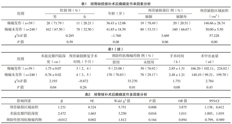
【摘要】 目的:探究颅骨缺损修复术后继发性癫痫的危险因素。方法:回顾性分析2014年10月-2017年12月于笔者所在医院神经外科因创伤行颅骨缺损修复术患者279例的临床资料。采用二元Logistic回归分析对颅骨缺损修复术后继发性癫痫的危险因素进行探讨。结果:279例患者中,颅骨缺损修复术后继发性癫痫39例(13.98%)。颅骨缺损区域面积、术前皮瓣凹陷深度均是颅骨缺损修补术后癫痫发作的独立危险因素[OR=2.079,95%CI(1.138,6.612);OR=1.011,95%CI(1.003,1.019)],差异均有统计学意义(P<0.05)。结论:对于颅骨缺损区域面积大,术前皮瓣凹陷深度深的患者,术后应密切观察并预防癫痫发作。
【关键词】 颅骨缺损修复术 继发性癫痫 危险因素
doi:10.14033/j.cnki.cfmr.2019.29.004 文献标识码 A 文章编号 1674-6805(2019)29-000-03
[Abstract] Objective: To analyze the risk factors of the secondary epilepsy after cranial defect repair. Method: The clinical data of 279 patients underwent cranial defect repair due to trauma in the neurosurgery department of the hospital from October 2014 to December 2017 were retrospectively analyzed. Binary Logistic regression analysis was used to explore the risk factors of the secondary epilepsy after cranial defect repair. Result: Among the 279 patients, 39 cases (13.98%) had secondary epilepsy after cranial defect repair. The area of skull defect area and depth of preoperative skin flap depression were the independent risk factors of the secondary epilepsy after cranial defect repair [OR=2.079, 95%CI (1.138, 6.612); OR=1.011, 95%CI (1.003, 1.019)], and the differences were statistically significant (P<0.05). Conclusion: For patients with larger of the skull defect area and deeper of preoperative skin flap depression should be closely observed and prevent epilepsy seizures after operation.
[Key words] Cranial defect repair Secondary epilepsy Risk factors
First-authors address: Linyi Central Hospital, Linyi 276400, China
重型顱脑损伤(severe craniocerebral injury)常伴有恶性颅内高压,保守治疗往往无效,需进行外科手术减压[1]。去骨瓣减压术是一种有效降低颅内压、抢救患者生命的手术方式,但术后会造成大范围颅骨缺损,导致不同的神经功能缺失,给患者造成一定的心理负担。为进一步改善患者神经功能及外观,可行颅骨缺损修复术。颅骨缺损修复术已较为成熟,但术后易引发癫痫发作、出血、感染、脑积水等并发症,应引起神经外科医生的重视[2-4]。癫痫是颅骨缺损修复术后最为常见的并发症,可影响患者生活质量,甚至危及生命。据相关文献[5-6]报道,颅骨缺损修复术后癫痫发作会极大增加患者心理负担、增加医疗费用等,严重影响患者工作及生活质量。本研究回顾性分析2014年10月-2018年12月于笔者所在医院因创伤行颅骨缺损修复术患者279例的临床资料,以探讨影响术后继发性癫痫的临床相关因素。……