Lesson Eighty-eight How to perform permanentHis bundle pacing in routine clinical practice
2019-04-10童鸿
Clinical permanent His bundle pacing(PHBP)was first described in 2000.Since then,several investigators acrossthe world have published the safety and feasibility of PHBP.Benefits include ability to achieve physiological pacing by recruiting the native His Purkinje system and avoiding electrical dyssynchrony,potentially translating into reduced HF incidence.Also,limited clinical observations have demonstrated that the lead does not cross the tricuspid valve and this may result in lower incidence of valvular regurgitation.
Equipment requirements
Following are the equipment requirements:
1.Medtronic(Minneapolis,MN)383069-cm His lead with 1.8-mm exposed helix,lead outer diameter(OD)4.2F(Figure 1A).It is a solid core pacing lead with no central lumen to deliver a stylet,requiring a long sheath for delivery.
2.Medtronic C315 His nondeflectable sheath(43cm),inner diameter(ID) 5.5F,OD 7.0F(see asterisk in Figure1B),primary curve to reach the superior aspect of the tricuspid annulus,secondary curve to reach the septum.A Medtronic deflectable sheath (C304-69;ID5.7F and OD 8.4F) is also available with unidirectional deflection(Figure1C);it can be helpful in challenging anatomical situations(dilated right atrium[RA] ,an inferiorly displaced His bundle[HB] ,etc.).
3.Short 7F peel-away sheath to place the C315 His sheath through it.It allows continued vascular access after the His sheath is split(guide wire can be retained as well).
4.Pace-sense analyzer(PSA)to record intracardiac electrograms(EGMs).Because of the inherent sensing algorithms built into the Medtronic PSA,we usually connect the pacing lead to the atrial channel(higher gain setting of 0.05 mV/mm).For PSAs of other manufacturers,the lead can be connected to the ventricular channel.
5.Unipolar connection to map the HB EGMs with the pacing lead.We use the pacing lead to map the HB with PSA EGMs,without the need for a mapping catheter.Also,an electrophysiology(EP) recording system can be used(PSA EGMs are adequate).
6.Twelve-lead electrocardiogram for the procedure(critical to analyze pacing morphologies to confirm recruitment of the HB).
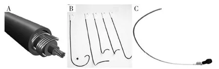
Figure 1 A:Select Secure 3830 lead.B:Several nondeflectable sheaths used for the 3830 lead.The first sheath marked by the asterisk is a C315 His nondeflectable sheath used for His bundle mapping and lead placement.All other sheaths are used for either right atrialor rightventricular lead placement.C:Deflectable sheath used occasionally to place the His bundle lead
Procedure description
Once vascular access is obtained(cephalic,axillary,or subclavian vein),a guidewire is advanced into the RA or RV.A short 7F sheath can be advanced over the wire to retain access.Otherwise,the C315 His sheath is advanced into the RA or RV and the guidewire removed.The pacing lead is advanced to the tip of the sheath,with the distal tip of the lead exposed minimally.Unipolar connections are made with the tip of the lead and cardiac tissue.
If the sheath and the lead tip are in the RV,the apparatus is gently pulled back to the atrioventricular grove with minimal counterclockwise rotation to ensure that the lead tip is abutting the septum.If the sheath and the lead are in the RA,gentle forward clockwise rotation tends to move the apparatus to the summit of the tricuspid annulus.
While this is being performed,it is important for both the operator and the person operating the PSA to pay careful attention to intracardiac EGMs.Small movements are encouraged as HB deflections can be easily missed.We usually set the sweep speed to 50 or 100 mm/s to allow better separation of atrial,HB,and local ventricular EGMs.
Once HB EGMs are obtained,unipolar pacing is performed since the proximal pole of the lead is within the sheath.We start pacing at 5 V@1 ms and assess 12-lead QRS morphologies.The following patterns can be observed:
1.Pure HB pacing where stimulus to ventricular activation is equal to the intrinsic HV interval and paced QRS morphology is identical to the intrinsic QRS complex.We term this as selective HB pacing(Figure 2,Figure 3).
2.HB capture with local ventricular fusion:stimulus-ventricular capture is shorter than the HV interval.The pacing output is decremented to assess the changing QRS morphologies(akin to para-His pacing performed to assess septal accessory pathways).We term this as nonselective HB pacing(Figure 2,Figure 3).Various responses can be observed:
(a)At high output,the HB is preferentially recruited with progressive widening as output is reduced,resulting in more local ventricular capture.
(b)At high output,more fusion is encountered because of local ventricular capture,and at lower output,HB is preferentially activated,resulting in less fusion(Figure 2,Figure 3).
(c)Sometimes just before loss of capture,pure HB pacing can be seen.Also,selective RBB or left bundle branch(LBB)capture can be demonstrated(Figure 3).
(d)Sometimes because of the proximity of RBB and more ventricular placement of the lead,RBB capture can occur.This results in an LBB block pattern-wide QRS complex.It may be difficult to determine whether local myocardial capture occurs along with RBB capture.If intrinsic conduction is present,measuring the HV interval can help determine RBB capture.Typically,a local HV interval would be short(<30 ms),with no far-field atrial EGM seen on the PSA.
We believe that these responses are due to anatomical and functional aspects of the HB system.Anatomy of the HB has been well characterized.Preferential pacing of RBB and LBB can be explained in part by the phenomenon of longitudinal dissociation of the His-Purkinje conduction system.Other properties such as favorable source-sink1ratios and virtual electrode polarization may play a part as well.
Once HB capture is ascertained,the lead is turned 4-5 times in a clockwise direction.Usually,torque is built up and transmitted to the end of the lead.Once it is released,the lead may unwind 1-2 rotations.It is important to use the sheath to support the lead and make good contact with the septum.The sheath is pulled back,and the lead is advanced to allow for slack.Current of injury,either of the HB or local ventricular tissue,is usually seen(Figure 4).However,there are cases where the lead is well affixed with good pacing thresholds and minimal local tissue injury.Testing is performed in both unipolar and bipolar configurations.Starting at 5V@1 ms,output is decremented to assess response to pacing.Either selective or nonselective HB pacing is accepted,and pacing thresholds less than 2V@1 ms are deemed acceptable.If the patient is pacemaker dependent,lower pacing thresholds are sought after before accepting the final lead position.If the pacing threshold margins are not deemed acceptable by the operator after multiple attempts,the lead is placed slightly more anteriorly,with largerventricularsensingand pacingmorphology consistent with septal pacing.
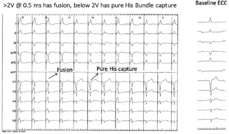
Figure 2 At pacing outputs above 2V@0.5ms,there is fusion with His bundle(HB)and local ventricular capture(nonselective HB capture).Below 2V@0.5ms,there is pure HB capture with paced QRS morphology identical to the native QRS complex(selective HB pacing).Note that once pure His capture is obtained as the pacing threshold,inter-mittent fusion is still high enough to intermittently recruit the local venticular myocardium.ECG=electrocardiogram.

Figure 3 At higher output(up to 1.5V@1 ms),there is fusion between His bundle and local myocardial capture(nonselective response).At lower outputs(up to 0.5 V@1ms),there is pure His bundle capture with stimulus to QRS onset(S-QRS)equal to the native HV interval(selective response,*).Below this output,there is selective right bundle branch(RBB)capture.
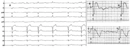
Figure 4 A:Ten-lead electrocardiogram of a 74-year-old man with permanent AF,incomplete left bundle branch block pattern,and bradycardia.B:Pace-sense analyzer electrograms as the His bundle is mapped in a unipolar configuration (connected to the atrial channel).C:Current of injury of the His bundle as the lead is screwed into place.Asterisk denotes current of injury of the His bundle as the lead is screwed into place.
Acute ventricular sensing issues
Because of the location of the lead tip,we see a wide range of ventricular sensing values(1.2-10 mV).We compare ventricular EGMs with atrial EGMs to assess sensing margins.Typically,the lead tip records small atrial EGMs and atrial oversensing is not a major issue.Unipolar and bipolar sensing is assessed,and if no far-field sensing is seen,unipolar sensing can be used if it offers better sensing safety margins.However,it is rare that we program unipolar ventricular sensing.In patients who are pacemaker dependent,we do not use unipolar sensing and ventricular sensitivity is usually set to>1.2 mV on the basis of measured values and care is taken to assess far-field atrial sensing before accepting the final location of the ventricular lead.
Acute pacing issues
The HB is encased in a fibrous sheath with various anatomical variations.Pacing thresholds are expected to be inherently higherbecause ofthe lead-tissue interface.Therefore,if pacing thresholds are lower than 2V@1 ms at implantation with good HB recruitment,we accept the lead position.Typically,as the pacing output decreases,there is more fusion with ventricular capture,thereby providing good safety margins(nonselective HB pacing cases only).The pacing output is set to 5V@1 ms at implantation and decreased at
2-month check.In dependent patients,we look for at least 2-3 times safety margin.We aim for nonselective HB pacing in such cases and determine safety margins from the lowest output capture of either the HB or local ventricular tissue.If selective HB pacing is desired in the setting of higher pacing thresholds,we place a backup lead in the RV.This necessitates placement of a BiV pacemaker with the HB lead plugged into the LV port.We program an LV to RV offset of 80 ms.If HB capture occurs,the RV stimulus falls in the refractory period,resulting in functional noncapture.If HB capture does not occur,the RV stimulus acts as safety pacing.
词 汇
recruit n.&v.新兵、心成员、新手;招募、组织、雇佣
dyssynchrony n.非同时、非同步、非共时性
helix n.螺旋、耳轮
unidirectional adj.单向的
inherent adj.内在的
algorithms adj.算法
grove n.沟、树丛、果树林
abut v.邻接、与 邻接,与 紧靠
intrinsic adj.固有的、内部的、内在的
decrement n.&v.减少、减量、减缩两;减少,减缩
akin adj.同类的、同族的、同源的
torque n.&v.扭转力、金属饰环、转距;加转距于
slack n.&adj.&adv.&v.松弛、萧条;松弛的、懈怠的、松闲
的;松弛地、松闲地;放松、懈怠、减缓
affix n.&v.附加物、词缀;黏上、盖、添上
deem n.&v.评价;认为、认为 是
margin n.&v.边缘、边、利润额、余地;加边于
encase v.把 装入箱子、围住、包装
offset n.&adj.&v.补偿、支管;补偿的、错断的、胶印的;补
偿、把 并列
注 释
1.source-sink(ratio)指“源 -库(比)”,最初用于描述生物物种的迁移现象。心脏电生理学上用source-sink来阐述一些心脏传现象,在此,source-sink指的是电流负荷(current-load),传导系统局部的容量变化可加快或减慢激动传导速度。When the source volume is larger compared with the sink volume,activation proceeds because there is sufficient electrical current to activate myocardial cells in the distal direction.当源容量大于库容量时激动进展,因有足够的电流激动远端心肌细胞。Local source-sink relationships determine the formation of conduction heterogeneities and provide conditions for the development of slow conduction,unidirectional block,and reentry.局部源-库关系决定传导异质性并提供发生慢传导、单向阻滞和折返的条件。
参考译文
第88课 临床实践中如何实施希氏束起搏
2000年首次提出希氏束起搏,自那以后,国际上多位研究者发表了希氏束起搏的安全性和可行性的报道。希氏束起搏的优点在于通过自身的希氏束系统达到生理性起搏,避免电不同步,从而减少心力衰竭的发生率。另外,有限的临床观察证实电极不跨过三尖瓣,三尖瓣反流的发生率较低。
设备要求
1.美敦力(Minneapolis,M)383069-cm 希氏束电极,突出的螺旋1.8-mm,电极外径(OD)4.2F(图1A)是一无实心起搏电极,无内腔供定型钢丝通过,而需长鞘输送。
2.美敦力C315希氏束非可调弯鞘(43cm),内径(ID)5.5F,OD 7.0F(见图1B星标),第一弯到达三尖瓣环上方,第二弯到达间隔。……