肺动脉高压影像学临床研究
2018-03-31张鹏
张鹏
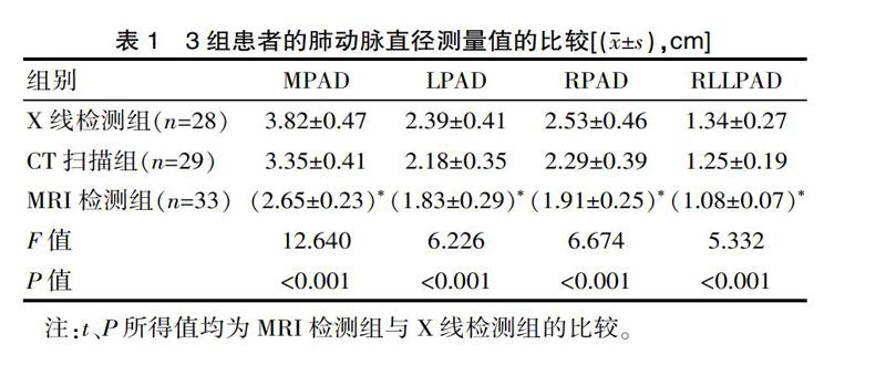
[摘要] 目的 總结肺动脉高压影像学研究进展。方法 随机抽取该组2014年2月—2016年11月90例肺动脉高压患者,其中行X线检查的患者28例,行CT扫描检查的患者29例,行MRI检查的患者33例。分别观察3组患者的主肺动脉直径及主肺动脉与主动脉直径比值,并且观察MRI组患者的顺应性测量值。除此之外,观察3种检测方法的特异性与敏感度。结果 MRI检测结果表明,主肺动脉直径为(2.65±0.23)cm,明显小于CT检查的(3.35±0.41)cm与X线检查的(3.82±0.47)cm,并且CT扫描效果明显优于X线检查效果;同时MRI检测的主肺动脉与主动脉直径比值为(1.18±0.16)cm,明显大于CT检查的(1.31±0.21)cm与X线检查的(1.59±0.33)cm,差异有统计学意义(P<0.05)。除此之外,MRI检测组的特异性与敏感度分别为92.47%与90.35%,明显高于CT扫描组的87.26%、89.37%与X线检测组的51.32%、47.29%,差异有统计学意义(P<0.05)。结论 利用MRI检查法可有效的对肺动脉高压患者进行病情检测,为医生提供更为准确的检测信息,以便采取更为有效的应对策略,具有较高的临床推广价值。
[关键词] 肺动脉高压;X线检测;CT检测;MRI检测
[中图分类号] R5 [文献标识码] A [文章编号] 1674-0742(2018)01(a)-0177-04
Imaging Clinical Study of Pulmonary Hypertension
ZHANG Peng
B Ultrasonic Room, Heze Municiple Hospital, Heze, Shandong Province, 274000 China
[Abstract] Objective This paper tries to summarize the imaging progress of pulmonary arterial hypertension. Methods 90 patients with pulmonary hypertension were randomly selected from February 2014 to November 2016 in the hospital. Among them, 28 patients underwent X-ray examination, 29 patients underwent CT scan, and 33 patients underwent MRI examination. The diameter of the main pulmonary artery and the ratio of the main pulmonary artery to the aortic diameter were observed in the three groups, and the compliance measurements of the patients in the MRI group were observed. In addition, the specificity and sensitivity of the three detection methods were observed. Results MRI results showed that the main pulmonary artery diameter(2.65±0.23) cm, significantly less than the CT examination (3.35±0.41) cm and X-ray examination (3.82±0.47) cm, and CT scan was superior to X-ray effect; while the main pulmonary artery and aortic diameter ratio for MRI detection (1.18±0.16)cm, was significantly higher than that of CT examination (1.31±0.21)cm and X-ray examination (1.59±0.33)cm, with statistical significance(P<0.05). In addition, the specificity and sensitivity of the MRI detection group were 92.47% and 90.35%, which was significantly higher than 87.26% and 89.37% in the CT scan group and 51.32% and 47.29% in the X-ray examination group, respectively (P<0.05). Conclusion Using the MRI test method can effectively carry out disease detection in patients with pulmonary hypertension, for doctors to provide more accurate detection information, so as to adopt more effective coping strategies, which has a high clinical value.
[Key words] Pulmonary hypertension; X ray detection; CT detection; MRI detection
所谓的肺动脉高压主要是指当人们休息时的肺动脉平均压超过3.33 kPa或者在运动时的肺动脉平均压超过4 kPa[1]。临床中肺动脉平均压的计算方法为肺静脉平均压与肺血管阻力之和,并于肺血流量相乘,因此不论是其中哪一指标的数值增大,都将引起肺动脉高压[2]。其主要的临床症状为劳力性呼吸困难,乏力腹胀,心绞痛,晕厥等,因此一旦患者的肺动脉压值升高,将产生较为严重的不良后果。在日常生活中患者可多食用降压或者软化血管的食物。目前临床中随着医疗技术的进步,对肺动脉高压患者的影像学检查逐渐由X线发展为CT扫描,并发展为现今的MRI检测,对于肺动脉高压的检测结果逐渐趋近准确[3]。现将2014年2月—2016年11月90例肺动脉高压患者的影像学临床研究进行如下报道,现报道如下。……