早期食管癌放射诊断的临床价值分析
2018-03-31余鎏袁超
余鎏 袁超
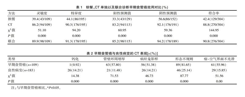
[摘要] 目的 分析早期食管癌放射诊断价值。方法 2013年1月—2017年2月,医院共采用钡餐造影、CT诊断怀疑为早期食管癌的患者304例。对比CT、钡餐的诊断灵敏度、特异度、阳性预测值、阴性预测值、符合率。对比食管癌与良性病变的典型表现,不同类型病变CT值。 结果 109例早期食管癌。CT的灵敏度、特异度、阳性预测值、阴性预测值、符合率分别为86.2%、90.3%、83.2%、92.1%、88.8%高于钡餐,差异有统计学意义(P<0.05);早期食管癌钙化率0.92%低于良性病变14.21%,管壁环周增厚、病灶龛影样、形态不规则、瘤-空气界面不光滑率分别为57.80 %、51.38%、81.65%、55.96% 高于良性病变11.48%、14.21%、25.14%、15.85%,食管癌的CT值(25.61±12.46)Hu高于间质瘤(18.6±11.5)Hu、平滑肌瘤(10.6±8.62)Hu、癌前病变(18.0±8.6)Hu,差异有统计学意义(P<0.05)。结论 以CT为主的放射诊断技术可作为早期食管癌的诊断、分期技术。
[关键词] 食管癌;放射;临床价值
[中图分类号] R735 [文献标识码] A [文章编号] 1674-0742(2018)01(a)-0196-03
Clinical Value of Radiation Diagnosis in Early Esophageal Cancer
YU Liu, YUAN Chao
Department of Radiology, Kaizhou Peoples Hospital, Chongqing, 405400 China
[Abstract] Objective This paper tries to analyze the value of radiotherapy in early diagnosis of esophageal cancer. Methods 304 cases of early esophageal cancer patients from January 2013 to February 2017 in the hospital using barium meal, CT diagnosis were selected. CT, the diagnostic sensitivity, GI specificity, positive predictive value, negative predictive value and accuracy of the two groups were compared. The typical contrast of esophageal cancer and benign lesions, lesions of different types of CT value. Results The sensitivity of the 109 cases of early esophageal cancer. CT specificity, positive predictive value, negative predictive value and accuracy were 86.2%, 90.3%, 83.2%, 92.1%, 88.8%, higher than that of barium meal, the difference was statistically significant(P<0.05); calcification rate was 0.92%, which was lower than 14.21% of early esophageal cancer and benign lesions, thickening of the wall of the ring, the shadow niche lesions, irregular shape, tumor - air interface of not smooth rate were 57.80%, 51.38%, 81.65%, 55.96% higher than that of benign lesions of 11.48%, 14.21%, 25.14%,15.85%, the CT value of esophagus cancer(25.61±12.46)Hu, was higher than that of interstitial tumor of (18.6±11.5)Hu, (10.6±8.62)Hu of leiomyoma, (18.0±8.6) Hu of precancerous lesion, the difference was statistically significant (P<0.05). Conclusion CT based diagnostic techniques can be used as a staging and diagnostic technique for early stage esophageal cancer.
[Key words] Esophageal carcinoma; Radiotherapy; Clinical value
食管癌是一種起源于食管黏膜上皮常见的消化系统恶性肿瘤,居人类恶性肿瘤第8位。我国是食管癌高发地区,这可能与我国的饮食特殊有关,食管癌是第5大恶性肿瘤,发病率高达22~23人/10万,年增长6/万人,5年生存率仅为15%~20%[1]。近年来,因肥胖、胃食管反流疾病发生率上升,烟草的泛滥,其发生率呈逐年上升趋势[2]。准确的诊断早期食管癌,有助于改善患者的预后。影像学是诊断食管癌的主要方法,目前可供选择的方法为X线、钡餐造影、CT,不同技术的诊断效用存在一定的差异。该次研究采用回顾性分析,以2013年1月—2017年2月,医院共采用钡餐造影、CT诊断怀疑为早期食管癌的患者304例作为研究对象,评价钡餐造影、CT、X线诊断早期食管癌的效用,现报道如下。
1 资料与方法
1.1 一般资料
该院共采用钡餐造影、CT诊断怀疑为早期食管癌的患者304例,其中男222例、女82例,年龄31~78岁,平均(56.1±6.2)岁。纳入标准:①最终经内镜活检、手术等方法确诊;②均经过X线、钡餐、CT诊断;③若诊断为食管癌,参照NCCN病理分期标准诊断为早期Ⅰ期;……