Wernicke脑病
2017-11-30韩彤
·临床医学图像·
Wernicke脑病
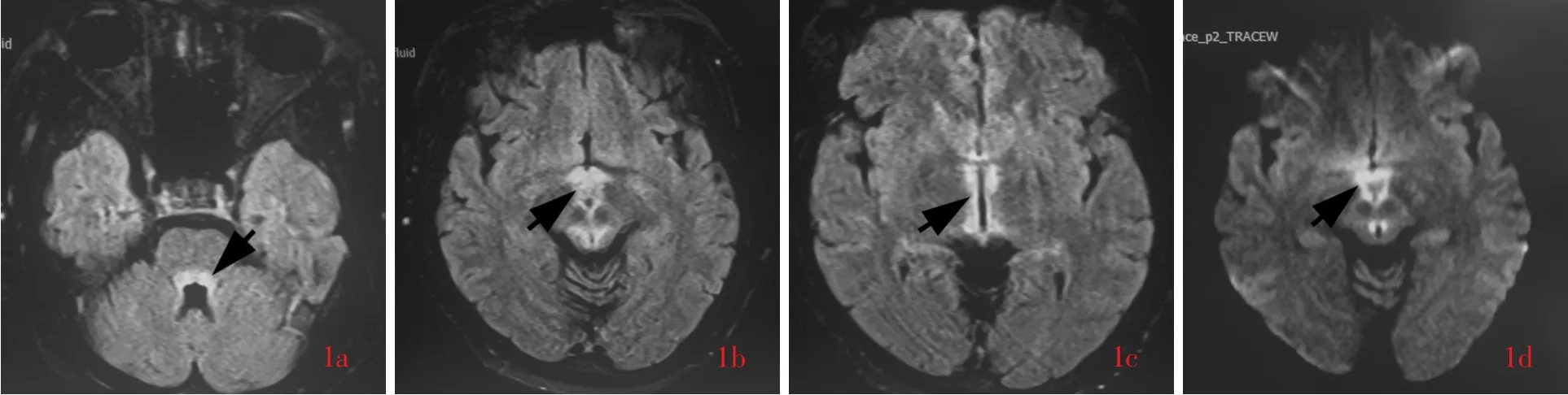
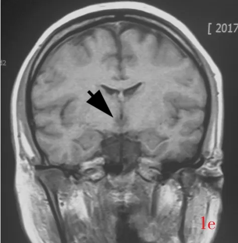
图1 女性患者,27岁,因头晕、行走不稳7 d并进行性加重,嗜睡2 d入院。既往有糖尿病病史,近2个月减肥,严格控制饮食摄入量(150 g/d)。体格检查双眼内收和外展功能不良,左右视和上视可见眼震,双侧跟⁃膝⁃胫试验欠稳准。临床诊断为Wernicke脑病。头部MRI检查所见1a 横断面FLAIR成像显示,第四脑室周围异常高信号影,强度均匀(箭头所示) 1b 横断面FLAIR成像显示,中脑导水管周围、四叠体、双侧乳头体和灰结节异常高信号影(箭头所示)1c 横断面FALIR成像显示,第三脑室周围异常高信号影(箭头所示) 1d 横断面DWI(图1b相同层面)显示,病变呈明显高信号(箭头所示) 1e 通过乳头体的冠状位T1WI显示,第三脑室旁异常低信号影,强度均匀(箭头所示);双侧乳头体未见明显萎缩Figure 1 A 27⁃year⁃old female patient suffered from progressive dizziness and walking unstable for 7 d and drowsiness for 2 d.Past medical history:diabetes.She had been on her special diet(principal food intake 150 g/d)to lose weight for 2 months.Physical examination showed bilateral ocular palsy,nystagmus in the side view(left and right)and front view.She could not complete both heel⁃knee⁃tibia test correctly and stalely.She was clinically diagnosed as having Wernicke's encephalopathy.Axial FLAIR image through pons revealed an abnormal heterogeneous hyperintensity mainly located at the tegmental part of pons around the forth ventricle(arrow indicates,Panel 1a).Axial FLAIR image through midbrain showed multiple heterogeneous hyperintensity lesions distributed symmetrically on the left and right sides located at periaqueductal area,corpus quadrigemina,bilateral mammillary bodies and tuberculum cinereum(arrow indicates,Panel 1b).Axial FLAIR image through thalamus showed abnormal hyperintensity located around the third ventricle(arrow indicates,Panel 1c).Axial DWI located at the same level as 1b revealed abnormal hyperintensity lesions(arrow indicates,Panel 1d).Coronal T1WI through mammillary body showed markedly heterogeneous slightly hypointensity in bilateral wall of the third ventricle(arrow indicates).There was no significant atrophy of bilateral mammillary bodies(Panel 1e).
Wernicke脑病是维生素B1缺乏导致的代谢性脑病,1881年由Wernicke首先描述,1940年Campbell和Russell提出其诱因是维生素B1缺乏。该病系维生素B1缺乏致特定区域神经细胞能量代谢障碍所致,呈急性或亚急性起病,临床主要表现为眼外肌瘫痪、共济失调和意识障碍,最常见于慢性酒精中毒和妊娠性呕吐;非维生素B1缺乏性Wernicke脑病系转酮醇酶(TK)基因缺陷所致。MRI对早期诊断和疾病分期有重要价值,是首选影像学方法。典型特征为特定部位出现特定分布的病变,特定部位包括第四脑室旁(脑区被盖和中脑顶盖,图1a)、中脑导水管周围、乳头体、四叠体(图1b)、第三脑室侧壁(丘脑内侧,图1c),其中乳头体最易受累,也可发生于延髓、小脑齿状核、红核、中脑顶盖、尾状核和大脑皮质等少见部位;……
