CT与MRI在宫颈癌术前分期诊断中的应用价值分析
2017-08-26高忠博苏丽梅梁淇星钱喜学
高忠博++苏丽梅++梁淇星++钱喜学
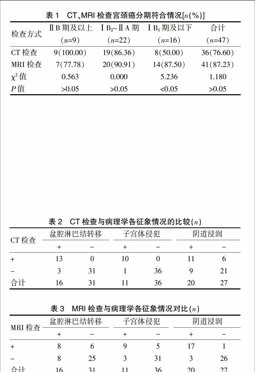
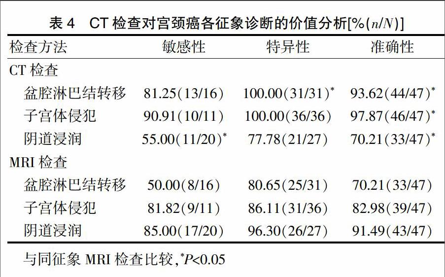
[摘要]目的 探討X线计算机体层(CT)与磁共振成像(MRI)在宫颈癌术前分期诊断中的应用价值。方法 选取2013年3月~2016年12月在我院就诊的47例宫颈癌患者,于术前1周行CT、MRI检查,并以病理诊断结果为金标准,分析CT和MRI在宫颈癌术前分期诊断中的应用价值。结果 经病理确诊,47例宫颈癌患者中ⅠB1期及以下16例,ⅠB2~ⅡA期22例,ⅡB期及以上9例,且盆腔淋巴结转移阳性16例,子宫体侵犯阳性11例,阴道浸润阳性20例;MRI诊断ⅠB1期及以下的符合率明显高于CT诊断,差异有统计学意义(P<0.05);CT诊断盆腔淋巴结转移的特异性、准确性及子宫体侵犯的准确性均显著高于MRI诊断,差异有统计学意义(P<0.05);MRI诊断阴道浸润的敏感性及准确性均高于CT诊断,差异有统计学意义(P<0.05)。结论 MRI诊断ⅠB1期及以下宫颈癌及阴道浸润情况效果显著,而CT诊断盆腔淋巴转移及子宫侵犯情况效果较好。
[关键词]X线计算机体层;磁共振成像;宫颈癌;术前分期诊断;应用价值
[中图分类号] R445.2 [文献标识码] A [文章编号] 1674-4721(2017)07(c)-0062-03
Application value of CT and MRI in the preoperative staging diagnosis of cervical cancer
GAO Zhong-bo SU Li-mei LIANG Qi-xing QIAN Xi-xue
Department of Imaging,People′s Hospital of Huaiji County in Guangdong Province,Huaiji 526400,China
[Abstract]Objective To investigate the application value of computed tomography (CT) and magnetic resonance imaging (MRI) in the preoperative staging diagnosis of cervical cancer.Methods 47 patients with cervical cancer who admitted into our hospital and diagnosed from March 2013 to December 2016 were selected as the research object.All the patients were given CT and MRI examination before operation for one week,and the results of pathological diagnosis were taken as gold standard.The application value of CT and MRI in the preoperative staging diagnosis of cervical cancer was analyzed.Results According to the pathological results,there were 16 cases of ⅠB1 stage and below,22 cases of ⅠB2-ⅡA stage,9 cases of ⅡB stage and above among 47 cases of patients with cervical cancer.At the same time,there were 16 cases of pelvic lymph node metastasis positive,11 cases of uterine invasion positive and 20 cases of vaginal infiltration.The accordance rate of ⅠB1 stage and below diagnosed by MRI was higher than that diagnosed by CT,and the difference had statistical significance (P<0.05).The specificity and accuracy of pelvic lymph node metastasis,the accuracy of uterine invasion diagnosed by CT were higher than those diagnosed by MRI,and the difference had statistical significance (P<0.05).The sensitivity and accuracy of vaginal infiltration diagnosed by MRI were higher than those diagnosed by CT,and the difference had statistical significance (P<0.05).Conclusion MRI in the diagnosis of cervical cancer (ⅠB1 stage and below) and vaginal infiltration has significant diagnostic effects,while the CT in the diagnosis of pelvic lymph node metastasis and uterine invasion has better diagnostic effects.
[Key words]Computed tomography;Magnetic resonance imaging;Cervical cancer;Preoperative staging diagnosis;Application value
宫颈癌是一种常见的妇科恶性腫瘤,其发病率位居女性恶性肿瘤第2位,且有年轻化趋势[1],严重影响女性生活质量,因此,早期诊断及治疗宫颈癌对改善患者预后显得尤为重要。同时,病变临床分期对治疗方案的制订有决定作用,而宫旁浸润诊断临床分期具有较低的准确率,故常需联合影像学检查进行诊断[2-3]。CT、MRI是临床常用的影像学方法,均具有较高的组织分辨率,其中MRI可多方面、多序列成像,而CT具有较为强大的处理技术,故哪一种诊断方式更为有效存在一定的争议性[4]。基于此,本研究对我院47例宫颈癌患者进行研究,旨在探讨CT与MRI在宫颈癌术前分期诊断中的应用价值,现报道如下。
1资料与方法
1.1一般资料
将2013年3月~2016年12月在我院就诊的47例宫颈癌患者纳入研究,其中年龄24~69岁,平均(38.67±8.79)岁;主要临床表现:分泌物异常增多1例,子宫颈糜烂加息肉2例,接触性出血12例,阴道不规则出血32例;……