胃癌组织中IL-33的表达变化及意义
2017-05-04夏兵祥李凡王轩袁振华郑苏文张业伟南京医科大学附属肿瘤医院南京210009
夏兵祥,李凡,王轩,袁振华,郑苏文,张业伟 (南京医科大学附属肿瘤医院,南京 210009)
胃癌组织中IL-33的表达变化及意义
夏兵祥,李凡,王轩,袁振华,郑苏文,张业伟
(南京医科大学附属肿瘤医院,南京 210009)
目的 探讨 IL-33在胃癌组织中的表达变化及其意义。方法 选择98例胃癌患者的癌组织、癌旁组织。患者中,早期胃癌16例,进展期胃癌82例;淋巴结转移76例、无淋巴结转移22例;另取同期36例健康体检者胃组织标本作为对照。应用免疫组化SP法和Real-time PCR法检测胃癌组织和正常胃组织中IL-33及其mRNA的表达情况,分析其与患者临床病理特征的关系,采用Kaplan-Meier法绘制IL-33表达阳性与阴性患者生存曲线,比较其中位生存时间。结果 胃癌组织中IL-33表达阳性率高于正常胃组织(P均<0.05)。早期胃癌以及淋巴结转移阴性胃癌组织与正常胃组织IL-33表达阳性率比较,P均>0.05,而进展期胃癌以及有淋巴结转移胃癌组织与正常胃组织比较,P均<0.05。胃癌组织中IL-33 mRNA相对表达量高于正常胃组织(P<0.05);有无淋巴结转移胃癌组织相比,P<0.05。IL-33表达与胃癌患者性别、年龄、肿瘤生长部位及肿瘤大小无关(P均>0.05),但与肿瘤分化程度、组织浸润程度、淋巴结转移、远处转移以及临床分期有关(P均<0.05)。IL-33表达阴性患者中位生存时间为30.6个月,IL-33表达阳性患者中位生存时间为22.7个月,两者比较,P=0.001。结论 胃癌组织中IL-33水平升高,其可能参与了胃癌的发生发展,并提示预后不良。
胃肿瘤;白细胞介素33;淋巴结转移;预后
胃癌是消化道最常见的恶性肿瘤,居人类肿瘤相关致死率第2位[1]。其最主要死亡原因是肿瘤转移和复发,其中,最常见的转移途径为淋巴结转移。临床上有超过80%的进展期胃癌患者出现淋巴结转移。有研究证明,淋巴结转移是胃癌患者预后不良的独立危险因素[2],但至今为止关于肿瘤淋巴结转移的机制尚不明确。IL-33是近年发现的具有多种生物学功能的细胞因子,可通过猪同线蛋白2/细胞外信号调节激酶(ST2-ERK1/2)通路促进胃癌细胞侵袭和转移[3]。2012年1月~2014年12月,本研究检测了IL-33在胃癌组织中的表达,并探讨了其临床意义。
1 资料与方法
1.1 临床资料 收集接受手术治疗的98例胃癌患者手术切除标本,标本经病理学证实为胃癌。术前未接受任何放、化疗等治疗手段。纳入标准:初诊经病理诊断为胃癌患者;入院一般情况良好,行D2淋巴结清扫的胃切除术患者;术后辅助SOX方案化疗6~8个疗程。排除标准:合并严重心脑血管疾病;有合并多种肿瘤病史;外科手术前接受放疗、化疗、靶向药物治疗等其他治疗手段;研究者认为影响预后、评估以及难以完成临床观察的其他严重疾病或残疾者。患者男50例、女48例,年龄42~86岁、中位年龄60.8岁;肿瘤直径1~10 cm、中位直径4.6 cm。根据WHO(2010年)胃癌组织学分类标准[4],管状腺癌(高、中分化)35例,低分化腺癌40例,黏液腺癌14例,印戒细胞癌9例。根据UICC胃癌TNM分期标准[5],Ⅰ期20例,Ⅱ期27例,Ⅲ期38例,Ⅳ期13例;早期胃癌16例,进展期胃癌82例;淋巴结转移76例,远处转移11例。另取同期36例健康体检者胃组织标本为对照组。男20例、女16例,年龄35~60岁。两组年龄、性别有可比性。
1.2 胃癌组织中IL-33表达阳性率的检测 采用免疫组化SP法。标本均经10%甲醛固定24 h,行常规石蜡包埋,连续切取4 μm厚切片。具体步骤按说明书进行。兔抗人IL-33多克隆抗体及S-P试剂盒均购自Dako公司。每批染色过程中均设PBS代替一抗的阴性对照组。以胞质呈清晰的棕黄色颗粒且其染色强度高于背景非特异染色者为阳性细胞,由两位经验丰富的病理科医生在相同条件下观察,并计数500个细胞中的阳性细胞数,以百分率表示。无阳性反应细胞或阳性反应细胞数<10%者为阴性,阳性反应细胞数≥10%者为阳性。
1.3 胃癌组织中IL-33 mRNA相对表达量的检测 采用Real-time PCR法。RNA提取试剂盒提取胃癌组织RNA,紫外分光光度仪检测其纯度,电泳检测其完整性。然后使用逆转录试剂盒把RNA逆转录为cDNA,扩增试剂盒扩增cDNA。Real-time PCR试剂盒购自上海生物工程技术服务有限公司。各基因引物序列分别为: IL-33上游引物5′-GACTCCTCCGAACACAGAGC-3′,下游引物5′-CCCAGCTTGAAACACAAGGC-3′,产物长度159 bp;β-actin上游引物5′-GAGCACAGAGCCTCGCCTTT-3′,下游引物5′-GGTGAGCTGCGAGAATAGCC-3′,产物长度129 bp。PCR反应条件:95 ℃预变性2 min,95 ℃ 、45 s,60 ℃、30 s,75 ℃、30 s,循环40次,72 ℃、5 min终止反应。采用2-ΔΔCT法计算相对基因表达量[5]。
1.4 随访 以门诊、电话、短信、家访等方式获得90例胃癌患者随访资料,失访8例。随访资料包括患者生存时间、有无病情进展及恶化、有无肝转移等。

2 结果
2.1 胃癌组织及正常胃组织中IL-33表达比较 光镜下正常胃黏膜上皮胞质中IL-33染色很弱或阴性。胃癌组织细胞内IL-33呈弥漫性或簇状分布的棕黄色颗粒,瘤间质中有时可见IL-33呈簇状的浅棕色染色。低分化腺癌中IL-33染色程度较高、中分化腺癌略深。正常胃组织、胃癌组织中IL-33表达阳性率分别为47.22%(17/36)、67.35%(66/98),正常胃组织中IL-33表达阳性率低于胃癌组织(P=0.012);其中早期胃癌和进展期胃癌组织中IL-33表达阳性率分别为52.00%(13/25)、72.60%(53/73),淋巴结转移阴性胃癌和淋巴结转移阳性胃癌组织中IL-33表达阳性率分别为54.55%(12/22)、71.05(54/76),二者比较,P分别为0.015、0.044;早期胃癌以及淋巴结转移阴性胃癌组织与正常胃组织比较,P分别为0.910、0.792,而进展期胃癌以及淋巴结转移阳性胃癌组织与正常胃组织比较,P分别为0.004、0.008。正常胃组织中IL-33 mRNA相对表达量为2.19±0.044,无淋巴结转移胃癌组织中IL-33 mRNA相对表达量为2.98±0.072,较正常胃组织升高了36.07%(P=0.006);有淋巴结转移胃癌组织中IL-33 mRNA相对表达量为3.88±0.031,较正常胃组织升高了77.17%(P=0.044);淋巴结转移者和未转移者相比,P=0.025。
2.2 胃癌组织 IL-33表达与患者临床病理参数的关系 见表1。

表1 胃癌组织IL-33表达与患者临床病理参数的关系
2.3 IL-33表达与胃癌患者生存率的关系 本研究98例胃癌患者,90例获得随访资料。其中36例患者至2015年12月仍存活(截尾资料),54例患者在随访期内死亡(完整资料),平均随访19.2个月(3~36月)。 IL-33阳性组术后中位生存期为22.7个月;IL-33阴性组术后中位生存期为30.6个月,两组比较,P=0.001 (见图1)。
3 讨论
胃癌是我国最常见的恶性肿瘤,近年来,随着医疗水平的逐步提高,胃癌的治疗取得重大进展,但是由于胃癌早期诊断率不高,大多数患者发现时已属中晚期,失去手术机会,治疗效果不令人满意[6],已经构成我国病死率最高的一个恶性肿瘤。肿瘤转移和复发是患者死亡的主要原因,也是胃癌治疗中的最大障碍,其中以淋巴结转移途径最常见。一般认为有淋巴结转移的术后5年生存率为11.3%~22%,无淋巴结转移者为36.4%~75%,两者相差约3倍[7]。肿瘤转移是一个复杂的、多步骤的、肿瘤细胞与宿主细胞相互作用的连续性选择过程,基本包括以下几个方面[8]:①原发肿瘤生长和增大,肿瘤新生血管生成;②携带转移表型的细胞亚群脱离原发病灶位置,侵犯邻近组织;③肿瘤细胞穿透基底膜进入脉管;④肿瘤细胞逃避机体的免疫监视,在血液循环中形成癌栓,随着血液流动到远离原发病灶的脏器,黏附在血管内皮上;⑤肿瘤细胞再次穿透血管基底膜进入组织实质;⑥肿瘤细胞在靶器官中生长、增殖并形成转移病灶。其中,肿瘤新生血管增生是肿瘤细胞增殖的最重要因素。
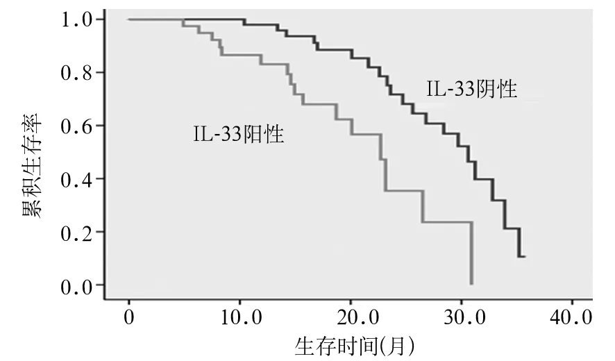
图1 IL-33表达阳性与表达阴性的胃癌患者生存曲线
IL-33是IL-1家族新成员[9],由270个氨基酸组成,相对分子质量为30 kD,具有抑制和(或)促进炎性反应的双重生理效应[10,11],在炎症、肿瘤等多种疾病中发挥重要的调控作用[12,13]。近年研究结果表明,体内IL-33及其受体ST2表达水平的变化对胃癌、肝癌、肺癌以及卵巢癌患者的病情进展、诊断及预后具有重要的参考价值[14~17]。IL-33能诱导内皮细胞增生、迁移和组织分化,从而促进新生血管生成[18];并且与上皮细胞浸润密切相关,参与肿瘤的发生和发展[19]。研究表明,IL-33可通过ST2-ERK1/2信号通路促进胃癌细胞侵袭和转移,作为胃癌诊断和治疗的有效标志物。
本研究结果显示,与正常胃组织相比,在胃癌组织中IL-33表达阳性率显著升高,而且其升高程度在进展期胃癌、淋巴结转移胃癌、UICC分期Ⅲ~Ⅳ期以及低分化胃癌和有远处转移的胃癌患者中更为明显,而与患者的性别、年龄、肿瘤部位以及肿瘤大小无关。IL-33表达阳性患者生存率明显低于IL-33表达阴性患者。提示IL-33表达升高可能与胃癌的淋巴结转移和恶性进展相关,可作为胃癌预后不良的标志。
IL-33是最近发现的IL-1家族细胞因子,关于其与肿瘤性疾病的研究才刚起步,因此进一步研究IL-33与胃癌侵袭转移的关系,揭示其在胃癌侵袭转移过程中的作用机制,有助于了解其生物学行为,为胃癌的靶向治疗提供潜在的作用靶点,同时也为胃癌的早期诊断和预后判断提供可靠的标志物。
[1] Jemal A, Bray F, Center MM, et al. Global cancer statistics[J]. CA Cancer J Clin, 2011,61(2):69-90.
[2] Degiuli M, Borasi A, Forchino F, et al. Lymph-nodal ratio in gastric cancer staging system[J]. Minerva Chir, 2011,66(3):177-182.
[3] Yu XX, Hu Z, Shen X, et al. IL-33 promotes gastric cancer cell invasion and migration via ST2-ERK1/2 pathway[J]. Dig Dis Sci, 2015,60(5):1265-1272.
[4] Li ZS, Li Q. The latest 2010 WHO classification of tumors of digestive system[J]. Zhonghua Bing Li Xue Za Zhi, 2011,40(5):351-354.
[5] Livak KJ, Schmittgen TD. Analysis of relative gene expression data using real-time quantitative PCR and the 2(-Delta Delta C(T)) Method[J]. Methods, 2001,25(4):402-408.
[6] Mohsen-Kanson T, Hafner AL, Wdziekonski B, et al. Expression of cell surface markers during self-renewal and differentiation of human adipose-derived stem cells[J]. Biochem Biophys Res Commun, 2013,430(3):871-875.
[7] 郑芝田.胃肠病学[M].北京:人民卫生出版社,2000:11.
[8] 王兴鹏. 现代胃肠病学高级进修教程[M].上海:上海科学技术文献出版社,2000:12.
[9] Castellani ML, Kempuraj D, Salini V, et al. The latest interleukin: IL-33 the novel IL-1-family member is a potent mast cell activator[J]. J Biol Regul Homeost Agents, 2009,23(1):11-14.
[10] Sattler S, Smits HH, Xu D, et al. The evolutionary role of the IL-33/ST2 system in host immune defence[J]. Arch Immunol Ther Exp (Warsz), 2013,61(2):107-117.
[11] Yang Q, Li G, Zhu Y, et al. IL-33 synergizes with TCR and IL-12 signaling to promote the effector function of CD8+T cells[J]. Eur J Immunol, 2011,41(11):3351-3360.
[12] Gangemi S, Allegra A, Profita M, et al. Decreased plasma levels of IL-33 could contribute to the altered function of Th2 lymphocytes in patients with polycythemia vera and essential thrombocythemia[J]. Cancer Invest, 2013,31(3):212-213.
[13] Pichery M, Mirey E, Mercier P, et al. Endogenous IL-33 is highly expressed in mouse epithelial barrier tissues, lymphoid organs, brain, embryos, and inflamed tissues: in situ analysis using a novel Il-33-LacZ gene trap reporter strain[J]. J Immunol, 2012,188(7):3488-3495.
[14] Bergis D, Kassis V, Ranglack A, et al. High serum levels of the interleukin-33 receptor soluble ST2 as a negative prognostic factor in hepatocellular carcinoma[J]. Transl Oncol, 2013,6(3):311-318.
[15] Hu LA, Fu Y, Zhang DN, et al. Serum IL-33 as a diagnostic and prognostic marker in non- small cell lung cancer[J]. Asian Pac J Cancer Prev, 2013,14(4):2563-2566.
[16] Ye XL, Zhao YR, Weng GB, et al. IL-33-induced JNK pathway activation confers gastric cancer chemotherapy resistance[J]. Oncol Rep, 2015,33(6):2746-2752.
[17] Tong X, Barbour M, Hou K, et al. Interleukin-33 predicts poor prognosis and promotes ovarian cancer cell growth and metastasis through regulating ERK and JNK signaling pathways[J]. Mol Oncol, 2016,10(1):113-125.
[18] Choi YS, Choi HJ, Min JK, et al. Interleukin-33 induces angiogenesis and vascular permeability through ST2/TRAF6-mediated endothelial nitric oxide production[J]. Blood, 2009,114(14):3117-3126.
[19] Sundlisaeter E, Edelmann RJ, Hol J, et al. The alarmin IL-33 is a notch target in quiescentendothelial cells[J]. Am J Patho, 2012,181(3):1099-1111.
Expression of IL-33 in gastric carcinoma and its significance
XIABingxiang,LIFan,WANGXuan,YUANZhenhua,ZHENGSuwen,ZHANGYewei
(TumorHospitalAffiliatedtoNanjingMedicalUniversity,Nanjing210009,China)
Objective To investigate the expression of IL-33 in gastric carcinoma tissues and its significance. Methods Cancer tissues and adjacent tissues of 98 patients with gastric cancer were selected, among whom, there were 16 cases with early gastric cancer, 82 cases with advanced gastric cancer, 76 cases with lymph node metastasis and 22 cases without lymph node metastasis. Meanwhile, we selected 36 cases of normal gastric tissues from healthy people as the control group. Immunohistochemical S-P method and real-time PCR were applied to detect the expression of IL-33 and its mRNA in gastric carcinoma tissues as well as in normal gastric tissues. We analyzed the relationship between the expression of IL-33 and clinicopathological features of gastric carcinoma tissues. Survival curves between IL-33 expression positive and negative patients were made using the Kaplan Meier method, and the median survival time was compared. Results The positive rate of IL-33 expression in gastric cancer tissues was higher than that in normal gastric tissues (P<0.05). No significant difference was found in the positive rate of IL-33 expression between in early gastric cancer and lymph node metastasis-negative gastric cancer tissues and in the normal gastric tissues (allP>0.05), while significant difference was found between in advanced gastric cancer and lymph node metastasis-positive gastric cancer tissues and in the normal gastric tissues (allP<0.05). The relative expression of IL-33 mRNA in gastric cancer tissues was higher than that in the normal gastric tissues (P<0.05). After the comparison of those between gastric cancer tissues with and without lymph node metastasis, significant difference was found (P<0.05). The expression of IL-33 was not related with gender, age, tumor growth and tumor size (allP>0.05), but it was correlated with tumor differentiation, tissue infiltration, lymph node metastasis, distant metastasis and clinical stage (allP<0.05). The median survival time was 30.6 months in IL-33 negative patients and 22.7 months in patients with positive IL-33 expression,P=0.001. Conclusion The increment on the level of IL-33 in gastric cancer tissues indicates that IL-33 may be involved in the occurrence and development of gastric cancer and predict poor prognosis.
stomach neoplasms; interleukin-33; lymph node metastasis; prognosis
国家自然科学基金面上项目(61371066);江苏省医学重点人才资助项目( RC2011090);江苏省重点研发计划(社会发展)(BE2015720);江苏省六大高峰人才项目(WSW-041)。
夏兵祥(1988-),男,在读硕士研究生,主要从事普外肿瘤基础与临床研究。E-mail:1065424827@qq.com
张业伟(1976-),男,教授,主任医师,主要从事肝胆胰肿瘤的基础与临床研究。E-mail: zhangyewei@njmu.edu.cn
10.3969/j.issn.1002-266X.2017.13.002
R735.2
A
1002-266X(2017)13-0005-04
2016-04-03)
