Catecholaminergic Polymorphic Ventricular Tachycardia
2014-06-07童鸿
●心电学英语
Catecholaminergic Polymorphic Ventricular Tachycardia
The primary electric disorders responsible for polymorphic ventricular tachycardia(VT)or ventricular fibrillation are long-QT syndrome,Brugada syndrome, the short-coupled variant of torsades de pointes, short-QT syndrome,and catecholaminergic polymorphic VT(CPVT).CPVT is a rare arrhythmogenic disorder characterized by adrenergic-induced bidirectional and polymorphic VT.The prevalence of the disease is estimated to be 1:10 000 in Europe.The first case was reported in 1975.Key features include polymorphic VT reproducibly induced during exercise tests,isoproterenol infusion,or emotion and exercise.CPVT occurs in children and adolescents and causes syncope and sudden cardiac death at a young age,in the absence of structural heart disease.The resting ECG,including the QTc interval,is normal.The mortality of CPVT is extremely high,reaching 31%by the age of 30 years when untreated.The estimated 4-and 8-year cardiac event rates were 33%and 58%,respectively,in our series of patients without β-blockers.There is a clear correlation between the age of the first syncope and the severity of the disease,with a worse prognosis in the case of early occurrence.β-blockers without sympathomimetic activity are clinically effective in reducing syncope.However,arrhythmic event rate with β-blocker therapy remains significant,suggesting the need for alternate pharmacological and nonpharmacological therapies.
With the advancements of molecular genetics and the identification of mutations in the genes encoding the cardiac ryanodine receptor and cardiac calsequestrin 21in patients with CPVT,the central role of the intracellular calcium dysregulation in myocardial cells is progressively better understood through expression studies and murine models.
Genetic Background
Priori et al and Laitinen et al identified the first mutations in the cardiac ryanodine receptor gene (RYR2)in families suffering of this type of CPVT,now known as CPVT1.Lahat et al.identified a homozygous missense mutation in the cardiac calsequestrin gene (CASQ2)as the cause of this recessive form,now known as CPVT2.RYR2 mutations are frequent, whereas CASQ2 mutations are rare;altogether,mutations are only found in 50%to 60%of patients with CPVT,which suggests that other genes are involved.
Ryanodine Receptor
The cardiac ryanodine receptors(RyR2)are calcium(Ca2+)release channels present in the sarcoplamic reticulum(SR),an intracellular vesicular network playing a major role in the regulation of Ca2+homeostasis in the heart.The mechanism of their activation is called calcium-induced calcium release because it requires that Ca2+provided by the activated L-type Ca2+channel (Cav1.2).Calcium binds to RyR2 and triggers opening of a high-conductance channel,allowing rapid Ca2+efflux from the SR.The consecutive high cytoplasmic Ca2+induces myocardial contraction,then Ca2+is reuptaken in the SR,where it is stored at high concentrations.
RYR2 Mutations
The 4967-amino acid RyR2 channel is encoded by one of the largest genes in the human genome,containing 105 exons.To date,>50 mutations have been reported,most of them for CPVT1 and unexplained or exercise-induced sudden death.Most of the RYR2 mutations are missense mutations,occurring in 3 hot-spotregions:the N-terminal region,the central region where the calstabin-21binding domain is localized,and the C-terminal domain,including the channel region.These 3 regions are well conserved among the RyR gene family and are involved in the regulation of RyR channels.
High variability of the phenotypic expression among subjects of the same family or unrelated families was demonstrated and estimates of the penetrance range from 25%to 100%.It is noteworthy that there are asymptomatic RyR2 mutation carriers with normal exercise stress tests.Some of them can further present with exercise-induced arrhythmia during a subsequent stress test,but more importantly may die suddenly as the first manifestation of the disease.
CASQ2 Mutations
The399-amino acid CASQ2 protein is encoded by a gene containing 11 exons.Twenty-one distinct CASQ2 mutations have been reported,either homozygous or compound heterozygous mutations transmitted under a recessive mode of inheritance.Half of them are missense mutations localized in different exons.The others lead to truncated proteins by various mechanisms,nonsense codon, small deletion,and abnormal splicing leading to premature stop codon.Interestingly,a synonymous c.381C>T variation in exon 3,recently identified in a family with CPVT2,was shown to induce abnormal splicing and a premature stop codon using a splicing minigene assay.
The phenotype is similar among the patients with 2 CASQ2 mutations and the patients with an RyR2 mutation.Most of the carriers of a single CASQ2 mutation are healthy.Nevertheless,several clinical investigations suggested that a single CASQ2 mutation could represent a potential susceptibility for ventricular arrhythmias in some subjects.The origin of the variability among subjects of a same family is still unknown.
Clinical Presentation
CVPT is extremely uncommon before the age of 2 years.The first episode of syncope usually occurs during the first or second decade of life.The symptoms are always triggered by exercise or emotional stress.Often,epilepsy is diagnosed and children are inappropriately treated with long-term antiepileptic therapy.A mean delay in diagnosis of≥2 years is usually reported in patients with syncope initially attributed to vasovagal or neurological causes.A family history of exercise-related syncope,seizure,or sudden death is reported in 30% of the patients.
Diagnosis
A history of exercise-induced or emotional stress-induced syncope with polymorphic ventricular arrhythmia in a child is highly suggestive of CPVT. The heart is structurally normal.The arrhythmia is reproducibly induced during an exercise test as well as during isoproterenol infusion.Holter monitoring or an exercise test can document CPVT by showing the ventricular arrhythmia progressively appearing after a heart rate threshold(around 120-130 beats per minute).Polymorphic VT is usually not inducible by programmed ventricular stimulation.Implantable loop recorders can be useful to record CPVT in children with adrenergically triggered,unexplained syncope.
Electrocardiographic key features in CVPT
The resting ECG is usually normal,and there is progressive ventricular ectopy as heart rate increases during exercise or isoproterenol infusion.Frequency and complexity increase as heart rate increases,first monomorphic ventricular premature beats(VPBs)followed by bidirectional VT(Figure 1).VPBs usually have a right bundle branch block pattern with alternating right and left axis deviation,suggesting a left ventricular origin.If the exercise is continued,salvos of polymorphic VT may appear and become more sustained and rapid,leading to syncope.Usually,the arrhythmia is self-terminating,but in some cases it can degenerate into ventricular fibrillation and sudden death(Figure 2). The arrhythmia disappears with the discontinuation of the exercise or after cessation of the isoproterenol infusion. The reverse heart rate-dependent sequence is usually observed during recovery.Some individuals expressing bidirectional VT during exercise may not have CPVT.Instead,clinical consideration of either Andersen-Tawil syndrome2or long-QT syndrome and appropriate genetic testing may be warranted for individuals without a RyR2 mutation but considered as patients with CPVT,particu-larly women.Careful inspection of the TU-wave morphology may assist in distinguishing between CPVT and Andersen-Tawil syndrome in a patient exhibiting exercise-induced bidirectional VT.Atrial arrhythmias,including atrial fibrillation,are not uncommon during exercise tests and have been described in some adult patients.
Current Management
β-Blockers
The first-line therapeutic option for patients with CPVT is β-blockers without sympathomimetic activity, in accordance with the arrhythmia's catecholaminergic mechanism,combined with exercise restriction.Nadolol, a long-acting drug,is preferred for prophylactic therapy and has been found to be effective clinically.In our experience,the dosage used to provide adequate prevention of CVPT and syncope is usually high(1.8 mg/kg).We reported in 2009 the long-term follow-up results of 101 patients with CPVT with an estimated 8-year cardiac event rate of 27%,even in those taking β-blockers.
The apparent discrepancy in the efficacy of β-blocker treatment between the various studies probably reflects differences in genetic background and in β-blocker dosages or a poor drug compliance.This discrepancy in β-blocker efficacy may also be because of the presence of polymorphisms influencing their metabolism.
Meanwhile,the maximal well-tolerated dosages of β-blockers should be prescribed and Holter recordings and exercise tests should be repeated periodically to ensure that the degree of sinus tachycardia that precedes the onset of arrhythmias is never reached.Furthermore, once the diagnosis is established,it is crucial to make the patients aware of the necessity of faultless compliance with the β-blocker therapy,given the number ofnoncompliance-related sudden cardiac deaths.It is strongly suggested that genetically positive family members should receive β-blockers even after a negative exercise test.
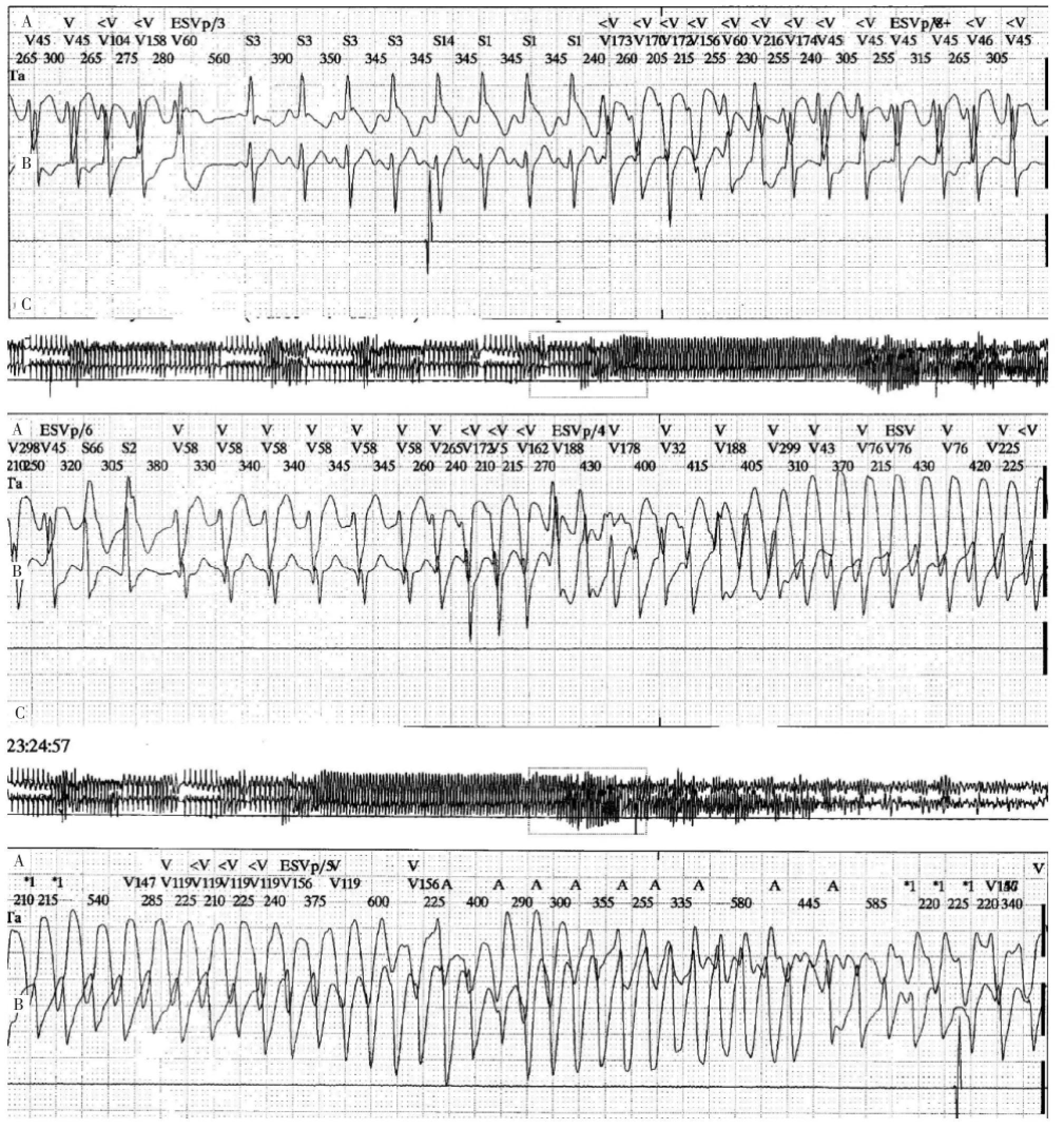
Figure 2 Holter tracings showing pleiomorphic and polymorphic ventricular tachycardia preceding the occurrence of ventricular fibrillation in a patient with catecholaminergic polymorphic ventricular tachycardia.
Implantable Cardioverter Defibrillator
An implantable cardioverter defibrillator(ICD)implantation is recommended in patients with CPVT and syncope or documented sustained VT,despite β-blocker therapy.Nevertheless,ICDs can potentially have proarrhythmic effects in patients with CPVT because stress caused by appropriate or inappropriate discharges could prove disastrous by evoking a self-induced vicious circle.However,a combination therapy involving both an ICD and an optimized dosage of β-blocker should safeguard against any such adverse effects and provide ultimate protection in nonresponsive patients.
Left Cardiac Sympathetic Denervation
The first publication reported the efficacy of LCSD in 3 young patients with CPVT,with a long follow-up in 2(aged 20 and 10 years)in whom ventricular arrhythmias were not controlled by β-blocker therapy.The following series reported results of LCSD in patients with resistant and symptomatic ventricular arrhythmias, despite optimal pharmacological therapy.Although the short-term results seem encouraging,more data from a long-term follow-up are needed.LCSD is not available in many centers worldwide because it requires well-trained surgeons and dedicated techniques.
词汇
Murine n.&adj.鼠;鼠的
Calsequestrin n.集钙蛋白
Salvo n.&v.一阵,一片,爆发,齐射;齐射
Exon n.外显子
Truncate v.截去,删节
Codon n.密码子
Faultless adj.无错误的,完美的
Disastrous adj.灾难性的,灾难性,极坏的
注释
1.Calstabin-2是一种小的调节蛋白即FKBP12.6,能与RyR2结合并稳定其,阻止这种结合会增加室性心律失常危险。
2.Andersen-Tawil syndrome又称长QT综合征7,是一种罕见的常染色体显性遗传疾病,60%与KCNJ2基因有关,除长Q-T间期,室性心律失常外,合并存在发作性或持续性肌无力及躯体异常,如小下颌、低耳位、指弯曲等。
参考译文
第59课儿茶酚胺源性多形性室性心动过速
与多形性室性心动过速(VT)或心室颤动相关的原发性电子病有长QT综合征、Brugada综合征、短偶联间期尖端扭转型室性心动过速、短QT综合征和儿茶酚胺源性多形性室性心动过速(CPVT)。CPVT是一种罕见的致心律失常疾病,特征表现为肾上腺素诱发的双向性和多形性VT。在欧洲该疾病发病率估计为1:10 000。1975年首次报道。关键特征包括运动试验、滴注异丙肾上腺素、情绪激动和运动能重复诱发多形性VT。CPVT发生于儿童和青少年,导致无结构性心脏病的少年晕厥和心脏性猝死。静息心电图包括Q-TC间期正常。CPVT死亡率极高,如不治疗,到30岁时可达31%。本文作者报道系列患者非β受体阻滞剂治疗下4年和8年的心脏事件发生率分别达33%和58%。首次晕厥发作的年龄与疾病严重性之间存在明确的相关性,早发者预后差。临床上,无拟交感活性的β受体阻滞剂能有效减少晕厥发生。然而,β受体阻滞剂治疗下心律失常事件仍然明显,提示需要更换药物和非药物治疗。……
