成人创伤后应激障碍海马体积研究的Meta分析☆
2011-04-19文敏周波张春林焦玲
文敏周波张春林焦玲
·论 著·
成人创伤后应激障碍海马体积研究的Meta分析☆
文敏*周波*张春林**焦玲△
目的 综合评价成人创伤后应激障碍(Posttraumatic stress disorder,PTSD)的左、右侧海马体积。方法 利用Meta分析方法对国内外公开发表的关于成人PTSD左、右侧海马体积的研究文献进行综合定量分析。结果 共收集到符合纳入标准的文献23篇,累计病例共337例,累计对照共389名。PTSD组与健康对照组左侧海马体积分析显示,总体效应检验有统计学意义(Z=4.77,P<0.01),95%CI横线位于无效竖线左侧,提示PTSD的左侧海马体积减小;PTSD组与健康对照组右侧海马体积比较分析显示,总体效应检验有统计学意义(Z=5.01,P<0.01),95%CI横线位于无效竖线左侧,提示PTSD的右侧海马体积减小。漏斗图分布呈倒漏斗形,近似对称,Begg检验证实发表无偏倚。结论 成人PTSD患者左、右侧海马体积均减小。
海马 创伤后应激障碍 核磁共振成像 META分析
近年来,随着严重自然灾害、严重交通事故和重大传染疾病流行等严重创伤性应激事件的频繁发生,创伤后应激障碍 (posttraumatic stress disorder,PTSD)已经成为危害人类身心健康的常见疾病。研究表明,PTSD的发生与脑解剖结构异常密切相关[1],其中与学习、记忆、情绪密切相关的海马体积异常备受关注[2-4]。有的报道PTSD患者双侧海马体积均较正常组减少[5],但也有阴性结果[6]。关于PTSD是否存在海马体积的变化,国内外研究结果仍存在很多争议[7]。因此,本研究通过对国内外公开发表的有关成人创伤后应激障碍海马体积的研究文献进行Meta分析,以期进行综合评价。
1 对象与方法
1.1 研究对象 在国内外期刊上已经公开发表的有关成人创伤后应激障碍左、右侧海马体积研究的文献。文献纳入标准:①1990~2010年之间国内外发表的一次文献;②独立性研究,目的一致;③研究对象来源和样本量明确,诊断明确;④原始数据提供了样本量、均数、标准差;⑤资料收集的方法科学,分析方法正确。文献排除标准:①动物实验;②非成年人研究(年龄低于18岁);③参考Lichtensiein等[8]提出的标准,剔除重复报告、质量差、报道信息太少(未给出海马体积均数和标准差、未设置健康对照组)等无法利用的文献。
1.2 检索方法 分别以 “创伤后应激障碍/posttraumatic stress disorder/post-traumatic stress Disorde/PTSD”、“海马/hippocampus”与“核磁共振成像/magnetic resonance imaging/MRI”结合以“主题、关键词、主题词”途径,联合检索中国生物医学文献数据库(CMB)和中国学术期刊全文数据库(CNKI)获得全部相关的中文文献,运用 Endnote检索Medline、Pubmed、LISTA(EBSCO)外文数据库,查询1990~2010年国内外公开发表的有关成人创伤后应激障碍海马体积的研究文献。
1.3 统计分析 运用RevMan 5.0分析软件进行异质性检验,如果各研究结果之间差异无统计学意义则用固定效应模型(fixed effects model),若有差异则采用随机效应模型 (random effects model),计算合并效应指标值及95%可信区间(95%CI),画出森林图,并进行敏感度分析和漏斗图分析。
2 结果
2.1 文献检索结果 共收集到符合纳入标准的文献23篇,累计患者共337例,累计正常对照共389名;排除5篇未给出正常对照组数据的研究。Bossini等[5、9-18]11篇研究显示PTSD患者双侧海马体积均较正常对照组减少,而 Jatzko等[6、19-23]6篇研究则显示PTSD患者双侧海马体积与正常对照组比较无差异。Pavic等[24-27]4篇研究显示PTSD患者右侧海马体积较正常组显著减少,左侧海马体积无差异。Bremner等[28]研究显示PTSD患者左侧海马体积较正常组显著减少,右侧海马体积无差异。见表1。
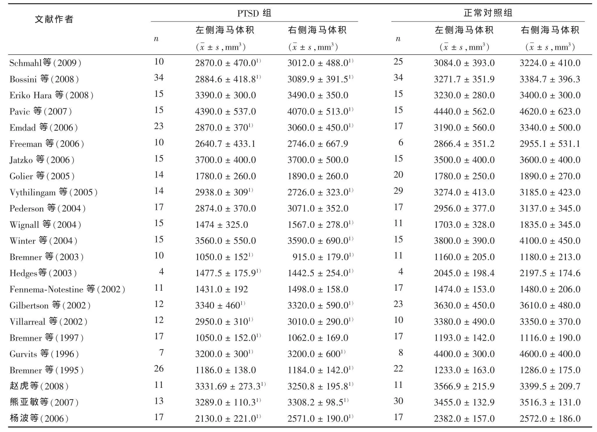
表1 纳入Meta分析文献的基本情况
2.2 PTSD的左侧海马体积分析 结果显示,各研究结果间存在异质性 (χ2=52.03,P<0.01,I2=58%),故采用随机效应模型对效应指标值进行合并统计分析。森林图结果显示,总体效应检验有统计学意义(Z=4.77,P<0.01),95%CI横线位于无效竖线左侧,显示PTSD组左侧海马体积小于正常对照组。见图1。
2.3 PTSD的右侧海马体积分析 结果显示,各研究结果间存在异质性 (χ2=47.72,P<0.01,I2=54%),故采用随机效应模型对效应指标值进行合并统计分析。森林图结果显示,总体效应检验有统计学意义(Z=5.01,P<0.01),95%CI横线位于无效竖线左侧,显示PTSD组右侧海马体积小于正常对照组。见图2。
2.4 敏感性分析 逐一剔除其中权重较大或较小数据后,再行Meta分析。结果显示,合并后95%CI横线仍位于无效竖线左侧,与上述结果一致,说明敏感性低,研究结果较为稳定可信。
2.5 发表偏倚的评估 RevMan软件绘制漏斗图,分布呈倒漏斗形,除了两小样本研究呈散在分布外(可能与其样本量小,权重较低,增加了各研究间异质性有关),近似对称,发表偏倚较小。Stata软件行Begg检验,进一步证实左右侧海马体积的研究的发表偏倚均较小 (P均大于0.05)。见图3A、3B。
3 讨论
本研究运用Meta分析方法,对23篇国内外PTSD海马体积的研究文献进行了综合分析,结果表明,PTSD患者双侧海马体积均明显减少。排除权重较大或较小的文献后,Meta分析结果合并效应量变化不大,说明结果较为稳定可信。Kitayama等[29]对1995-2003年间133例PTSD与133正常对照之间进行Meta分析,结果显示PTSD患者双侧海马体积均缩小,与本文结果一致。Woon等[7]对2008年以前的17篇英文研究进行合并分析,也得到了同样结果。
对于成年PTSD与海马体积关系的研究,国内外已有大量的研究,但迄今也没有得到一个统一的结论,各研究之间存在很大差异的原因可能有以下两个方面:①磁共振扫描条件不统一,如扫描设备、扫描厚度、体积分析软件、海马位置的界定标准等,均可能影响到实验结论;Stein等[30]在海马的MRI研究中发现,海马体积变化范围很大(2.1-6.4 cm2),提示海马体积测量中存在很多方法学干扰。Bremner等[27]在海马体积对比中,只比较了中段海马体积,而其它研究均比较了全海马体积。这些因素均可能影响海马体积,从而影响到实验结论;②研究对象不统一,患者的年龄、性别、疾病严重程度及持续时间的不同,可能导致海马损伤程度不同,从而影响实验结论。Hara等[21]在对肿瘤诊断后1年的PTSD患者双侧海马进行MRI检测时发现海马体积并未减小,提示海马体积减小可能与PTSD持续时间、PTSD严重程度有关。Bonne等[31]在对早期的PTSD患者的研究中也未发现双侧海马体积减小。Kitayama等[29]总结以前的研究也证实了海马体积减小出现在长期、严重的PTSD患者。同样,年龄也可能是影响实验结果的关键因素,Carrion等[32]研究发现未成年人PTSD患者双侧海马体积无明显变化,提示年龄是影响海马体积变化的因素,这可能与未成年人海马发育掩盖了海马的损伤有关。
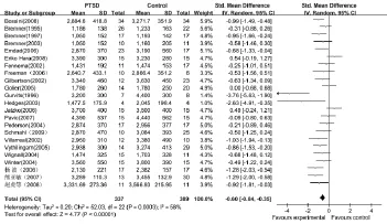
图1 PTSD组与正常对照组左侧海马体积比较的随机效应森林图
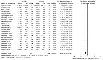
图2 PTSD组与正常对照组右侧海马体积比较的随机效应森林图
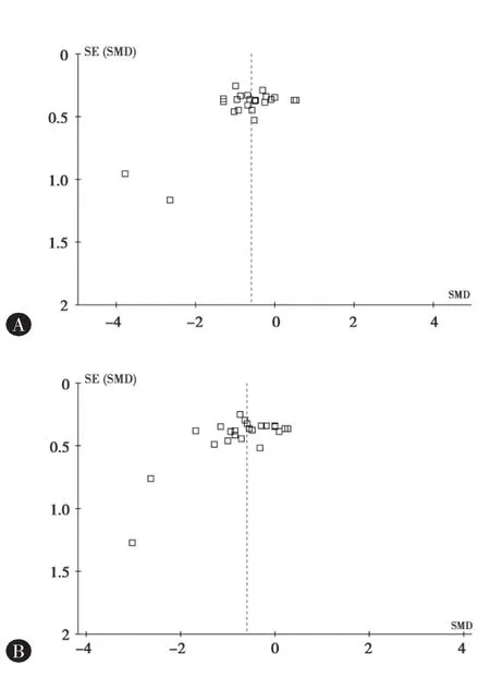
图3 A:左侧海马体积比较漏斗图;B:右侧海马体积比较漏斗图
本文的系统分析支持PTSD双侧海马体积均降低。然而,海马是一个结构复杂的脑区,从组织学角度来看,海马可以分为CA1、CA2、CA3、CA4和齿状回等亚区,PTSD患者海马体积减小究竟是均匀发生于各个亚区,还是集中于某一亚区,有待于进一步研究。海马体积减小的病理机制仍不清楚。Wang等[33]对PTSD患者海马亚区的MRI研究显示,CA3亚区与齿状回体积均显著减小。Neylan等[34]在严重睡眠障碍的PTSD患者MRI研究中也发现同样的结论,这可能与创伤后应激诱导神经毒性抑制海马齿状回神经再生[35]和增加CA3区神经元死亡[36]有关,但相关的影像学研究较少,尚需大量研究证实。
[1]Karl A, Schaefer M, Malta LS, et al.A meta-analysis of structural brain abnormalities in PTSD[J].Neurosci Biobehav Rev,2006,30(7):1004-1031.
[2]Brewin CR,Kleiner JS,Vasterling JJ,et al.Memory for emotionally neutral information in posttraumatic stress disorder: a meta-analytic investigation[J].J Abnorm Psychol,2007,116(3):448-463.
[3]McEwen BS.Physiology and neurobiology of stress and adaptation:central role of the brain[J].Physiol Rev,2007,87(3):873-904.
[4]Buchanan TW,Kern S,Allen JS,et al.Circadian regulation of cortisol after hippocampal damage in humans[J].Biol Psychiatry,2004,56(9):651-656.
[5]Bossini L,Tavanti M,Calossi S,et al.Magnetic resonance imaging volumes of the hippocampus in drug-naive patients with post-traumatic stress disorder without comorbidity conditions[J].J Psychiatr Res,2008,42(9):752-762.
[6]Jatzko A,Rothenhofer S,Schmitt A,et al.Hippocampal volume in chronic posttraumatic stress disorder(PTSD):MRI study using two different evaluation methods[J].J Affect Disord,2006,94(1-3):121-126.
[7]Woon FL,Sood S,Hedges DW.Hippocampal volume deficits associated with exposure to psychological trauma and posttraumatic stress disorder in adults:a meta-analysis[J].Prog Neuropsychopharmacol Biol Psychiatry,2010,34(7):1181-1188.
[8]Lichtenstein MJ,Mulrow CD,Elwood PC,et al.Guidelines for reading case-control studies[J].J Chronic Dis,1987,40(9):893-903.
[9]Emdad R,Bonekamp D,Sondergaard HP,et al.Morphometric and psychometric comparisons between non-substance-abusing patients with posttraumatic stress disorder and normal controls[J].Psychother Psychosom,2006,75(2):122-132.
[10]Vythilingam M,Luckenbaugh DA,Lam T,et al.Smaller head of the hippocampus in Gulf War-related posttraumatic stress disorder[J].Psychiatry Res,2005,139(20):89-99.
[11]Villarreal G,Hamilton DA,Petropoulos H,et al.Reduced hippocampal volume and total white matter volume in posttraumatic stress disorder[J].Biol Psychiatry,2002,52(2):119-125.
[12]Gurvits TV,Shenton ME,Hokama H,et al.Magnetic resonance imaging study of hippocampal volume in chronic,combat-related posttraumatic stress disorder[J].Biol Psychiatry,1996,40(11):1091-1099.
[13]杨波,周义成,夏军,等.创伤后应激障碍的海马容积和氢质子波谱研究[J].中华放射学杂志,2006,40(1):36-40.
[14]熊亚敏,胡章勇,周义等.不同类型创伤后应激障碍患者海马容积的MRI研究[J].神经损伤与功能重建,2007,2(2):96-99.
[15]Hedges DW,Allen S,Tate DF,et al.Reduced hippocampal volume in alcohol and substance naive Vietnam combat veterans with posttraumatic stress disorder[J].Am J Psychiatry,2003,16(4):219-224.
[16]Schmahl C, Berne K, Krause A, et al.Hippocampus and amygdala volumes in patients with borderline personality disor-der with or without posttraumatic stress disorder[J].J Psychiatry Neurosci,2009,34(4):289-295.
[17]Bremner JD,Vythilingam M,Vermetten E,et al.MRI and PET study of deficits in hippocampal structure and function in women with childhood sexual abuse and posttraumatic stress disorder[J].Cogn Behav Neurol,2003,160():924-932.
[18]Gilbertson MW,Shenton ME,Ciszewski A,et al.Smaller hippocampal volume predicts pathologic vulnerability to psychological trauma[J].Nat Neurosci,2002,5(11):1242-1247.
[19]Freeman TW,Kimbrell T,Booe L,et al.Evidence of resilience:neuroimaging in former prisoners of war[J].Psychiatry Res,2006,146(1):59-64.
[20]Golier JA,Yehuda R,De Santi S,et al.Absence of hippocampal volume differences in survivors of the Nazi Holocaust with and without posttraumatic stress disorder[J].Psychiatry Res,2005,139(1):53-64.
[21]Hara E,Matsuoka Y,Hakamata Y.Hippocampal and amygdalar volumes in breast cancer survivors with posttraumatic stress disorder[J].J Neuropsychiatry Clin Neurosci,2008,20(3):302-308.
[22]Fennema-Notestine C,Stein MB,Kennedy CM,et al.Brain morphometry in female victims of intimate partner violence with and without posttraumatic stress disorder[J].Biol Psychiatry,2002,52(11):1089-1101.
[23]Pederson CL,Maurer SH,Kaminski PL,et al.Hippocampal volume and memory performance in a community-based sample of women with posttraumatic stress disorder secondary to child abuse[J].J Trauma Stress,2004,17(1):37-40.
[24]Winter H,Irle E.Hippocampal volume in adult burn patients with and without posttraumatic stress disorder[J].Am J Psychiatry,2004,161(12):2194-2200.
[25]Pavic L, Gregurek R, Rados M, et al.Smaller right hippocampus in war veterans with posttraumatic stress disorder[J].Psychiatry Res,2007,154(2):191-198.
[26]Wignall EL, Dickson JM, Vaughan P, et al.Smaller hippocampal volume in patients with recent-onset posttraumatic stress disorder[J].Biol Psychiatry,2004,56(11):832-836.
[27]Bremner JD,Randall P,Scott TM,et al.MRI-based measurement of hippocampal volume in patients with combat-related posttraumatic stress disorder[J].Am J Psychiatry,1995,152(7):973-981.
[28]Bremner JD,Randall P,Vermetten E,et al.Magnetic resonance imaging-based measurement of hippocampal volume in posttraumatic stress disorder related to childhood physical and sexual abuse-a preliminary report[J].Biol Psychiatry,1997,41(1):23-32.
[29]Kitayama N,Vaccarino V,Kutner M,et al.Magnetic resonance imaging(MRI)measurement of hippocampal volume in posttraumatic stress disorder:a meta-analysis[J].Journal of Affective Disorders,2005,88(1):79-86.
[30]Stein MB,Koverola C,Hanna C,et al.Hippocampal volume in women victimized by childhood sexual abuse[J].Psychological Med,1997,27(4):951-959.
[31]Bonne O, Brandes D, Gilboa A, et al.Longitudinal MRI study of hippocampal volume in trauma survivors with PTSD[J].Am J Psychiatry,2001,158(8):1248-1251.
[32]Carrion VG,Weems CF,Eliez S,et al.Attenuation of frontal asymmetry in pediatric posttraumatic stress disorder[J].Biological Psychiatry,2001,50(12):943-951.
[33]Wang Z,Neylan TC,Mueller SG,et al.Magnetic resonance imaging of hippocampal subfields in posttraumatic stress disorder[J].Arch Gen Psychiatry,2010,67(3):296-303.
[34]Neylan TC,Mueller SG,Wang Z,et al.Insomnia severity is associated with a decreased volume of the CA3/dentate gyrus hippocampal subfield[J].Biological Psychiatry,2010,68(5):494-496.
[35]Nagata K,Nakashima-Kamimura N,Mikami T,et al.Consumption of molecular hydrogen prevents the stress-induced impairments in hippocampus-dependent learning tasks during chronic physical restraint in mice[J].Neuropsychopharmacology,2009,34(2):501-508.
[36]Zhao H,Xu H,Xu XH,et al.Predatory stress induces hippocampal cell death by apoptosis in rats[J].Neuroscience Letters,2007,421(2):115-120.
(责任编辑:曹莉萍)
The hippocampus volumes in adults with posttraumatic stress disorder: a meta-analysis.
WEN Min,ZHOU Bo,ZHANG Chunlin,JIAO Ling.The department of Anatomy,Guiyang Medical University,4 BeiJing Road,Guiyang 550004.China.Tel:0851-6909571.
Objective To comprehensively evaluate the association of hippocampal volumes in adults with posttraumatic stress disorder.Methods All the published studies on the hippocampal volumes and posttraumatic stress disorder were retrieved and standard techniques of meta-analysis to combine with the results of these studies were used to produce a more precise estimate.Results Based on the criteria.23 studies were selected with a total of 337 accumulative cases and 389 controls.Meta-analysis results showed that compare the hippocampal volumes of controls,the overall effect value of the left(z=5.01)and right(z=4.77)in PTSD was statistical significant(P<0.01).Inverted funnel plot and Begg's test showed the publication bias was not significant(P>0.05).Conclusions The right and left hippocampal volumes in adults were smaller in the posttraumatic stress disorder group compared to the control group.
Hippocampus PTSD MRIMeta-analysis
R749.5
A
2011-05-09)
☆ 贵州省社会发展科技攻关计划项目 [编号:黔科合SY字[2008]3046号]
* 贵阳医学院人体解剖学教研室(贵阳 550004)通讯作者(E-mail:jiaoling5151@sina.com)
** 贵阳医学院生物学教研室
△ 贵阳医学院第一附属医院神经内科