Tuberculosis Verrucosa Cutis on the Buttocks:A Case Report
2023-10-20YuXiaoShanShanPengLyuYaZhangJieLinYouHongHuMuPingFang
Yu Xiao, Shan-Shan Peng,2, Lyu-Ya Zhang, Jie Lin, You-Hong Hu, Mu-Ping Fang,*
1 Department of Dermatology, The Central Hospital of Xiaogan, Xiaogan, Hubei 432000, China; 2 Department of Respiration, The Central Hospital of Xiaogan, Xiaogan, Hubei 432000, China.
Abstract
Keywords: tuberculosis, buttocks, case report
Introduction
Tuberculosis is an infectious disease caused byMycobacterium tuberculosis.1About 1.5% of all extrapulmonary tuberculosis cases show cutaneous involvement.1Tuberculosis verrucosa cutis is a rare form of cutaneous tuberculosis that is caused by exogenous reinfection in previously sensitized individuals with a relatively high degree of immunity.2The incidence of tuberculosis verrucosa cutis is more frequent in Asia than in western countries, and the incidence is especially high in developing countries.1We present a rare case of tuberculosis verrucosa cutis on the buttocks that remained misdiagnosed for 9 years.The information presented herein will help dermatologists to promptly diagnose similar conditions.
Case report
A 54-year-old Chinese man presented with a 9-year history of asymptomatic verrucous lesions on both buttocks.The initial lesion was a small raised red papule on the right buttock that was not associated with itching or pain.Over the course of several months, the lesion gradually enlarged,spread to the left buttock, and developed a verrucosa appearance.A rural clinic had prescribed various topical treatments including corticosteroids and antibacterial ointments, which did not lead to any substantial changes.One week before the patient’s referral to our hospital, the lesions had become mildly painful with yellowish purulent discharge.The patient reported neither any injury to the buttocks nor history of contact with tuberculosis.
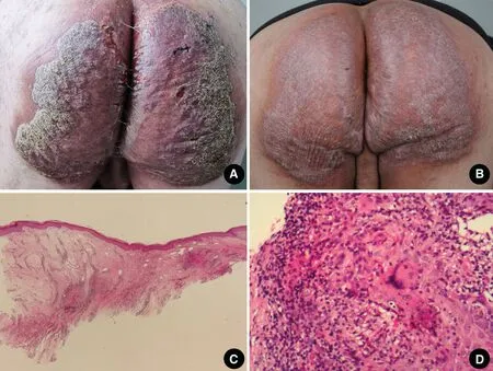
Figure 1.The clinical features and histopathological results of the lesion from the patient with tuberculosis verrucosa cutis.(A) A well-defined plaque, verrucous lesion with yellowish purulent discharge on both buttocks at the time of presentation.(B) Substantial thinning of the hyperkeratosis plaque after 1 month of antituberculosis treatment.(C and D) Histopathological results show caseating granulomatous changes within the papillary and reticular dermis, and the presence of Langerhans giant cells within the dermis (C: H&E, × 25; D: H&E, × 400).
Systemic examinations were normal.There was no regional or generalized lymphadenopathy.Examination of the lesion at the time of presentation revealed a welldefined plaque, verrucous lesion, and yellowish purulent discharge on both buttocks (Fig.1A).Routine laboratory tests showed no abnormalities in blood,blood coagulation function, erythrocyte sedimentation rate, urine, stool, liver function, and renal function.The patient was negative for human immunodeficiency virus antibody, hepatitis C virus antibody, treponemal antibody, and antinuclear antibodies.There were no abnormalities detected on microscopic examination for fungus and fungal culture, chest radiography, and abdominal computed tomography.The patient was positive for SS-A/Ro52 antibody.The tuberculin test was negative, but the T-spot test was positive.Histological analysis of a biopsy from the lesion showed prominent pseudoepitheliomatous hyperplasia of the epidermis with hyperkeratosis and dermal granulomatous inflammation composed of epithelioid histiocytes and mixed inflammatory cells.Numerous Langerhans giant cells were identified in the granulomas,with foci of caseous necrosis (Fig.1C and 1D).Special histological stains forMycobacterium tuberculosis,atypical mycobacteria, and fungi were negative.Polymerase chain reaction (PCR) testing forMycobacterium tuberculosiswas also negative.
The patient was started on triple antituberculosis therapy, including triple antituberculosis (rifampicin 0.6 g/d,isoniazide 0.3 g/d, ethambutol 0.75 g/d).After 1 month of treatment, the lesions had substantially improved(Fig.1B) and the patient was well tolerated.The patient was satisfied with its good effect, and no further follow-up organized.
Patient consent declaration
The patient gave the written informed consent about her case publication, including the images and other disease details.
Discussion
Tuberculosis verrucosa cutis is the third-most common form of cutaneous tuberculosis, and results from direct inoculation of the mycobacteria into the skin of previously infected patients.2Tuberculosis verrucosa cutis has a predilection for the lower extremities,for example hands in adults, and lesions commonly start as small painless papules or papulo-pustules that progress into verrucous or hyperkeratotic plaques.The plaques are firm, verrucous, or fissures and may produce purulent discharge, but the regional lymph nodes are not usually enlarged.1Tuberculosis verrucosa cutis is easily misdiagnosed and has a long course.Eunet al.3reported a similar case of a 43-year-old Asian woman with tuberculosis verrucosa cutis on the left buttock that had been present for more than 20 years.Because of long-term misdiagnosis and untimely treatment, tuberculosis verrucosa cutis can develop into squamous cell carcinoma and elephantiasis nostras verrucosa, which triggers chronic lymphedema and severely debilitating physical disability.4Therefore,it is important to accurately and promptly diagnose tuberculosis verrucosa cutis.The diagnosis of tuberculosis verrucosa cutis is mainly based on history, laboratory findings, and histological features.
The histological features of tuberculosis verrucosa cutis are prominent pseudoepitheliomatous hyperplasia of the epidermis with hyperkeratosis and a dense inflammatory cell infiltrate consisting of neutrophils, lymphocytes, and giant cells.Acid-fast staining for mycobacteria is rarely positive, and typical tuberculous foci with caseating necrosis are uncommon.1
Some studies have reported that the sensitivity of PCR is better than that of microscopic examination and is comparable with that of culture.3Unfortunately, PCR testing is often negative in cutaneous tuberculosis, which could be due to a low bacillary load in the specimen.Hence, the sensitivity of PCR can be improved by the selection of good quality, appropriate specimens.5
The differential diagnoses for tuberculosis verrucosa cutis include verrucous porokeratosis, discoid lupus erythema-tosus hypertrophicus lichen planus, chromoblastomycosis, and atypical mycobacterial infections.1Antitubercular therapy is recommended for most of these diagnoses.
The present patient was a farmer with a rare case of tuberculosis verrucosa cutis on both buttocks that had been misdiagnosed for 9 years; typical laboratory findings were negative, which resulted in a diagnostic dilemma.Finally,histopathological characteristics and response to antitubercular therapy confirmed the diagnosis of tuberculosis verrucosa cutis.The limitation was the short-term follow-up.In conclusion, this case can provide clinician with more experience on diagnosis for such a questioned disease.
杂志排行
国际皮肤性病学杂志的其它文章
- Transcription and Secretion of lnterleukin-1β and HMGB1 in Keratinocytes Exposed to Stimulations Mimicking Common lnflammatory Damages
- Dermoscopic Assessment of Pityriasis Versicolor: A Cross-Sectional Observational Study
- Evaluation of Serum Vitamin D Concentration and Blood Eosinophil and Basophil Counts in Patients With Vitiligo: A Cross-sectional Study From Rafsanjan and Zarand, Iran
- Teledermatology During the COVID-19 Pandemic in a Developing Country: Could This Be the Answer to Improving the Reach of Dermatology Care?
- Basal Cell Carcinoma Excision Guided by Dermoscopy: A Retrospective Study in Macau
- The Role of Endoplasmic Reticulum Stress in Melanoma