Subcortical Visual Pathway and Topological Perception*
2023-07-19YUZhaoQianZHAOXuDongYANGLiChuanMENGQianLiQIRenLi
YU Zhao-Qian, ZHAO Xu-Dong, YANG Li-Chuan, MENG Qian-Li, QI Ren-Li
(1)First Affiliated Hospital of Kunming Medical University, Kunming 650032, China;2)Graduate School, Kunming Medical University, Kunming 650500, China;3)State Key Laboratory of Brain and Cognitive Science, Institute of Biophysics, Chinese Academy of Sciences, Beijing 100101, China;4)Department of Psychological and Brain Sciences, University of Delaware, Delaware 19701, USA)
Abstract Research has shown that in addition to the classical visual cortical pathway, there is an “ancient”, rapid subcortical pathway responsible for the rapid processing of emotion-related information both consciously and unconsciously. This pathway consists of the superior colliculus, pulvinar, and amygdala and bypasses the primary visual cortex. Our study shows that this pathway is also responsible for processing visual topological information, and that the visual cortex is not related to topological information processing. Based on these findings, we believe that in early stage of vision, the brain detects signals involving life or safety, putting the brain into an alert state, which is essential for the survival of the species. Therefore, in the early stage of vision, the objects to be detected are only “appearing” and “disappearing”, not “texture”, “shape”, etc. Both “appearance” and “disappearance” are changes in topological features. The presence of topological perception and subcortical pathways may be the neural basis for early warning.In primates, the peripheral area of retina is distributed mainly with rod cells, the visual information processing in this area is mainly processed by subcortical visual pathway, this type of retinal structure appeared in more than one hundred million years ago. The area close to the center of the retina is the fovea, the visual information processing in this area is mainly processed by visual cortex, cone cell density increased greatly, the visual spatial resolution becomes very high. This structure emerged only 50 million years ago, so our eyes span at least 50 million years from the peripheral region to the central region. It is a Mosaic of an older structure and a younger one, and their coexistence gives us perfect vision. When we consider the significance of the existence of subcortical visual pathway, we should also consider the function of cortical pathways.
Key words subcortical visual pathway, topological perception, visual information processing, cortex, evolution
In primates, cortical processing of visual information is the dominant mode of visual information processing. The first step in cortical visual information processing occurs in the occipital lobe, which receives input from the ipsilateral lateral geniculus of thalamus. The dominant view is that visual information from the thalamus must first pass through the primary visual cortex and then be processed further in the other areas of brain. However,some studies have found that some visual information in the brain does not necessarily come from the primary visual cortex. To illustrate this, let us start with the following experiment.
1 Subcortical pathways and unconsciously visual information processing
In some patients, part of the primary visual cortex has lost its function due to stroke and other causes. They reported that they could not see external objects, but if they were allowed to walk freely and then some obstacles were placed in front of them,even though the subjects claimed that they could not see anything, they would unconsciously avoid the obstacles. This showed that visual information entering outside the primary visual cortex could still be processed in the brain and could still guide subject’s behavior, even if he could not perceive it[1].How is this information processed in the brain?
Recent studies have shown that in addition to classical cortical visual pathways, there are also several so-called subcortical pathways in the brain.One of which starts in the retina, and the information is transmitted from the retina to the superior colliculus of the midbrain and then through the pulvinar to the amygdala[1-6](Figure 1).

Fig. 1 Subcortical visual pathway linking the superior colliculus (SC), the pulvinar (PULV), and the amygdala(AMG)[7]
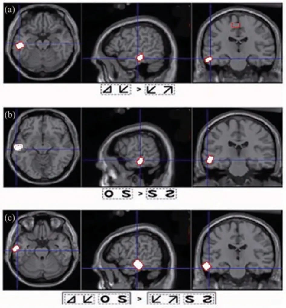
Fig. 2 Excitatory of anterior temporal lobe induced by perceptual discrimination of topological properties[83]
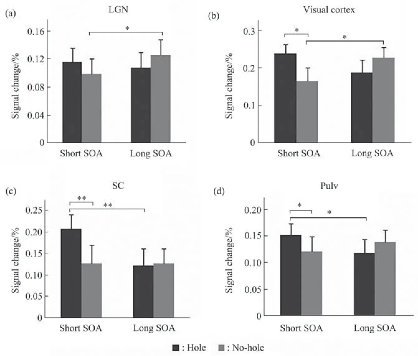
Fig. 3 The mean peak blood oxygen level-dependent (BOLD) signal intensity in LGN (a), visual cortex (b), SC (c) and pulvinar (d)[84]
The superior colliculus is an ancient structure located in the midbrain, which is the first nucleus of visual information processing behind the retina.Classical theory of vision believes that superior colliculus is related to oculomotor control, but in recent years, it has been found that the function of this pathway is much more than that. The superior colliculus receives information directly from the retina and is activated primarily by low spatial frequency and emotion-related visual stimuli,particularly sensitive to unconscious emotional visual stimuli[2,8-11].
In recent years, with the development of new neurobiological technologies, such as brain imaging and photogenetics, we have gained a better understanding of the neural circuits and functions of the superior colliculus[12-17]. In rodents, the superior colliculus, like the amygdala, is associated with fear emotion[18].
The pulvinar nucleus is the second nucleus of visual information processing in this pathway. The pulvinar is involved in the regulation of attention[2,19-21]. When the pulvinar is impaired, the attention capture, induced by emotional visual stimuli on consciousness will be affected. Compared with the superior colliculus, the neural circuit of pulvinar is relatively complex, and it has complex fiber connections with some nuclei of thalamus and cerebral cortex. In humans, with the development of brain imaging technology, more detailed understanding of the anatomy and function of this structure has been achieved[22-28].
The amygdala is the last stage of this pathway and the most widely studied brain structure in this pathway. These studies of amygdala have been mainly focused on following fields: (1) the phylogeny[29-32]and ontogeny of amygdala[33-35]; (2) anatomical and functional connectivity of amygdala with limbic system and prefrontal lobe[36-38]; (3) the functional relationship of amygdala and emotional regulation with some mental disorders, such as autism[39-40];(4) the neural information processing of amygdala during conscious and unconscious state[41-46].
Amygdala is mainly responsible for the information processing of emotions (mainly negative emotions, such as fear), especially emotional processing under consciousness[4,47-54]. The amygdala mainly receive information from two brain regions:one is from the sensory cortex, and the other is from subcortical nuclei[49]. It is generally believed that the amygdala can quickly process some danger information in the environment[9,55], for example,amygdala responds to visual signals from complex objects within 60 ms in monkey[55].
During early perception, the amygdala receives emotion-related information and projects this processed information to the sensory cortex to further regulate attention and perception. Some studies have found that the visual cortex is activated when new emotional stimuli are presented. This excitation is related to the activation of the amygdala. There is a high correlation between the visual cortex and amygdala. Clinical studies have found that this phenomenon disappears in patients with amygdala damage[52].
Subcortical pathways have been extensively studied in humans and some animals[8,56-61]. In one study, magnetic resonance imaging (MRI) was used to track the direction of white matter fibers in the brain of patients with cortical impairment, and it was found that the number of nerve fibers in the subcortical pathway increased, and the three nuclei formed a loop structure[56]. This is strong evidence for the existence of subcortical pathways. Many brain imaging and clinical studies have also confirmed that subcortical pathway plays an important role in the rapid processing of consciously and unconsciously visual information[62-64]. One of the features of this pathway is that it can process non-fine visual information under consciousness, whereas the fine visual information processing relies on classical cortical visual pathways[65]. Studies have shown that, in most cases, the amygdala rapidly analyzes incoming information to see if there are risk factors in the external environment that require a rapid behavioral response[51,53,64,66-71]. It is important for survival.Many emotion-related visual studies, especially the studies of brain response to fearful faces, also suggest that this subcortical pathway mainly processes emotion-related visual information, such as facial expression[3,56,72-76]. Garvertet al.[77]used magnetoencephalography to record the brain activity of people during rapid facial expression analysis, and the results showed that the amygdala played an important role in such analysis. In fact, such facial information processing not only occurs in the amygdala but also in the superior colliculus and the pulvinar[78]. They found that, unlike the face neurons in the cortex, which have very fine encoding for facial features, the superior colliculus and the pulvinar have no extensive encoding for face details. In primates,facial expressions are important for survival, and recent research suggests that fetuses develop a preference for face-like shapes early in life. Newborns can recognize faces several hours after birth[79], at this time the primary visual cortex is far from mature and the recognition of local features of objects is still poor.
2 Topological visual perception
There is an old and unsolved question: “Where does vision begin?” Different from the traditional theory, “from the part to the whole”, the “global first”theory proposed by prof. CHEN Lin[80]suggests that visual perception is “from the whole to the part”.Compared with the perception of other local geometric properties (projective, affine, Euclidean geometry), the perception of topological properties is the most basic and earliest perception. Vision starts with extracting topology properties. However, there is little research on the neural pathways that regulate the topological properties of perception.
2.1 Subcortical processing of visual topological information
In 2003, Zhuoet al.[80-81]suggested that in humans, the anterior temporal lobe may be the region responsible for processing visual topological information. Subsequently, some studies have shown that topological information processing may not require the primary visual cortex[60,82-83]. Notably, the last stop of the subcortical visual pathway, the amygdala, is located ventrally in the anterior temporal lobe. Our series of studies have shown that this subcortical visual pathway is involved in the processing of topological visual information[80-87]and another study suggested that since the subcortical visual pathway is involved in emotion processing, fear emotion strongly interferes with topological processing but has no effect on other visual local features[83], suggesting that emotional and topological processing share common neural circuits (Figure 2).
“Hole” is an important concept in topology. In general, topological differences are most likely to occur in “hole”, such as “with or without hole”.Understanding the brain information processing of hole is very helpful for understanding the topological information processing of the brain[81]. Studies on infants have found that infants are able to distinguish the nature of “hole” by the time they are two to three days old, their visual cortex is far from welldeveloped at this time[79].
In our research, we want to know if the information processing of hole involves the primary visual cortex. To address this issue, (1) we first investigated the response speed of subjects to hole perception and found that the response speed to hole was much faster than the response speed to local features such as line segment direction. (2) With the electrophysiological ERP method, we studied the EEG characteristics induced by hole and line segment.The results showed that the N1 component of ERP induced by hole were smaller than that induced by line segment. N1 components in the primary visual cortex are activated more by local geometric visual stimuli than by hole[82]. (3) If the information processing of hole also involves the subcortical pathway? To address this issue, a backward masking paradigm combined with functional magnetic resonance imaging (fMRI) was applied to measure neural responses to hole and no-hole stimuli in anatomically defined cortical and subcortical regions of interest (ROIs) under different visual awareness levels by modulating masking delays. The results showed that, for no-hole stimuli, the neural activation of cortical sites was greatly attenuated when the nohole perception was impaired by strong masking,whereas an enhanced neural response to hole stimuli in subcortical sites was obtained when the stimulus was rendered invisible. The results suggested that whereas the cortical pathway was required to drive a perceptual response for no-hole stimuli, a subcortical pathway might be involved in coding the hole feature,resulting in a rapid hole perception in primitive vision[84](Figure 3).
Also we want to know if primary visual cortex is involved in topological information processing. Visual studies have found that visual function decline in elderly people is mainly manifested by decreased visual resolution, decreased visual spatial/depth perception and decreased visual acuity[70]. Therefore,we selected people with different ages as research subjects. The objective is to compare the differences in topological perception and other local geometric perception between the elderly and normal subjects.
In all groups, there was no difference in the average response time in the topological property discrimination task. However, there was a significant difference in the response time of the local geometric property discrimination task in the group of subjects aged over 70 years. This result supports the hypothesis that functional degradation of the visual cortex does not affect topological perception but affects local geometric perception. With the functional degradation of the visual cortex, the perception of topological properties in elderly people were not affected, indicating that topological perception does not require the involvement of the primary visual cortex[85].
To directly verify whether the primary visual cortex participates in the processing of visual topological information, we inhibited the function of the primary visual cortex to determine whether it has the same effect on topological information and nontopological information processing. In this experiment, we used transcranial magnetic stimulation(TMS) to inhibit the primary visual cortex. The results show that non-hole perception was impaired, but not hole perception, strongly suggesting that information processing of hole is independent of the primary visual cortex[86].
2.2 The brain still has advantages in unconscious topological information processing
Previous studies have demonstrated that hole features have an advantage over non-hole features in conscious states. However, does the brain still have an advantage in unconsciously processing visual information? The binocular competition paradigm has become a common approach in consciousness research in recent years[45]. We used this paradigm to study this problem. Our results show that hole stimulation also has advantages over non-hole stimulation in unconsciously processing visual information. These results suggest that hole features can be processed unconsciously and that hole are more likely to break through the inhibition induced by binocular competition and enter consciousness than non-holes[87].
The above experiments consistently prove that the perception of topological properties is not carried out through classical visual pathways.
This evidence clearly indicates that local feature perception and topological feature perception are separated in the brain. Therefore, we believe that the functions of subcortical pathways are not limited to emotion-related visual information processing but have many more functions, such as early and rapid processing of the overall nature of visual information.
3 Topological perception and subcortical pathway may be the neural basis of early warning
As mentioned above, subcortical pathway process both fear emotion, and topological information. Can we have a unified explanation about this? For this problem, our explanation is as following:
(1) Topology change and warning
We know that the vision in non-primate mammals is underdeveloped[75]. Most non-primate mammals do not have full trichromatic vision and are even color blind. In addition, the retina of non-primate mammals lacks a macular and has more rods and fewer cones than the retina of primates[88-91]. Thus, in addition to lacking complete color vision, these mammals also lack fine vision. Non-primate mammals receive olfactory, auditory and visual information from the outside world. For the warning response of external risk factors, the most effective information should be visual information, however,without color vision and fine vision, the external world in their eyes is very dim. This raises a question,what is the warning information extracted by the visual system?
In topological mathematics, the calculated objects have no concept of size, length and shapes. In fact, topology studies the invariance and continuity of objects in the process of spatiotemporal change. When the eyes detect an object appears or disappears in the background, the environment will undergo topological changes according to the definition of topology. In the actual situation, a change in the environment may mean danger coming. Therefore, the ability to visually detect topological transformation is an innate defense and self-protection ability of animals. In non-primate mammals, although they cannot detect the shape,texture and other geometric features of objects, but their vision is competent for topological information processing. Newborns are sensitive to the topological changes, it is a defensive mechanism. Subcortical pathways in mammals may be responsible for this early warning function.
(2) Hole and face perception
Our study suggests that primates, possibly all mammals, have an innate discrimination advantage for hole. We consider that this advantage may be related to fear. From the perspective of the evolution of the visual pathway, the eye of non-primate mammalian has no cortical visual pathway, lacks fine vision and color vision, so it does not fit to see the landscape, but fits to detect changes in the environment , these changes may bring death to the animal.
In order to avoid danger, species have developed a set of defense mechanisms in evolution. One of these mechanisms is the establishment of fear which produces fear emotional response. Many of us have an inexplicable fear of holes. Why do we have such a fear? Several year ago, we have speculated that in a living environment, a hole, like a cave, might harbor a deadly killer, so animals are instinctively wary of holes and caves[92]. However, after our further analysis later, we believe that the sensitivity to holes is more likely to be the sensitivity to faces. Our face is made up of 7 holes, and the 3 holes, the eyes and mouth are the most distinctive features of the face. Of the 3 holes, the eye is the most sensitive.
When tigers are hunting, they always attack from behind of prey rather than from front of prey. Bengal tigers are often seen in the mountains of Southeast Asia. In order to prevent the tigers from attacking people from behind, local people always put a picture of a human face on their backs.
So how does an animal know if it’s face to face with an opponent? Obviously, by looking at the opponent’s face, if the eyes and mouth can be seen, it means face to face and danger, it should immediately adjust the strategy to avoid danger and damage. When fighting face to face, animals always open their eyes and mouths wide to show the “holes” in their faces and intimidate each other. Almost all mammals fear eye-to-eye contact. When someone stares into another one’s eyes, it often causes alarm in this one being stared at.
In addition to the eyes, the animal’s mouth is also an important organ to convey information. When an animal sees a hole, it may mean a killer with a gaping mouth coming towards it. Usually to display power, animals are often open “big mouth” to give a warning to the opponent.
As mentioned above, newborns can distinguish faces and holes/non-holes clearly, in this time, their vision is still very blurred, and a study has reported that human fetuses show a preference for face-like shapes. Evolutionary principles suggest that these first developed functions must be relevant for individual survival.
In summary, in the early stage of vision, the brain detects signals that involve life or safety, which warns the brain that something appears and puts the brain into a state of alert that is essential to the survival of the species. Therefore, in the early stage of vision, the objects that need to be detected are only“appearance” and “disappearance”, rather than“texture”, “shape”,etc. Both “appearance” and“disappearance” are changes in topological features.The existence of topological perception and subcortical pathways may be the neural basis of early warning. “Early warning” may be one of the biological mechanisms of the existence of subcortical pathways.
4 The eye of primates is a Mosaic of an older visual system and a younger one
When talking about the evolution of the nervous system, it is generally believe that as the cerebral cortex develops, the visual function of the midbrain was replaced by the function of cerebral visual cortex.The visual function of the midbrain became less important. However, from the above analysis, we can see that the subcortical visual pathway is very important for the survival of individuals, and its warning function enables individual to avoid risks.However, this important function does not exist in the cerebral visual cortex. We often say that in primates,the eye is a perfect structure, and not just because we have perfect fine and color vision, both of which are done by cortical visual pathways. But we may not notice that the peripheral area of retina is distributed mainly with rod cells, the visual information processing in this area is mainly processed by subcortical visual pathway, this type of retinal structure existed more than one hundred million years ago. The area close to the center of the retina is the macular, the visual information processing in this area is mainly processed by visual cortex, cone cell density increased greatly, the visual spatial resolution becomes very high. This structure appeared only 50 million years ago, so our eyes span at least 50 million years from the peripheral region to the central region,it is a Mosaic of an older structure and a younger one,and their coexistence gives us perfect vision. When we consider the significance of the existence of subcortical pathway, we should also consider the function of cortical pathways.
Even though we have the above findings, we do believe that we are still very shallow understanding of subcortical pathways. Besides the functions we mentioned above, are there more functions than those mentioned above? For example, some studies have found that many diseases, such as epilepsy, chronic pain, depression, autism are related to the amygdala[93-94],we do not know the relationship between these diseases and the amygdala function. Is there a unified mechanism behind these phenomenas and are the functions discussed above are only subfunction? This is also something we need to further study.