八宝丹通过AKT与ERK信号通路抑制乳腺癌4T1细胞增殖
2021-08-23蓝兆何嘉陈进晓沃达马恩彭军朱伟东任丹妮
蓝兆 何嘉 陈进晓 沃达 马恩 彭军 朱伟东 任丹妮
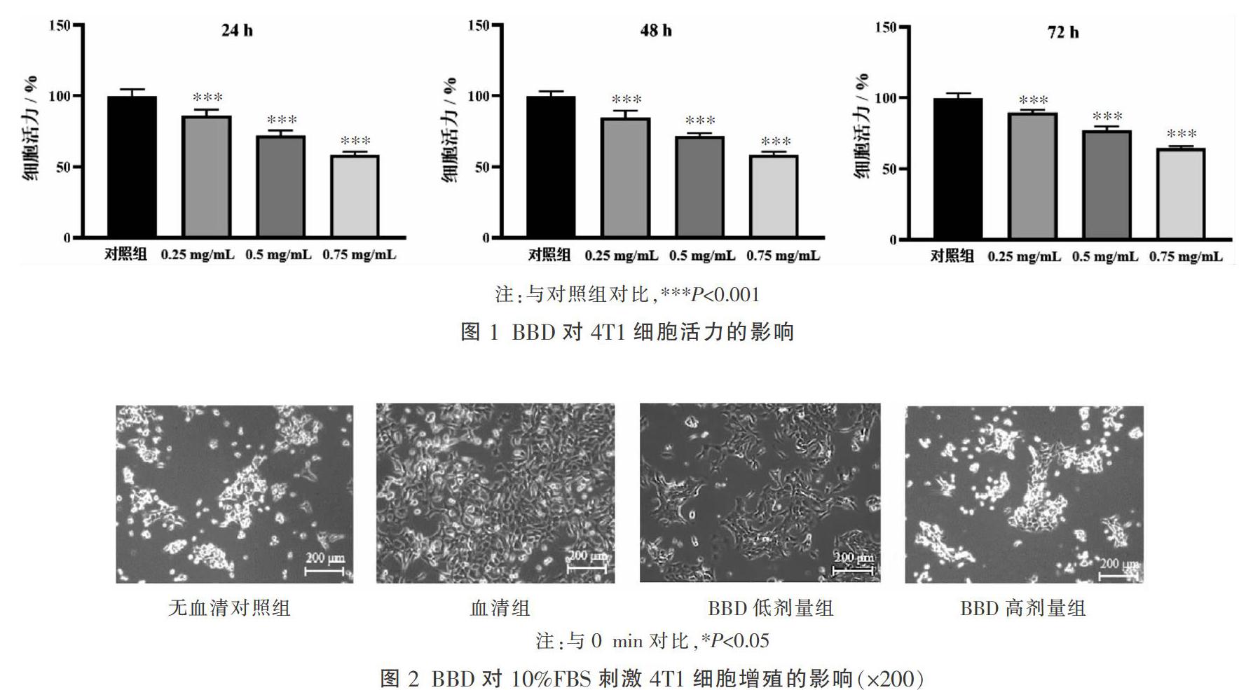
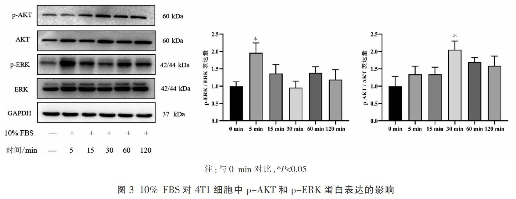
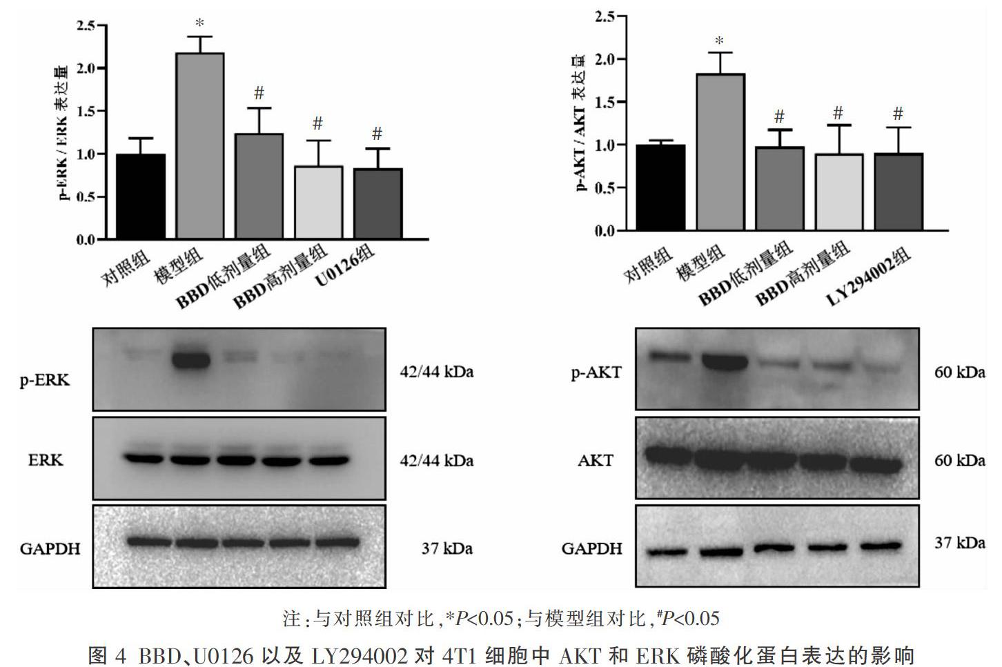
〔摘要〕 目的 探讨八宝丹(Babao Dan, BBD)通过AKT与ERK信号通路对小鼠乳腺癌(4T1)细胞增殖的影响。方法 CCK8法检测不同浓度(0.25、0.5、0.75 mg/mL) BBD干预不同时间点(24、48、72 h)的4T1细胞活力;镜下观察无血清对照组、血清组、BBD低剂量组(0.5 mg/mL)、BBD高剂量组(0.75 mg/mL)中4T1细胞的增殖变化;Western blot法检测10%胎牛血清(FBS)刺激不同时间点(0、5、15、30、60、120 min)后4T1细胞中p-AKT、p-ERK的蛋白表达水平,以确认信号通路激活的最佳时间。与此同时,将4T1细胞随机分为6组:对照组、模型组、BBD低剂量组、BBD高剂量组、U0126组(10 μmol/L)和LY294002组(20 μmol/L),采用Western blot法检测p-AKT、p-ERK的蛋白表达水平;最后将对数增长期的4T1细胞随机分为对照组、BBD组、U0126组(ERK信号通路抑制剂)、LY294002组(AKT信号通路抑制剂)、BBD+U0126组、BBD+LY294002组、U0126+LY294002、BBD+U0126+LY294002组8组,通过台盼蓝染色法检测以上各组干预24、48、72 h后对4T1细胞增殖的影响。结果 CCK8法结果表明,与对照组相比,不同浓度BBD干预24、48、72 h后细胞活力均显著下降,差异具有统计学意义(P<0.001)。镜下观察可见,与无血清对照组相比,血清组细胞形态饱满且生长迅速;与血清组相比,BBD低剂量组以及BBD高剂量组细胞生长缓慢。Western blot结果显示,与0 min比较,10% FBS干预5 min时p-ERK蛋白表达显著升高(P<0.05),同时30 min p-AKT蛋白表达明显上调并且均达到峰值(P<0.05)。与对照组相比,模型组p-ERK、p-AKT蛋白表达均显著上调(P<0.05);与模型组相比,BBD低劑量组、BBD高剂量组p-ERK、p-AKT蛋白表达均显著下调(P<0.05),与U0126组和LY294002组抑制效果相当。台盼蓝染色法结果可见,与对照组相比,BBD组、U0126组以及LY294002组均可显著抑制4T1细胞增殖(P<0.001),其中BBD+U0126+LY294002组与U0126+LY294002组抑制效果相似,均可发挥显著增殖抑制作用(P<0.001),且24、48、72 h结果一致。结论 BBD可显著抑制4T1细胞增殖,其机制可能与抑制AKT与ERK信号通路表达有关。
〔关键词〕 八宝丹;乳腺癌;细胞增殖;ERK;AKT;磷酸化
〔中图分类号〕R285.5 〔文献标志码〕A 〔文章编号〕doi:10.3969/j.issn.1674-070X.2021.07.009
〔Abstract〕 Objective To investigate the effect of Babao Dan (BBD) on the proliferation of mouse breast cancer 4T1 cells through AKT and ERK signaling pathways. Methods CCK8 assay was used to detect the 4T1 cells viability treated with different concentrations BBD (0, 0.25, 0.5, 0.75 mg/mL) in different time points (24, 48, 72 hours); the proliferation of 4T1 cells in serum-free control group, serum group, low-dose BBD group (0.5 mg/mL) and high-dose BBD group (0.75 mg/mL) were observed under microscope. Western blot was used to detect the activation of AKT and ERK signaling pathways in 4T1 cells treated with fetal bovine serum at different time points (0, 5, 15, 30, 60, 120 minutes) to confirm the optimal time to activate the signaling pathway. In addition, the cells were divided into 6 groups: control group, model group, BBD low-dose group, BBD high-dose group, U0126 group (10 μmol/L), LY294002 group (20 μmol/L), Western blot was used to detect the protein expression of p-AKT, AKT, p-ERK and ERK. Finally, 4T1 cells in logarithmic growth phase were randomly divided into control group, BBD group, U0126 group (ERK signaling pathway inhibitor), LY294002 group (AKT signaling pathway inhibitor), BBD + U0126 group, BBD + LY294002 group, U0126 + LY294002 group, BBD + U0126 + LY294002 group, the effects of the above groups on the proliferation of 4T1 cells after 24, 48 and 72 hours of intervention were detected by trypan blue staining. Results CCK8 assay showed that compared with control group, the cell viability was significantly reduced treatment with different concentrations of BBD for 24, 48, 72 hours, the differences were statistically significant (P<0.001); microscopic observation showed that compared with the serum-free control group, the cells in the serum group were full in shape and grew rapidly, compared with the serum group, the cells in the BBD low-dose group and the BBD high-dose group grew slowly. Western blot showed that, compared with 0 minute, the protein expression of p-ERK was significantly increased at 5 minutes treated with 10% fetal bovine serum (P<0.05), and the protein expression of p-AKT was significantly increased and reached the peak value at 30 minutes simultaneously (P<0.05); compared with model group, the protein expression of p-ERK and p-AKT was significantly decreased in BBD low-dose group and BBD high-dose group (P<0.05), which was equivalent to U0126 group and LY294002 group. Trypan blue staining showed that compared with the control group, BBD group, U0126 group and LY294002 group could significantly inhibit the proliferation of 4T1 cells (P<0.001), and BBD+U0126+ LY294002 group and U0126+ LY294002 group had similar inhibitory effect, and they could play a significant inhibitory effect on proliferation (P<0.001), the results were consistent at 24, 48 and 72 hours. Conclusion BBD can inhibit the proliferation of mouse breast cancer cells 4T1, its mechanism may be related to the inhibition of the expression of AKT and ERK signaling pathways.
〔Keywords〕 Babao Dan; breast cancer; cell proliferation; ERK; AKT; phosphorylation
乳腺癌是世界上最常见的女性高发癌症,疾病呈现快速增长趋势,多趋于年轻化[1],发病率在全球范围逐步攀升,晚期乳腺癌致死率高[2]。恶性肿瘤是在多种病理因素作用之下产生的,主要表现为细胞异常增殖[3],其中,AKT信号通路是人类癌症最常见的信号通路之一,调控着细胞异常增殖以及肿瘤发生发展的关键环节[4]。在PI3K信号通路中,AKT的磷酸化过程发生在通路上游,能够活化下游诸多分子,促进乳腺癌的发生发展[5]。此外,ERK 1/2是丝裂原活化蛋白激酶超家族成员,主要介导细胞增……