18F-FDG PET/CT/MR异机融合显像联合MR增强扫描在肝癌中的应用价值
2021-03-24鲁力
鲁力
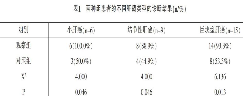
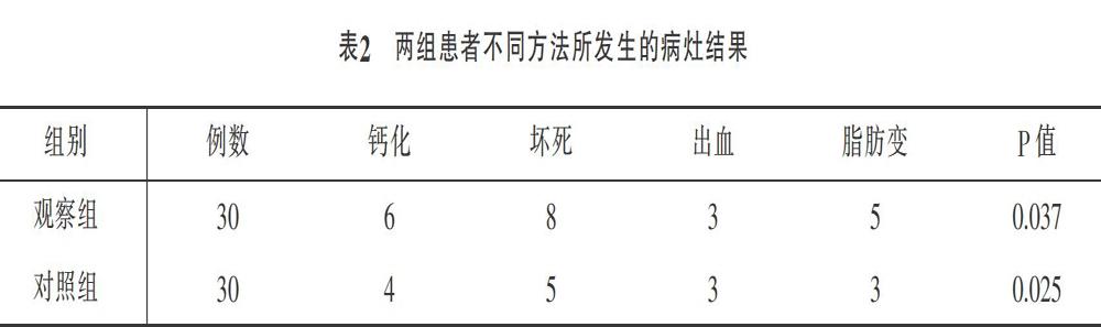
摘要:目的:肝癌患者通过18F -FDG PET/CT/MR异机融合显像技术和MR增强扫描技术进行联合诊断,对诊断价值进行研究分析。方法:选择60例肝癌患者,以随机数字表法分成观察组(n=30)和对照组(n=30),所有患者均接受病理检查。对照组患者通过MR增强扫描进行诊断,观察组患者通过18F-FDG PET/CT/MR异机融合显像技术联合MR增强扫描进行诊断,比较两组患者的小肝癌、结节性肝癌、巨块型肝癌的确诊概率,比较两组检出病灶的例数差异。结果:观察组中小肝癌、结节性肝癌、巨块型肝癌的诊断率均高于对照组[(100%,88.9%,93.3%)vs(50%,44.4%,53.3%)],P<0.05;观察组的检出病灶结果钙化、坏死、脂肪变的病例数均高于对照组患者,P<0.05,差异具有统计学意义。结论:肝癌患者通过18F-FDG PET/CT/MR异机融合显像技术和MR增强扫描进行联合诊断,可以有效诊断出各类型肝癌,诊断价值较高。
关键词:18F-FDG PET/MR显像;MR增强扫描;肝癌
Abstract: Objective: To study and analyze the diagnostic value of combined diagnosis of liver cancer patients by 18F -FDG PET/CT/MR hetero-computer fusion imaging technique and MR-enhanced scanning technique. Methods: 60 patients with liver cancer admitted to our hospital were selected and randomly divided into observation group (n=30) and control group (n=30) from January 2019 to July 2020. According to the pathological examination, there were 6 small and medium-sized liver cancer patients in the two groups, 9 nodular liver cancer patients, and 15 huge fast liver cancer patients. Patients in the control group were diagnosed by enhanced MR scanning, and patients in the observation group were diagnosed by 18F-FDG PET/CT/MR allo-fusion imaging combined with enhanced MR scanning. The two groups of patients were compared for small liver cancer, nodular liver cancer, the diagnosis probability of massive liver cancer was compared between the two groups in the number of cases with lesions detected. Results: The diagnosis rates of small and medium liver cancer, nodular liver cancer, and massive liver cancer in the observation group were higher than those in the control group [(100%, 88.9%, 93.3%) vs (50%, 44.4%, 53.3%)], P<0.05 ; The number of cases of calcification, necrosis, and steatosis detected in the observation group were higher than those in the control group, P<0.05. Conclusion: The combined diagnosis of 18F-FDG PET/CT/MR heterogeneous fusion imaging technology and MR enhanced scan in patients with liver cancer can effectively diagnose various types of liver cancer with high diagnostic value.
Key Words: 18F-FDG PET/MR Imaging; Enhanced MR Scan; Liver Cancer
临床中肝癌的发病率相对较高,据国家癌症中心发布的《2019年全国癌症报告》显示,肝癌的发病率居于我国高发恶性肿瘤的第四位,死亡率位居第二,严重威胁着人类的生命健康[1]。而早期肝癌患者的临床症状不显著,没有特异性[2],容易造成误诊,大多数患者在首次确诊时病情就已经进入中晚期,以致错过了最佳的手术根治时期,影响肝癌患者的预后效果,导致肝癌患者的生存期限普遍较短。因此,提高诊断准確率,做好早期筛查工作对于治疗效果的改善以及五年生存率的提高是十分重要的[3]。近年来通过F-FDG PET/CT成像技术进行检查,将人体生理代谢、分子信息以及解剖形态进行整合,可以有效诊断肝癌。但是,如果肝癌患者体内的肿瘤细胞发生分化,病灶密度低,或者患者存在脂肪肝,通过F-FDG PET/CT成像技术进行诊断,则容易发生误诊以及遗漏病灶。PET/CT/MR异机融合显像技术基于MRI技术,MRI技术具有成像清晰、分辨率高的特点,通过多序列成像可以提高肝癌的诊断准确率。……