彩色多普勒超声对乳腺髓样癌与乳腺纤维瘤的诊断价值
2019-12-09林燕明
林燕明
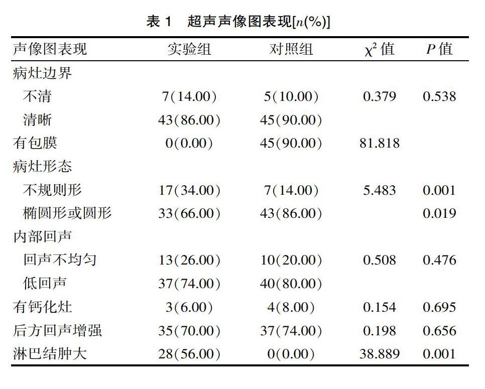

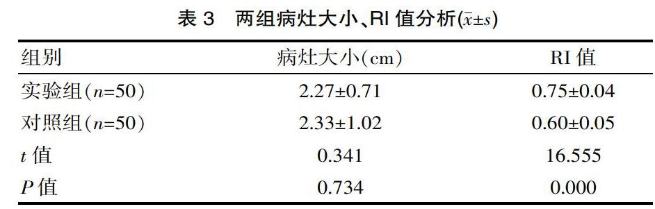
[摘要] 目的 探究乳腺髓樣癌和乳腺纤维瘤诊断中应用彩色多普勒超声的价值。方法 回顾性分析2016年5月—2018年5月福建省三明市永安总医院三明市第二医院超声影像科手术后经病理证实的乳腺髓样癌患者50例为实验组,同时选取乳腺纤维瘤患者50例为对照组,对病灶的二维及彩色多普勒声像图特征包括病灶的形态、边界、内部的回声、腋窝淋巴结转移及彩色血流的特点分析,比较其差异性。 结果 实验组诊断符合率为86.00%,对照组诊断符合率为94.00%;实验组病灶边缘清晰率(86.00%)、后方回声增强率(70.00%)、内部回声不均匀率(26.00%)、大小(2.27±0.71)cm和对照组相比,差异无统计学意义(病灶边缘清晰率相比,χ2=0.379,P=0.538;后方回声增强率相比,χ2值=0.198,P=0.656;内部回声不均匀率相比,χ2=0.508,P=0.476;大小相比,t=0.341,P=0.734);实验组其肿瘤有包膜的检出率(0.00%)明显低于对照组,腋窝淋巴结肿大的检出率(56.00%)、形态不规则检出率(34.00%)明显高于对照组(肿瘤包膜检出率相比,χ2=81.818,P=0.001;腋窝淋巴结肿大检出率相比,χ2=38.889,P=0.001;病灶边缘不规则形态检出率相比,χ2=5.483,P=0.019);实验组1~3级血流的检出率(94.00%)以及其RI值(0.75±0.04)明显高于对照组(1~3级血流的检出率相比,χ2=32.972,P=0.001;RI值相比,t=16.555,P=0.000)。 结论 根据彩色多普勒在乳腺髓样癌与乳腺纤维瘤超声图像差异,可较好对其进行鉴别诊断。
[关键词] 彩色多普勒超声;乳腺髓样癌;乳腺纤维瘤
[中图分类号] R445 [文献标识码] A [文章编号] 1674-0742(2019)09(c)-0184-03
[Abstract] Objective To investigate the value of color Doppler ultrasound in the diagnosis of breast medullary carcinoma and breast fibroma. Methods A retrospective analysis of 50 patients with breast medullary carcinoma confirmed by pathology after operation in the Department of Ultrasound Imaging, Second Hospital of Sanming City, Sanming City, Fujian Province from May 2016 to May 2018, selected breast fibroma 50 patients were in the control group. The two-dimensional and color Doppler sonographic features of the lesion included the morphology, boundary, internal echo, axillary lymph node metastasis and color flow characteristics of the lesion, and the differences were compared. Results The diagnostic coincidence rate of the experimental group was 86.00%, and the diagnostic coincidence rate of the control group was 94.00%. The clear margin of the lesions in the experimental group (86.00%), the enhancement rate of the posterior echo (70.00%), the internal echo heterogeneity rate (26.00%), and the size (2.27±0.71)cm. Compared with the control group, the difference was statistically significant(the lesion edge margin ratio was compared with,χ2=0.379,P=0.538;the posterior echo enhancement rate was compared with,χ2=0.198, P=0.656;internal echo unevenness ratio,χ2=0.508, P=0.476;size comparison,t=0.341, P=0.734); the detection rate of tumor envelope in the experimental group was significantly lower (0.00%). In the control group, the detection rate of axillary lymphadenopathy (56.00%) and the irregular shape detection rate (34.00%) were significantly higher than those of the control group the tumor capsule detestion rate,χ2=81.818, P=0.001. The detection rate of axillary lymph node enlargement was compared with,χ2=38.889, P=0.001; compared with the detection rate of irregular morphology of lesion margin,χ2=5.483, P=0.019; experimental group 1~3 blood detection rate of the flow (94.00%) and its RI value (0.75±0.04) were significantly higher than those of the control group(and the detection rate of 1~3 blood flow was compared,χ2=32.972, P=0.001; RI value compared,t=16.555,P=0.000). Conclusion According to the difference of color Doppler in the ultrasound images of breast medullary carcinoma and breast fibroma, it can be better diagnosed.