肝脏局灶性结节增生MRI平扫及Gd—EOB—DTPA增强扫描的影像表现分析
2017-12-15黄惠贞赵英杰洪天佑
黄惠贞++赵英杰++洪天佑
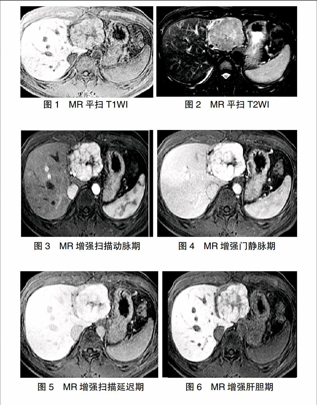
[摘要]目的 分析肝局灶性结节增生(FNH)MRI平扫及钆塞酸二钠注射液(Gd-EOB-DTPA)增强扫描的典型与非典型影像特征。方法 回顾性分析2012年7月~2016年12月我院收治的12例(15个病灶)FNH患者MR平扫及Gd-EOB-DTPA增强扫描的影像学资料。结果 ①MRI平扫显示,15个病灶在T1WI主要呈等信号或低信号,在DWI、T2WI-FS则以高信号为主。MRI平扫发现有7个病灶可见中央瘢痕,其中T1WI均呈低信号,但T2WI则6个呈高信号、1个呈低信号,而DWI则均呈高信号。②MRI增强扫描,动脉期有14个(93.33%)病灶呈明显均匀强化,而门脉期和延迟期的病灶均呈稍高信号。动脉期中央瘢痕均未见明显强化,但门脉期和延迟期则有6个病灶瘢痕延迟性强化。肝胆期FNH有多种强化形式,且其中央瘢痕均呈低信号。结论 MRI平扫及Gd-EOB-DTPA增强扫描能够清楚显示FNH内部结构特点及信号特征,因此掌握FNH的典型及不典型MR影像征象有助于临床疾病鉴别诊断,进而避免不必要的病理活检或手术,减轻患者的痛苦。
[关键词]磁共振成像;肝脏局灶性结节性增生;影像表现
[中图分类号] R735.7 [文献标识码] A [文章编号] 1674-4721(2017)11(a)-0128-04
Imaging analysis of focal nodular hyperplasia MRI and Gd-EOB-DTPA enhanced scanning
HUANG Hui-zhen ZHAO Ying-jie HONG Tian-you
Department of MRI,the Affiliated Xiamen Hospital of TCM of Fujian University of Traditional Chinese Medicine, Fujian Province,Xiamen 361009,China
[Abstract]Objective To analyze the typical and atypical imaging features of hepatic focal nodular hyperplasia by MRI plain scan and Gd-EOB-DTPA enhanced scan.Methods From July 2012 to December 2016,the imaging data of 12 patients(15 lesions) with focal nodular hyperplasia of MRI plain scan and Gd-EOB-DTPA enhanced scanning were retrospectively analyzed.Results ①MRI plain scan showed that 15 lesions were mainly low signal in T1WI,while the DWI and T2WI-FS were mainly high signal.In addition,MRI scan found that 7 lesions showed central scar,of which T1WI were low signal,but T2WI showed 6 high signals and 1 low signals,DWI showed high signal.②MRI enhanced scan showed that 14 lesions in the arterial phase with obvious uniform strengthening,but the lesion of portal and delayed phases showed slightly higher signals.The central scar of arterial period was not significantly enhanced,while there were 6 lesions with delayed enhancement of scar in the portal and delayed periods.The focal nodular hyperplasia of hepatobiliary phase has a variety of enhanced form,and the central scar were low signal.Conclusion MRI scan and Gd-EOB-DTPA enhancement scan can clearly show the internal structure and signal characteristics of the focal nodular hyperplasia.Therefore,mastering the typical and atypical MRI imaging features of focal nodular hyperplasia is helpful to the differential diagnosis of clinical diseases,avoiding unnecessary pathological biopsy or operation,and relieving the pain of patients.
[Key words]MRI;Hepatic focal nodular hyperplasia;Imaging findings
肝臟局灶性结节增生(FNH)是一种常见的肝脏良性病变,患者的预后情况较好,一般无需手术治疗[1]。因此,在临床上提高FNH的诊断准确率,避免非必要的侵入性治疗至关重要。MRI属于新型的临床影像诊断技术,具备较高的软组织分辨率,可以准确检测出病灶。而MRI新型肝脏特异性对比剂Gd-EOB-DTPA则能够对病灶进行动态增强成像,正确显示病灶组织的血供情况[2]。为进一步明确MRI平扫及Gd-EOB-DTPA增强扫描FNH的影像表现,本文回顾性分析12例(15个病灶)FNH的MRI影像学资料,以期为临床早期诊治FNH提供依据。……