肺尖孤立性结节的CT诊断与鉴别诊断
2017-05-08黄恩善陈鹏李瑞雄
黄恩善++陈鹏++李瑞雄
【摘要】目的探讨肺尖孤立性结节的CT影像表现与鉴别诊断。
方法回顾性分析56例经手术及病理证实的肺尖结节患者的CT影像资料。
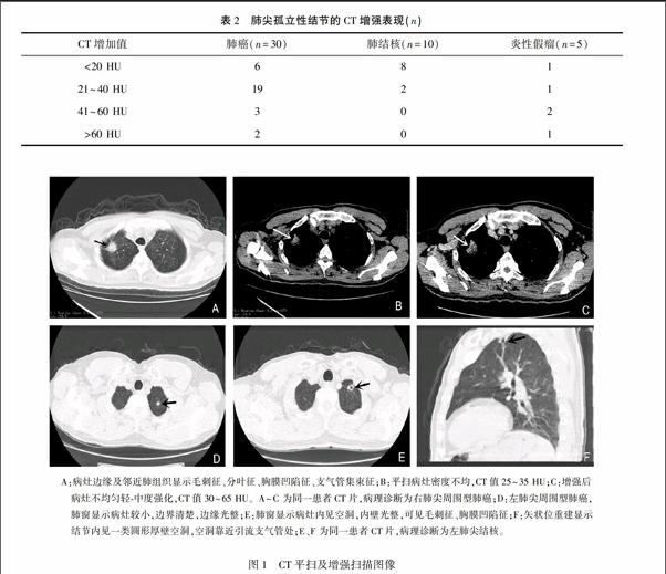
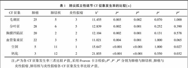
结果56个病灶中有32个肺癌,16个肺结核,8个炎性假瘤。发生率最高的是肺癌,占57%(32/56),其次為肺结核,占29%(16/56)。肺癌毛刺征、分叶征、胸膜凹陷征、支气管集束征发生率较肺结核及炎性假瘤高。而肺结核空洞、钙化的发生率较肺癌及炎性假瘤高。肺癌、肺结核、炎性假瘤三者的各CT平扫征象发生率差异有统计学意义(P<0.05)。CT增强扫描提示肺癌及炎性假瘤以轻中度强化为主,肺结核表现为轻度或无强化。肺癌、肺结核、炎性假瘤在CT增强扫描时,三者在CT增加值幅度<20 HU、21~40 HU、41~60 HU,>60 HU的发生率差异有统计学意义(H=12.463,P=0.002)。
结论CT在肺尖孤立性结节诊断与鉴别诊断中具有重要临床价值。
【关键词】肺尖;结节;体层摄影技术
中图分类号:R814.42文献标识码:ADOI:10.3969/j.issn.10031383.2016.06.020
CT diagnosis and differential diagnosis of solitary pulmonary apex nodules
[HJ2][HJ]
HUANG Enshan,CHEN Peng,LI Ruixiong
(Peoples Hospital of Wuzhou,Wuzhou 543000,China)
[HJ2][HJ]
【Abstract】ObjectiveTo investigate CT manifestations and differential diagnosis of solitary pulmonary apex nodules.
MethodsCT image data of 56 patients with solitary pulmonary apex nodules who were confirmed by surgery and pathology were retrospectively analyzed.
ResultsOf the 56 lesions,there were 32 lung cancer,16 pulmonary tuberculosis,and 8 inflammatory pseudotumor.Lung cancer had highest incidence,accounting for 57%(32/56),followed by tuberculosis,accounting for 29%(16/56).Incidence rates of burr sign,lobulated sign,pleural indentation sign and blood vessels cluster sign of lung cancer were higher than those of tuberculosis and pulmonary inflammatory pseudotumor.There was statistically significant differences in incidence rates of the CT signs of lung cancer,pulmonary tuberculosis and pulmonary inflammatory pseudotumor(P<0.05).CT enhanced scan showed mild to moderate enhancement of lung cancer and inflammatory pseudotumor,and mild or no enhancement to tuberculosis.In CT enhancement scan,difference of incidence rate of CT increase value rang<20 HU,21~40 HU,41~60 HU,and>60 HU was statistically significant(H=12.463,P=0.002).
ConclusionCT has an important clinical value in the diagnosis and differential diagnosis of solitary pulmonary apex nodules.
【Key words】pulmonary apex;nodule;tomography
肺部孤立性结节是诊断工作中常见的肺部病变,其一直是胸部影像研究的重点和难点[1]。肺孤立性结节CT表现为位于肺部的直径≤3 cm的圆形或类圆形结节,不伴肺门淋巴结肿大、肺不张和肺内转移灶。区分结节的良恶性是影像学的最终目的,充分认识结节的特征,正确地评价结节的性质既能使恶性结节患者得到及时的手术治疗,又能使良性结节患者避免不必要的手术。而发生于肺尖的孤立性结节相对少见,近年来较少有文献专门报道,本文研究目的在回顾性分析其CT影像特点,以进一步提高肺尖孤立性结节诊断的准确性。……
