Cripto—1介导的上皮间质转化在卵巢浆液性腺癌形成中的作用
2016-08-19步晓琳
步晓琳
【摘要】 目的 检测Cripto-1、E-钙粘蛋白(E-cadherin)、波形蛋白(Vimentin)在卵巢肿瘤中的表达, 探讨卵巢浆液性腺癌的发生发展机制, 以及Cripto-1介导的上皮间质转化(EMT)在其中的作用。方法 采用免疫组化SP法检测Cripto-1、E-cadherin、Vimentin在60例卵巢浆液性腺癌、30例卵巢交界性浆液性肿瘤、20例卵巢浆液性腺瘤中的表达, 并探讨其表达程度的相关性及与临床病理参数之间的关系。结果 Cripto-1在卵巢浆液性腺瘤中的表达20.0%低于浆液性腺癌76.7%和交界性浆液性肿瘤56.7%, 差异具有统计学意义(P<0.05), E-cadherin在卵巢浆液性腺瘤中的表达90.0%高于浆液性腺癌35.0%和交界性浆液性肿瘤53.3%, 差异具有统计学意义(P<0.05)。Cripto-1、Vimentin在卵巢浆液性腺癌中的表达与组织学分级、淋巴结转移有关(P<0.05);E-cadherin的表达与组织学分级有关(P<0.05), 与淋巴结转移无关(P>0.05)。在卵巢浆液性腺癌中Cripto-1与E-cadherin的表达呈负相关(r=-0.81, P<0.05), Cripto-1与Vimentin的表达呈正相关(r=0.42, P<0.05)。结论 在卵巢浆液性腺癌中Cripto-1、Vimentin表达上调, E-cadherin表达下调, 提示Cripto-1介导的EMT可能在卵巢浆液性腺癌侵袭转移过程中起作用。
【关键词】 Cripto-1;E-钙粘蛋白;波形蛋白;卵巢浆液性腺癌;上皮间质转化
DOI:10.14163/j.cnki.11-5547/r.2016.22.001
Effect by Cripto-1 mediated epithelial-mesenchymal transition in formation of ovarian serous adenocarcinoma BU Xiao-lin. Department of Pathology, Xinglin Branch of First Affiliated Hospital of Xiamen University, Xiamen 361000, China
【Abstract】 Objective To detect expression of Cripto-1, E-cadherin and Vimentin in ovarian tumor, and to investigate development mechanism of ovarian serous adenocarcinoma and effect by Cripto-1 mediated epithelial-mesenchymal transition (EMT). Methods Immunohistochemical SP test was applied to detect expression of Cripto-1, E-cadherin and Vimentin in 60 cases with ovarian serous adenocarcinoma, 30 cases with ovarian borderline serous tumor and 20 cases with ovarian serous adenoma. Investigation was made on relationship between expression degree and clinical pathological parameters. Results Expression of Cripto-1 was lower in ovarian serous adenoma as 20.0% than 76.7% in ovarian serous adenocarcinoma and 56.7% in ovarian borderline serous tumor, their difference had statistical significance (P<0.05). Expression of E-cadherin was higher in ovarian serous adenoma as 90.0% than 35.0% in ovarian serous adenocarcinoma and 53.3% in ovarian borderline serous tumor, their difference had statistical significance (P<0.05). Expression of Cripto-1 and Vimentin showed their relationship with histological grade and lymphatic metastasis in ovarian serous adenocarcinoma (P<0.05); while expression of E-cadherin was related with histological grade (P<0.05) and not related with lymphatic metastasis (P>0.05). Cripto-1 and E-cadherin showed negatively correlated expression in ovarian serous adenocarcinoma (r=-0.81, P<0.05), and expression of Cripto-1 and Vimentin showed their positive correlation (r=0.42, P<0.05). Conclusion Up-regulated expression of Cripto-1 and Vimentin and down-regulated expression of E-cadherin in ovarian serous adenocarcinoma suggest the effect by Cripto-1 mediated EMT in invasion and metastasis of ovarian serous adenocarcinoma.
【Key words】 Cripto-1; E-cadherin; Vimentin; Ovarian serous adenocarcinoma; Epithelial-mesenchymal transition
卵巢癌是女性生殖器官常见的恶性肿瘤之一, 是卵巢恶性肿瘤最常见的组织类型, 因为其组织学类型复杂, 早期病情隐匿, 大部分患者发现到确诊时肿瘤细胞已经播散和转移, 错过了最佳治疗时间, 治疗效果不佳, 生存期短, 是女性生殖系统中病死率最高的肿瘤。卵巢癌的侵袭转移方式主要有两种:①直接在腹腔内播散和血管淋巴管转移;②研究发现, EMT是癌细胞发生侵袭转移的重要机制之一[1]。在EMT的发生过程中, 肿瘤细胞不仅形态学上发生改变, 其表达的标志物也会随之发生变化。最近一项研究表明, Cripto-1可以促进EGFR基因突变的非小细胞肺癌患者的EMT[2]。本研究旨在探讨Cripto-1介导的EMT在卵巢浆液性腺癌发生发展过程中的作用, 现报告如下。
1 材料与方法
1. 1 标本来源 选取2010~2015年厦门大学附属第一医院病理确诊卵巢浆液性腺癌患者60例。年龄24~73岁, 中位年龄58岁, 平均年龄56.3岁。术前未经辅助治疗。选取同期病理确诊的卵巢浆液性腺瘤患者20例及交界性浆液性肿瘤患者30例作为对照。所有标本均经10%福尔马林固定, 常规脱水、石蜡包埋, 将蜡块制备3 μm厚度连续切片。
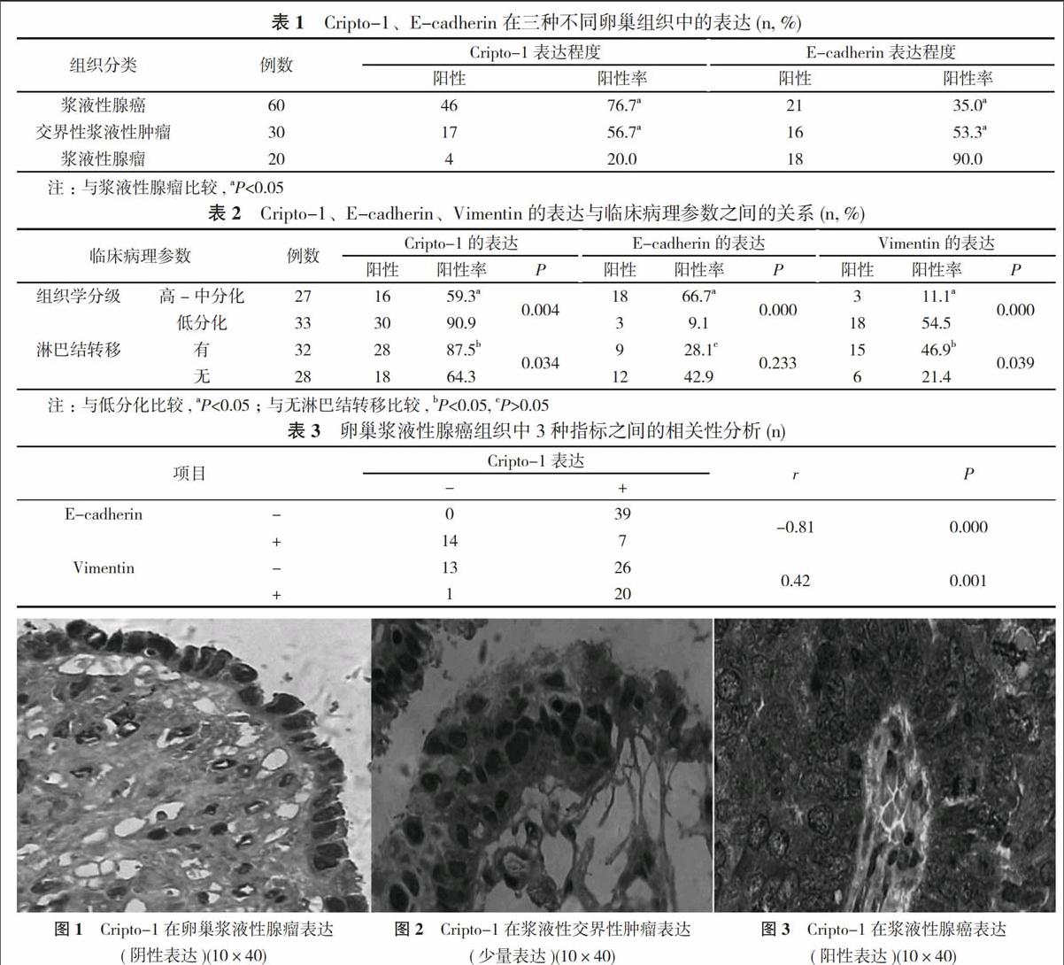
1. 2 结果判定 采用免疫组化SP法, 按试剂盒说明书操作, Cripto-1主要定位于细胞质, 出现棕黄色颗粒状染色为阳性;E-cadherin蛋白主要定位于细胞膜, 细胞膜呈棕黄色颗粒为阳性;Vimentin蛋白定位于胞浆, 胞质中呈棕黄色颗粒为阳性表达。
1. 3 统计学方法 采用SPSS17.0统计学软件处理数据。计数资料以率(%)表示, 采用χ2验;相关性采用Spearman相关分析。P<0.05表示差异具有统计学意义。
2 结果
Cripto-1在卵巢浆液性腺瘤中的表达低于浆液性腺癌和交界性浆液性肿瘤, 差异具有统计学意义(P<0.05), E-cadherin在卵巢浆液性腺瘤中的表达高于浆液性腺癌和交界性浆液性肿瘤, 差异具有统计学意义(P<0.05)。见表1。Cripto-1、Vimentin在卵巢浆液性腺癌中的表达与组织学分级、淋巴结转移有关(P<0.05);E-cadherin的表达与组织学分级有关(P<0.05), 与淋巴结转移无关(P>0.05)。见表2。在卵巢浆液性腺癌中Cripto-1与E-cadherin的表达呈负相关(r=-0.81, P<0.05), Cripto-1与Vimentin的表达呈正相关(r=0.42, P<0.05)。见表3。Cripto-1分别在卵巢浆液性腺瘤、浆液性交界性肿瘤、浆液性腺癌中的免疫组化染色见图1, 图2, 图3。
3 讨论
肿瘤细胞发生侵袭转移是卵巢癌死亡的主要原因, 其机制尚未十分明确, 一直以来是研究者关注的热点。肿瘤细胞由良性向恶性转变时, 为适应与周围基质的接触, 其形态学和分子机制也随之发生了变化, 上皮细胞经去分化转变为间叶细胞, 同时细胞的粘附力降低, 运动迁移能力增强, 即EMT, 既往有研究报道, EMT与获得干细胞样特性有关[3]。Luo等[4]发现, 卵巢癌EMT的发生与干细胞的形成密切相关, 而干细胞对传统治疗手段不敏感[5, 6]。积极的探索卵巢浆液性腺癌的发病机制, 从初始阶段抑制肿瘤细胞的发生发展, 对卵巢癌患者的防治尤为重要。
E-cadherin是跨膜糖蛋白, 主要位于上皮细胞, 能抑制基质金属蛋白酶的产生, 通过介导上皮细胞间的粘附, 促进同型细胞间连接的紧密性, 维持细胞形态、运动及粘附能力。发生EMT时, 正常上皮细胞表面的E-cadherin表达下调, 而代表间质细胞表达的标记蛋白表达升高, 导致癌细胞之间粘附能力下降, 加快细胞的侵袭转移[7, 8]。本研究中, 在浆液性腺瘤中E-cadherin的表达程度较高, 其表达程度与浆液性腺癌患者的组织学分级有关(P<0.05), 肿瘤从良性向交界性再向恶性转变过程中, 其表达程度越来越低, 并且表达程度与浆液性腺癌患者的分化有关, 在高-中分化组织中的表达高于低分化, 说明从良性到癌这个转变过程中其细胞间的粘附能力越来越差。Vimentin是间质表达的标记物, 但在一些低分化恶性肿瘤的上皮组织中也有表达。本研究中Vimentin的表达水平与卵巢浆液性腺癌患者的组织学分级和淋巴结转移有关(P<0.05), 分化越差的组织中其表达程度越高, 说明在一些肿瘤发生过程中, 其细胞形态和生长方式也发生变化, 上皮细胞向纤维细胞样形态转变, 更易移动, 进而引起EMT的发生。
Cripto-1基因为表皮生长因子家族的成员。在人几种不同类型的肿瘤中过表达, 但其生物学行为在恶性肿瘤中的机制不明。本研究发现, 浆液性腺瘤患者Cripto-1的表达水平较低, 其表达程度与浆液性腺癌患者的组织学分级和淋巴结转移有关(P<0.05)。提示其阳性的正常组织细胞存在有恶变的潜在风险, 高表达可能促进肿瘤生长。进一步行Spearman相关分析结果显示, 在癌组织中Cripto-1的表达程度较高, 而E-cadherin的表达程度较低, 二者之间呈负相关(r=-0.81, P<0.05), 而与Vimentin的表达程度呈正相关(r=0.42, P<0.05), 由此可见, 随着病变恶性程度的增高, Cripto-1的阳性表达增高, E-cadherin表达下调, Vimentin表达于上皮中。本次研究发现, 当Cripto-1表达较高时, E-cadherin的表达受到抑制, 而其表达程度降低说明细胞间粘附能力下调, 进而促进上皮间质转化的发生, 肿瘤细胞即播散种植于腹盆腔, 发生侵袭转移。其机制可能是, Cripto-1 通过激活Ras-Raf-MEK- ERK和PI3K-Akt信号通路刺激肿瘤细胞的增殖与存活。但EMT的发生是多因子共同作用的结果, 是肿瘤发生发展过程中的重要枢纽, 对其发生的分子机制还有待进一步研究, 早期阻断EMT的转变可以提高患者的远期生存率, 并且为治疗提供宝贵的机会。
综上所述, 在卵巢浆液性腺癌中Cripto-1、Vimentin表达上调, E-cadherin表达下调, 提示Cripto-1介导的EMT可能在卵巢浆液性腺癌侵袭转移过程中起作用。
参考文献
[1] Schaefer D, Somarelli JA, Hanna G, et al. Cellular migration and invasion uncoupled: increased migration is not an inexorable consequence of epithelial-to-mesenchymal transition. Mol Cell Biol, 2014, 34(18):3486-3499.
[2] Park KS, Raffeld M, Moon YW, et al. CRIPTO1 expression in EGFR-mutant NSCLC elicits intrinsic EGFR-inhibitor resistance. J Clin Invest, 2014, 124(7):3003-3015.
[3] Mani SA, Guo W, Liao MG, et al. The epithelial-mesenchynml transion generates cells with properties of stem cells.Cell, 2008, 133(4):704-715.
[4] Luo X, Dong Z, Chen Y, et al. Enrichment ofovarian cancer stem-like cells is associated with epithelial to mesenchymal transition through an miRNA-activated AKT pathway.Cell Prolif, 2013, 46(4):436-446.
[5] Blagosklonny MV. Cancer stem cell and cancer stemloids: from biology totherapy. Cancer Biol Ther, 2007, 6(11):1684-1690.
[6] Ishii H, Iwatsuki M, Ieta K, et al. Cancer stem cells and chemoradiation resistance. Cancer Science, 2008, 99(10):1871-1877.
[7] Ksiazkiewicz M, Markiewicz A, Zaczek AJ. Epithelial-mesenchy-mal transition:a hallmark in metastasis formation linking circulating tumor eels and cancer stem cells. Pathobiology, 2012, 79(4):195-208.
[8] Guo F, Parker Kerrigan BC, Yang D, et al. Post-transcriptional regulatory network of epithelial-to-mesenchymal and mesenchymal- to-epithelial transitions. J Hematol Oncol, 2014, 7(1):7-19.
[收稿日期:2016-03-25]