PFOS对东亚三角涡虫HSP70蛋白及基因表达的影响
2014-03-21宫晓宁袁佐清白芸
宫晓宁 袁佐清 白芸
(山东理工大学生命科学学院,淄博 255049)
PFOS对东亚三角涡虫HSP70蛋白及基因表达的影响
宫晓宁 袁佐清 白芸
(山东理工大学生命科学学院,淄博 255049)
全氟辛烷磺酸(Perfluorooctane sulfonate,PFOS)是一种持续的有机污染物,是全氟化合物(Perfluorinated chemicals,PFCs)在环境中的代表性物质。以低等三胚层动物涡虫作为试验材料,使用Western blot及RT-PCR技术探究PFOS对涡虫HSP70蛋白及基因表达的影响。结果表明,PFOS短时间处理能诱导涡虫hsp70 mRNA表达量升高,涡虫再生10 d mRNA表达受到抑制,HSP70蛋白表达量在涡虫再生4 d和7 d时随PFOS浓度显著上升,其它组变化不明显,PFOS对mRNA和蛋白表达量趋势影响不一致,可能少量PFOS能抑制HSP70蛋白翻译,药物在涡虫体内积累量增加,蛋白高表达抵御毒性伤害。
PFOS 三角涡虫 HSP70 RT-PCR Western blot
以全氟辛烷磺酸盐(Perflourooctane,PFOS)为代表的全氟化合物(PFcs),曾被作为无毒化合物使用了50多年,使用范围涉及生产生活的诸多方面[1-3]。PFOS具有稳定的C-F结构,不易降解,具有生物积累作用,能够通过食物链富集,从陆地到海洋,从低等藻类到高等哺乳类和鸟类都能检测到PFOS的存在[4]。已知PFOS 集中分布于肝脏、肾脏、肺脏、血液等部位[5,6],损害生物体的内脏器官[5,7-9],并对发育生殖过程具有毒害作用。毒性机理和药代动力学研究显示,PFOS能够对体外培养的细胞造成氧化压力,破坏细胞膜,改变基因表达,损伤基因结构等[9-12],但对其毒性作用原理仍不明确。
目前,对PFcs的研究多集中于小白鼠、鱼类等脊椎动物,在无脊椎动物方面的研究仍很匮乏。本试验以涡虫为对象,其作为低等扁形动物,对环境变化十分敏感,并且具有强大的再生能力,是研究再生与凋亡平衡的良好材料。涡虫暴露于高浓度PFOS中,产生一种外界环境压力,而hsp70正是一种生物体应对外界压力的标志基因[13]。本研究通过使用Western blot及RT-PCR技术检测PFOS诱导完整涡虫和再生涡虫HSP70蛋白及基因表达的变化,旨在初步探究PFOS对涡虫的毒性作用。
1 材料与方法
1.1 材料
东亚三角涡虫采自博山泉河头的泉水中,22℃凉开水培养,试验前饥饿7 d处理。PFOS产自ALORICH公司,DMSO(Dimethyl Sulfoxide)产自Klontech公司。RNA抽提液Trizol购自Invitrogen公司,反转录酶、RNase抑制剂、dNTP、Taq酶等均购自Thermo公司,一抗和二抗分别为Santa Cruz生物技术公司生产的HSP70/HSC70(sc-33575)和bovinerabbit IgG-HRP(sc-2370),Bioss生 产 的Anti-beta-Actin(Loading Control)(bs-0061R)。DAB显色液购自天根公司。
1.2 方法
1.2.1 RT-PCR用涡虫的处理 PFOS以0.005%的DMSO溶液溶解,配置为0、0.5、1、5、15和30 mg/L 6个浓度,每个浓度3个处理,每个处理10条涡虫,每天更换1次培养溶液。处理1、4和10 d后取涡虫提总RNA。再生涡虫自耳突下缘切去头部,PFOS处理方式与完整涡虫的相同。
1.2.2 涡虫总RNA的提取及cDNA的合成 将10条涡虫置于1.5 mL离心管中,吸干水,液氮中迅速冷冻,使用塑料杵子研磨成粉末,加入0.2 mL Trizol,继续研磨成糊状,4℃ 12 000 r/min离心5 min,取上清液加入1/5体积三氯甲烷,4℃ 12 000 r/min离心15 min,取上清加入等体积异丙醇,4℃ 12 000 r/min离心10 min,弃掉上清,加入1 mL 70%乙醇洗涤沉淀,4℃ 12 000 r/min离心5 min,弃乙醇,干燥皿中干燥1 h,加入适量无RNase水溶解。使用Reverse Transcriptase M-MLV进行涡虫总RNA反转录,合成第一链cDNA。
1.2.3 PCR及电泳 自NCBI数据库取得Djhsp70 mRNA全长序列,设计一对引物如下:hsp70 sense:5'-GGAGATACGCATTTAGGC-3',hsp70 anti-sense:5'-TCAACTGGTTCCAAGGTC-3',内参β-actin sense[14]:5'-GGATGATGAGATGCGATGTTG-3',β-actin antise-nse:5'-ATGCCAGGTCCAGATTCGTCA-3'。PCR反应程序为:94℃ 5 min;94℃ 50 s,52℃ 50 s,72℃60 s,30个循环;72℃ 10 min。保存电泳图片并使用软件进行亮度分析。
1.2.4 Western blot用涡虫的处理及蛋白提取 以0、0.5、1、5和10 mg/L 5个浓度的PFOS溶液处理涡虫,每个浓度3个处理,每个处理30条涡虫,每天更换一次培养液,在1、4和7 d三个时间点取涡虫提蛋白样品,试验分再生组与非再生组。提取蛋白过程为:涡虫放入1.5 mL EP管,洗净,将水吸干,液氮中瞬时冷冻,加适量PBS(M/V=1∶100,PBS 0.01 mol/L,pH7.4),用塑料杵子研磨至糊状,4℃10 000 r/min离心30 min,取上清,4℃ 10 000 r/min离心15 min,取上清分装保存。
1.2.5 Western blot检测 涡虫体内HSP70蛋白变化趋势以Western blot法进行检测,β-Actin作内参。蛋白样品以10% SDS-PAGE胶电泳(Bio-Rad100-120型电泳仪,Bio-Rad miniprotea垂直电泳槽),半干法转 膜(15 V,18 min,Bio-Rad Trans-Blot SD Cell),PBST漂洗一遍,5%脱脂奶粉室温封闭2 h,4℃一抗孵育12 h,PBST漂洗3遍,4℃二抗孵育10 h,PBST漂洗数遍,DAB显色。
2 结果
2.1 RT-PCR结果
PFOS诱导完整涡虫hsp70 mRNA相对表达量变化见图1和图2。完整涡虫的RT-PCR结果(图1)显示,在1 d时,1-30 mg/L组呈现高表达,表达量超过对照组的1.5倍,到4 d和10 d时这种高表达不明显。浓度为30 mg/L的PFOS超出涡虫的承受能力,在处理10 d时,涡虫大量死亡,无法完成RNA提取,此组涡虫hsp70表达量没有数据。再生涡虫的RT-PCR结果(图2)表明,再生涡虫对PFOS更加敏感,30 mg/L PFOS处理1 d造成涡虫大量死亡,10 mg/L PFOS处理10 d造成涡虫大量死亡,所以将PFOS最高浓度调为8 mg/L。诱导再生涡虫1 d时,与对照组相比除5 mg/L组以外,hsp70 mRNA表达量均下降,4 d时,各浓度PFOS对hsp70基因表达量无显著影响,10 d时,PFOS抑制hsp70的表达,并有一定浓度相关性。
2.2 Western blot结果
对于完整涡虫,HSP70蛋白表达量变化与PFOS处理浓度存在一定相关性(图3),多数处理条件下,PFOS诱导使涡虫HSP70蛋白表达量略有增加,仅0.5
mg/L处理1 d组以及10 mg/L处理1 d和4 d组表达量下降,最多下降50%。总体来说,PFOS诱导没有使涡虫HSP70蛋白表达明显变化。再生涡虫暴露于10 mg/L PFOS 7 d时毒害作用最大,死亡率达到50%。并且再生涡虫HSP70蛋白变化十分显著(图4),诱导1 d时各诱导浓度的涡虫HSP70蛋白表达变化不显著;诱导再生涡虫4 d时,各组HSP70的量随PFOS的浓度增加而升高,5 mg/L组达到最高,为对照组的8倍,再生7 d HSP70呈先下降后上升的趋势,1 mg/L PFOS处理组表达量极低,到5 mg/L PFOS处理组表达量最高,为对照组的5倍。
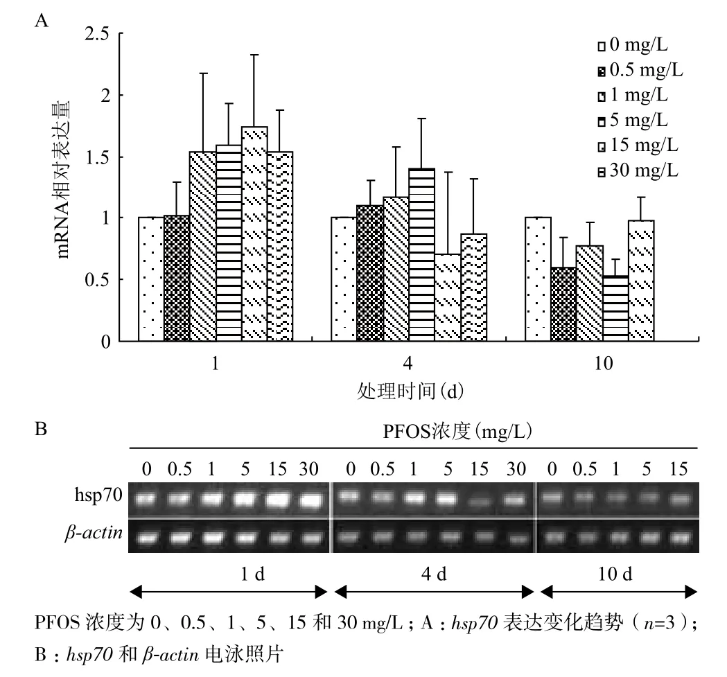
图1 完整涡虫hsp70 mRNA相对表达量变化
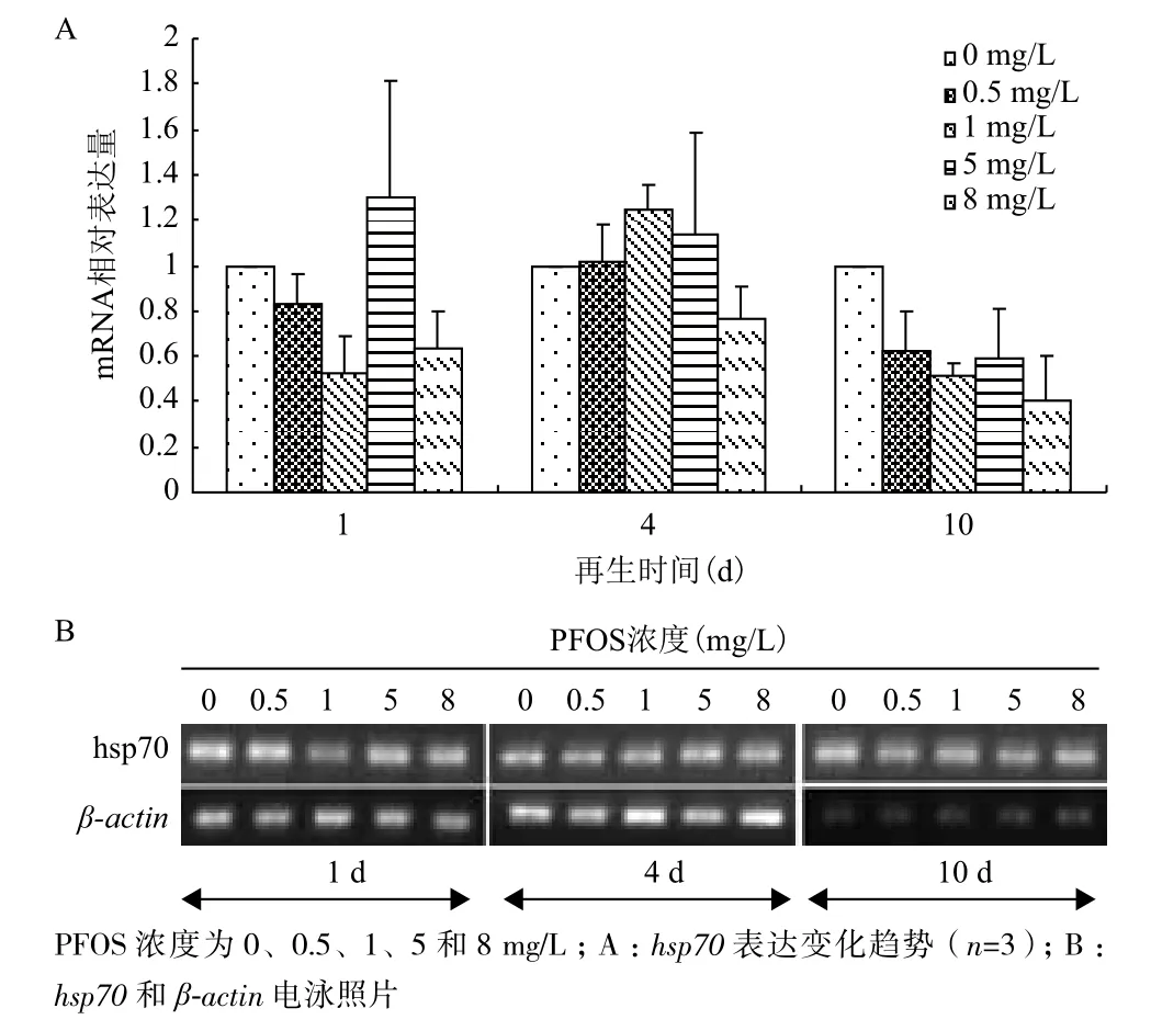
图2 涡虫再生过程中hsp70 mRNA相对表达量变

图3 完整涡虫暴露于各浓度PFOS中HSP70蛋白相对表达量变化
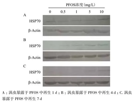
图4 再生涡虫暴露于各浓度PFOS中HSP70蛋白相对表达量变化
3 讨论
PFOS能引起众多基因表达量变化,Hu等[15]将小鼠癌细胞暴露于高浓度PFOS中,约400个基因表达受到显著影响,这些基因涉及脂肪酸同化酶、细胞色素450以及激素调节等。有试验表明PFOS能直接破坏DNA结构[16],但也有试验显示PFOS不造成基因的损伤[16],而是具有细胞毒性[10]。PFOS的毒性机制虽然还没有完全清楚,但
大量试验表明,其主要作用是造成细胞的氧化压力,体外培养的细胞和动植物个体试验均显示,PFOS诱导能造成活性氧(ROS)含量升高[17],过氧化氢酶,超氧化物歧化酶等抗氧化酶类表达量均显著增加[12,17-20]。hsp70是生物体应对外界压力的标志性基因,具有抗氧化的作用。本试验显示,完整涡虫短时间暴露于PFOS中,诱导hsp70表达,但暴露时间延长,逐步呈抑制表达状态,可能是PFOS在涡虫体内积累量少时,能诱导hsp70表达,随积累量增加,又存在抑制表达的作用。Anne等[21]的试验中大西洋鲑暴露于15.1 mg/L和25.0 mg/L PFOS中1 d,hsp70 mRNA表达量显著增加,暴露于PFOS中2 d时,25.0 mg/L组hsp70表达量较15.1 mg/L组下降,与本试验结果一致。
本试验分别检测了hsp70 mRNA和蛋白表达量变化,通过比较发现hsp70 mRNA和蛋白表达量变化趋势不一致,完整涡虫暴露于PFOS中1 d时,hsp70 mRNA表达量上升,但对应HSP70蛋白不存在显著增加,再生涡虫长时间暴露于高浓度的PFOS中hsp70 mRNA表达量受到抑制,但对应的蛋白表达量增加数倍。可能存在PFOS对蛋白表达的抑制作用,低浓度的PFOS能通过某种作用,在mRNA高表达的情况下降低蛋白表达量,而高浓度的PFOS不存在这种抑制作用。
本试验中观察到,高浓度长时间PFOS处理完整涡虫,会造成涡虫躯体出现孔洞、残缺以至完全解体,再生涡虫比完整涡虫更敏感,推测试验过程PFOS对涡虫的氧化压力是存在的,并且能引起涡虫的凋亡,hsp70基因受到少量PFOS诱导作用而激活转录,但在蛋白表达过程中被PFOS抑制,蛋白量没有显著变化,大量PFOS 抑制基因的转录,但对蛋白表达没有影响,所以HSP70蛋白表达量上升。另外,涡虫在再生过程中,hsp70的表达量会有显著升高,并且可能与再生早期干细胞的迁移聚集有关,而与细胞分裂分化无关,因此hsp70的表达在再生48 h时开始下降[22],可能是本试验中造成完整涡虫和再生涡虫基因表达不同的原因。
4 结论
PFOS刺激涡虫能够引起涡虫的应激反应,影响hsp70基因的转录,但mRNA量和蛋白量不成线性关系,可能存在PFOS对蛋白表达的抑制作用;低剂量PFOS对hsp70的影响大于高剂量,诱导转录抑制蛋白量,而高剂量PFOS作用正好相反。
[1]Sinclair E, Mayack DT, Roblee K, et al. Occurrence of perfluoroalkyl surfactants in water, fish, and birds from New York State[J]. Arch Environ Contam Toxicol, 2006, 50(3):398-410.
[2]Jin YH, Liu W, Sato I, et al. PFOS and PFOA in environmental and tap water in China[J]. Chemosphere, 2009, 77(5):605-611.
[3]Pan G, You C. Sediment-water distribution of perfluorooctane sulfonate(PFOS)in Yangtze River Estuary[J]. Environmental Pollution, 2010, 158(5):1363-1367.
[4]Houde M, Martin JW, Letcher RJ, et al. Biological monitoring of polyfluoroalkyl substances:a review[J]. Environmental Science & Technology, 2006, 40:3463-3473.
[5]Luebker DJ, Hansen KJ, Bass NM, et al. Interactions of fluorochemicals with rat liver fatty acid-binding protein[J]. Toxicology, 2002, 176(3):175-185.
[6]Andersden ME, Clewell HJ, Tan YM, et al. Pharmacokinetic modeling of saturable, renal resorption of perfluoroalkylacids in monkeys-probing the determinants of long plasma half-lives[J]. Toxicology, 2006, 227(1-3):156-164.
[7]Anne EL, Jerry LC, John LB, et al. Evaluation of placental and lactational pharmacokinetics of PFOA and PFOS in the pregnant, lactating, fetal and neonatal rat using a physiologically based pharmacokinetic model[J]. Reproductive Toxicology, 2012, 33(4):468-490.
[8]John LN, Katherine KC, Susan AB, et al. Effects of perfluorooctane sulfonate on mallard and northern bobwhite quail exposed chronically via the diet[J]. Environmental Toxicology and Pharmacology, 2007, 23(1):1-9.
[9]Eriksen KT, Raaschou-Nielsen O, Sørensen M, et al. Genotoxic potential of the perfluorinated chemicals PFOA, PFOS, PFBS, PFNA and PFHxA in human HepG2 cells[J]. Mutation Research, 2010, 700(1-2):39-43.
[10]Florentin A, Deblonde T, Diguio N, et al. Impacts of two peruorinated compounds(PFOS and PFOA)on human hepatoma cells:Cytotoxicity but no genotoxicity?[J]. International Journal
of Hygiene and Environmental Health, 2011, 214(6):493-499.
[11]Xie W, Ludewig G, Wang K, et al. Model and cell membrane partitioning of perfluorooctanesulfonate is independent of the lipid chain length[J]. Colloids and Surfaces B:Biointerfaces, 2010, 76(1):128-136.
[12]Hu WY, Jones PD, Celius T, et al. Identification of genes responsive to PFOS using gene expression profiling[J]. Environmental Toxicology and Pharmacology, 2005, 19(1):57-70.
[13]Basu N, Todgham AE, Ackerman PA, et al. Heat shock protein genes and their functional significance in fish[J]. Gene, 2002, 295(2):173-183.
[14]Yuan ZQ, Zhao BS, Zhang JY, Zhang SC. Characterization and expression of djpreb Gene in the planarian Dugesia jiaoniea[J]. Molecular Biology, 2010, 44(l):8-13.
[15]Hu WY, Jones PD, Coen WD, et al. Comparison of gene expression methods to identify genes responsive to perfluorooctane sulfonic acid[J]. Environmental Toxicology and Pharmacology, 2005, 19(1):153-160.
[16]Lu LP, Xu LH, Kang TF, Cheng SY. DNA damage due to perfluorooctane sulfonate based on nanogold embedded in nanoporous poly pyrrole film[J]. Applied Surface Science, 2013, 284:258-262.
[17]Liu CS, Yu K, Shi XJ, et al. Induction of oxidative stress and apoptosis by PFOS and PFOA in primary cultured hepatocytes of freshwater tilapia(Oreochromis niloticus)[J]. Aquatic Toxicology, 2007, 82(2):135-143.
[18]Panaretakis T, Shabalina IG, Grander D, et al. Reactive oxygen species and mitochondria mediate the induction of apoptosis in human hepatoma HepG2 Cells by the rodent peroxisome proliferator and hepatocarcinogen, perfluorooctanoic acid[J]. Toxicology and Applied Pharmacology, 2001, 173(1):56-64.
[19]Xu DM, Li CD, Chen H, Shao B. Cellular response of freshwater green algae to perfluorooctanoic acid toxicity[J]. Ecotoxicology and Environmental Safety, 2013, 88:103-107.
[20]Santos MA, Pacheco M, Ahmad I, Anguilla L. Antioxidants responses to in situ bleached kraft pulpmill effluent outlet exposure[J]. Environment International, 2004, 30(3):301-308.
[21]Krøvel AV, Søfteland L, Torstensen B, Olsvik PA. Transcriptional effects of PFOS in isolated hepatocytes from Atlantic salmon Salmo salar L[J]. Comparative Biochemistry and Physiology Part C:Toxicology & Pharmacology, 2008, 148(1):14-22.
[22]Ma KX, Chen GW, Lou H, F EI LN. Cloning and expression analysis of hsp70 gene from freshwater planarian Dugesia japonica[J]. Biologia, 2009.64(5):1018-1024.
(责任编辑 马鑫)
Effects of PFOS on Expression of HSP70 in Planarian Dugesia japonica
Gong Xiaoning Yuan Zuoqing Bai Yun
(School of Life Science,Shandong University of Technogy,Zibo 255049)
Perfluorooctane sulfonate(PFOS)is a persistent organic pollutant and has been found to be the predominant perfluorinated chemicals(PFCs)in the environment. In this study, we used planarians Dugesia japonica, which belong to the phylum Platyhelminthes, class Turbellaria, as an animal assay to evaluate the toxicological effects of PFOS on protein and mRNA expression of HSP70. The results indicated that the expression of hsp70 mRNA of planarians increased exposed to PFOS in short time and decreased when planarians regenerated of 10-days. However, protein expression of HSP70 increased significantly after regenerating planarians exposed to PFOS for 4- and 7-days, and other groups were not influenced significantly. The trend of mRNA expression were inconsistent with protern. It may be that little PFOS would inhibit the translation of HSP70, and the number of HSP70 increased to protect from toxicity of PFOS as that was accumulated in planarians.
PFOS Planatian HSP70 RT-PCR Western blot
2014-02-12
国家自然科学基金资助项目(31100377),山东省自然科学基金资助项目(ZR2011CQ018)
宫晓宁,男,硕士研究生,研究方向;毒理学;E-mail:gongxiaoning321@163. com
袁佐清,女,博士,副教授,研究方向:毒理学;E-mail:yuanzuoqing2008@126.com