Grain-sized moxibustion inhibits the progression of Alzheimer disease in 5XFAD transgenic mice
2022-12-28YUJing余静GONGXiaowei龚晓炜CHUJiamei楚佳梅ZHANGYongsheng张永生FANZhenyu樊振宇Shujian李书剑BAOYehua包烨华
YU Jing (余静), GONG Xiaowei (龚晓炜), CHU Jiamei (楚佳梅), ZHANG Yongsheng (张永生), FAN Zhenyu (樊振宇),LⅠ Shujian (李书剑), BAO Yehua (包烨华)
1 Department of Acupuncture and Moxibustion, Hangzhou TCM Hospital Affiliated to Zhejiang Chinese Medical University,Hangzhou 310007, China
2 Baiyang Community Health Service Center, Hangzhou City, Zhejiang Province, Hangzhou 310018, China
3 Zhejiang Chinese Medical University, Hangzhou 310053, China
4 Department of Tuina, Zhejiang Hospital, Hangzhou 310016, China
Abstract
Keywords: Moxibustion Therapy; Moxibustion with Grain-sized Moxa Cone; Alzheimer Disease; Amyloid β Deposit;Astrocyte; Microglia; Mice
Alzheimer disease (AD) is a primary neurodegenerative dementia characterized by a comprehensive disorder of acquired advanced functions in the cerebral cortex. A prominent pathologic manifestation of AD is senile plaques (SP) formed by abnormal accumulation of amyloid β (Aβ) protein and neurofibrillary tangles caused by tau protein hyperphosphorylation[1]. AD has a progressive clinical course. The major clinical manifestations are impairments in learning, memory and language,cognitive deficits, affective disorder, and visuospatial impairment, all of which affect activities of daily living in patients[2-3]. AD is the most common type of dementia,accounting for 60% to 70% of dementia. Age is the main risk factor for AD, whose incidence is increasing with the longer life expectancy of the world population.
The pathogenesis of AD is a long process, and the detailed underlying mechanisms are not fully understood, though it is known that pathologic alterations in the brain precede the emergence of clinical symptoms[4]. Current drugs provide only minor symptomatic relief in the middle and advanced stages of the disease[5]. Therefore, it is possible that intervention at a preclinical stage before the neurodegenerative process is established may be more effective[4]. In traditional Chinese medicine (TCM),prevention before disease onset is an important concept[6]. Acupuncture and moxibustion are TCM techniques that have multiple applications but few adverse effects. Both have shown clinical efficacy in the prevention and treatment of AD through the induction of apoptosis and modulation of aging-related gene expression. In particular, moxibustion has been shown to improve cognitive function in patients with mild cognitive impairment. A previous study found that moxibustion effectively improved clinical symptoms,affective state, and the ability to perform activities of daily living in patients with AD.
Amyloid precursor protein/presenilin-1 (APP/PS1)double-transgenic mice harbor AD-related gene mutations and are a useful model for investigating the pathogenesis of AD. One of these mouse models is the transgenic mice with 5 familial AD mutations (5XFAD),which co-express human amyloid precursor protein(APP) and presenilin-1 (PS1) with 5 familial AD (FAD)mutations[7]. The 5XFAD mice generate and assemble Aβ42almost exclusively and rapidly starting at the age of 1.5 months, and begin to produce visible amyloid deposits and gliosis at the age of 2 months[8].
The aim of the present study was to evaluate the efficacy of grain-sized moxibustion in the treatment of AD using 5XFAD transgenic mice and investigate the underlying mechanisms.
1 Materials and Methods
1.1 Experimental animals and groups
Four male 5XFAD [B6SJL-Tg (APPSwFlLon,PSEN1*M146L*L286V) 6799Vas/J] mice were obtained from JING Naihe, a professor of the State Key Laboratory of Cell Biology, Shanghai Institute of Biochemistry and Cell Biology, Chinese Academy of Sciences. Wild-type (WT) hybridization mice were received from Shanghai SLAC Laboratory Animal Co.,Ltd., China [SCXK (2007-0005)]. The heterozygous 5XFAD transgenic mice and WT controls were fed and bred in the specific-pathogen-free (SPF) grade transgenic animal laboratory [SYXK (2003-2003)] of Laboratory Animal Research Center, Zhejiang Chinese Medical University. The genotype of the newborn mice was carried out by polymerase chain reaction (PCR) of tail DNA biopsies to identify mice with transgenic genes at the age of 3 to 4 weeks. After verification, 40 5XFAD transgenic mice were randomly divided into an AD model group (5XFAD group,n=20) and a grain-sized moxibustion group (5XFAD + GM group,n=20), with 20 age-matched littermate wild C57BL/6J mice serving as a normal control group (WT group). The animal rearing environment was constant temperature(22±1) ℃ with 12-hour light/dark cycles. Animals had free access to water and food. All animal experiments were carried out according to the animal protection principle, animal welfare principle, ethical principle, and comprehensive scientific evaluation principle. This experiment was approved by the Animal Experiment Management Committee of Zhejiang Chinese Medical University (No. ZSLL-2013-92).
1.2 Genetic phenotype identification results of 5XFAD transgenic mice
Genotypes of the offspring mice were identified by PCR. There were totally 84 offspring mice, and 40 offspring mice were APP/PS1 double-transgenic mice.Representative PCR pictures are shown in Figure 1.

Figure 1 Genetic phenotype identification results of 5XFAD transgenic mice
1.3 Intervention methods
Mice in the 5XFAD + GM group received grain-sized moxibustion at Xinshu (BL15) and Shenshu (BL23) at the age of 1.5 months. The locations of points were based on the map of the experimental animal acupuncture points in theExperimental Acupuncture Science[9]and well-corresponded to the anatomical site of human points. Xinshu (BL15) is located in the intercostal space that is below and lateral to the spinous process of the fifth thoracic vertebra. Shenshu (BL23) is located below and lateral to the spinous process of the second lumbar vertebra. The hair on the treatment area was shaved before treatment. The moxa cones were made in the size of a grain of wheat (2 mm × 3 mm). The operator fixed the mice with one hand and located the moxa cones on Xinshu (BL15) and Shenshu (BL23) with another hand. Then ignited the cones. The cones were removed when burnt to 3/5. One moxa cone once. It took 25-30 s for every grain-sized moxibustion treatment, once a day for 10 consecutive days as a course of treatment. There was a 2-day rest between two courses, and the total course of treatment lasted for 5.5 months (all mice were 7 months old when treatment finished).
Mice in the WT and 5XFAD groups received no treatment but were fixed in the same way as the 5XFAD + GM group to ensure an equivalent condition.
1.4 Behavioral testing
1.4.1 Morris water maze test
The Morris water maze test includes orientation navigation and space exploration trials. It was carried out after the treatment of grain-sized moxibustion for evaluating spatial learning and memory ability. The water maze apparatus is a round stainless-steel water tank (80 cm in diameter and 50 cm in height). The surface area of the water tank was divided into 4 equal quadrants, and a hidden platform of 25 cm in diameter was placed in the 3rd quadrant. The hidden platform was submerged 1 cm below the surface of the water. A digital camera was connected to the computer monitoring screen above the water tank, and the mice movement track was tracked and recorded by computer software. Mice received orientation navigation trials from day 1 to day 6 for 6 d, and the probe trial on day 7.In the orientation navigation trials, mice were allowed 60 s to find and climb on the hidden platform. The mice would be guided to the hidden platform and stay on the platform for 10 s if they could not find the hidden platform within 60 s. The computer recorded the track,the length of the route, and the time the mice took to find the platform. Finally, 24 h after the last orientation navigation trial, a probe test, where the platform was removed, was performed. The percentage of residence time spent in the target quadrant (where the previous hidden platform was located) and the crossing number of the target area (where the previous hidden platform was located) were recorded to reflect the spatial memory retention of the mice.
1.4.2 Y-maze test
Y-maze test was performed as described in the previous research[10-11]. The Y-maze apparatus (BWMYM103, Panlab, Spain) with a conductive electric grid floor consisted of 3 identical arms (Ⅰ, Ⅱ, and Ⅲ arms)positioned at equal angles. Each arm had a 15 W lamp at the distal end of the arm, regarding light as a signal of electric shock. When the experiment was carried out,the signal lamp of one arm would emit bright light, and there was no electric shock at the floor of this arm,which was the safety zone. At the same time, the lamps of the other two arms were dark, and constant electric shocks (50-70 V) were applied at the floor of the arms,which were the non-safety zones. The safety and non-safety zones could be switched randomly. A correct reaction occurred when the mice moved to the safety zone within 10 s of an electric shock. This testing process was repeated 20 times a day. If the mice had 9 or more correct reactions in 10 consecutive training sessions (correct reaction rate ≥90%), they would be defined as having reached the learning criterion, and the experiment process would be terminated. The required times of reaching the learning criterion were recorded as the ability of learning spatial resolution responses. We took 100 times as the maximum number of electric shocks. If it failed to reach the learning criterion within 100 times, 100 times would be recorded. After a 24 h interval that the mice reached the learning criterion, the mice were tested 10 consecutive times as before, and the number of correct responses that the mice moved to the safety zone within 10 s of an electric shock was recorded as the memory ability.
1.5 Pathologic and biochemical analysis
1.5.1 Brain tissue collection
After the behavioral test, the mice were sacrificed.For sandwich enzyme-linked immunosorbent assay(ELISA), the brains of 6 mice from each group were frozen in liquid nitrogen and stored at -80 ℃. For histopathological examinations, 6 mice from each group were perfused with PBS and 4% paraformaldehyde via ascending aorta until the tail of the mice was cocked or the limbs twitched. After perfusion, the brains were taken out completely and fixed in 4% paraformaldehyde solution for 24 h. The brain was incised along the coronal plane on the optic cross plane, fixed in the embedding frame, and embedded in paraffin. The paraffin-embedded brain was sectioned continuously before and after the optic cross plane, and one piece was taken out from 3 pieces, at a thickness of 4 μm. The slices were baked in a 56 ℃ oven for 1 h and stored at room temperature.
1.5.2 Quantification of Aβ40and Aβ42levels by ELISA
The levels of Aβ40and Aβ42in the brain of 5XFAD transgenic mice were determined by ELISA. We used the mouse Aβ(1-40)ELISA kit (MAB96181R, R&D Systems,USA) and mouse Aβ(1-42)ELISA kit (MAB9618R, R&D Systems, USA) in this research. The operation of the experiment was in accordance with the manufacturer’s instruction. Half brain was homogenized in PBS,centrifuged at 3 000 r/min at 2-8 ℃ for 10 min, and then the supernatants were obtained. Next, the standards and samples were mixed with a specific first antibody in duplicate, and then HRP-conjugated secondary antibody (DAB140B, R&D Systems, USA) was added to the plates. Covered the sealing film, shaken gently, and incubated for 60 min at room temperature.After extensive washing, the chromogen solution was added to the plates, shaken gently, and developed with chromogen for 10 min at room temperature without light. Added the stop solution into the plates, and the enzymatic reaction was then terminated. Measured the optical density (OD) in sequence at 450 nm wavelength on a microplate spectrophotometer. The measurement carried out within 10 min after the stop solution was added. Calculated the concentrations according to the standard curve.
1.5.3 Thioflavin-S (Th-S) staining
Th-S is a common method used to stain senile plaques. Brain tissues of mice from each group (n=6)were first fixed in 4% paraformaldehyde, then dehydrated, and embedded in paraffin. The paraffinembedded brains were sectioned with a microtome at a thickness of 4 μm. The sections were deparaffinized in xylene, then treated in gradient ethanol solutions(100%, 95%, 90%, and 85%) for 1 min each, and washed with distilled water 3 times. Then, the sections were immersed in the Th-S solution (SC-215969, Santa Cruz Biotechnology, USA) for 5 min (1% Th-S in distilled water). Then washed with distilled water 3 times.Anti-fluorescence quenching sealing liquid (Beyotime Biotechnology, China) was used to hold the coverslips.All sections were observed by another investigator using a fluorescent microscope (Olympus IX71, Olympus,Japan). All slice counts from the same mouse were averaged to yield the number of amyloid plaque depositions per mouse. All counts of the number of Th-S positive Aβ deposits were performed using the blind method.
1.5.4 Immunohistochemical staining
For immunohistochemical staining, the paraffinembedded brains from each group (n=6) were sectioned with a microtome at a thickness of 4 μm. The sections were deparaffinized and then immersed in a citric acid buffer solution (10 mmol/L, pH 6.0).Microwaved for 15 min for antigen retrieval. Used a 3%hydrogen peroxide solution to block endogenous peroxidase activity for 10 min at room temperature.Then blocked with goat serum in PBS for 60 min and then incubated at 4 ℃ overnight with primary antibodies against glial fibrillary acidic protein (GFAP)(1:500, Beijing Biosynthesis Biotechnology, China),cluster of differentiation 11b (CD11b, 1:200, Beijing Biosynthesis Biotechnology, China), choline acetyltransferase (ChAT, 1:200, Beijing Biosynthesis Biotechnology, China), and brain-derived neurotrophic factor (BDNF, 1:200, Beijing Biosynthesis Biotechnology,China), followed by washing with PBS. Then incubated with secondary antibodies (1:200, Beijing Biosynthesis Biotechnology, China) and conjugated with horseradish peroxidase for 1 h at room temperature. The sections were visualized by the 3, 3-diaminobenzidine chromogen solution (60882801, Zhong Shan Golden Bridge Biotechnology, China) for a few seconds,followed by washing with PBS. All sections were observed by an optical microscope (Nikon eclipse 80i,40× magnification). Image-Pro Plus 5.0 software was used to quantify the integrated OD of the positive cells in the hippocampus and cortex regions.
1.6 Statistical analysis
All data were analyzed by the SPSS version 18.0 software. Data were presented as mean ± standard deviation (±s). Two-way analysis of variance (ANOVA)with repeated measures was used for analyzing data from the Morris water maze test. Other statistical tests were performed using one-way ANOVA test, and the least significant difference test for post hoc multiple comparisons. Non-normal data were subjected to nonparametric tests.P<0.05 indicated statistical significance.
2 Results
2.1 Grain-sized moxibustion ameliorates cognitive impairment in 5XFAD transgenic mice
2.1.1 Morris water maze test
Spatial learning was assessed based on the escape latency (the time that the mice took to find and climb on the hidden platform) in orientation navigation trials in the Morris water maze test. The 5XFAD group had a longer escape latency time compared with the WT group and the 5XFAD + GM group (P<0.05). There was no significant difference between the 5XFAD + GM group and the WT group (P>0.05). See Figure 2.

Figure 2 Escape latency in the orientation navigation test
Spatial memory retention was evaluated based on the percentage of residence time spent in the target quadrant and the number of target area crossings in the probe test. The 5XFAD group spent less time in the target quadrant than the WT group (P<0.05) and crossed the target area fewer times than the WT group(P<0.05). Mice in the 5XFAD + GM group spent more time in the target quadrant than the 5XFAD group(P<0.05) and had more target area crossings than the 5XFAD group (P<0.05). There were no significant differences between the 5XFAD + GM group and the WT group (P>0.05). See Figure 3 and Figure 4.

Figure 3 Percentage of residence time spent in the target quadrant in the probe test
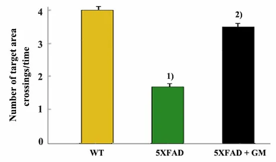
Figure 4 Number of target area crossings in the probe test
2.1.2 Y-maze test
The learning ability was assessed with the Y-maze test based on the number of training times of the mice before the mice had ≥9 correct reactions in 10 consecutive training sessions, with a lower number indicating better learning ability. Compared with the WT group, the 5XFAD group required more training times(P<0.05). Compared with the 5XFAD group, the number of training times of the 5XFAD + GM group was significantly decreased (P<0.05). There was no significant difference between the 5XFAD + GM group and the WT group (P>0.05). See Figure 5.
Memory ability was assessed by the number of correct responses in mice receiving 10 electric shocks,with a higher number indicating better memory ability.Compared with the WT group, mice in the 5XFAD group had fewer correct responses (P<0.05). However, the number was higher in the 5XFAD + GM group compared with the 5XFAD group (P<0.05). There was no significant difference between the 5XFAD + GM group and the WT group (P>0.05). See Figure 6.
Collectively, these results demonstrate that grainsized moxibustion at Xinshu (BL15) and Shenshu (BL23)enhances learning and memory performance in 5XFAD mice.

Figure 5 Training number in Y-maze test

Figure 6 Number of correct responses in Y-maze test
2.2 Grain-sized moxibustion decreases overexpression of Aβ40 and Aβ42 in the brain of 5XFAD transgenic mice
To investigate the effect of grain-sized moxibustion on Aβ40and Aβ42, ELISA was used in the present study.ELISA test results showed that the levels of Aβ40and Aβ42in the brain of mice in the 5XFAD group were significantly higher compared with the WT group(P<0.05). At the same time, the levels of Aβ40and Aβ42in the 5XFAD + GM group were significantly lower than those in the 5XFAD group (P<0.05). There were no significant differences in the levels of Aβ40and Aβ42between the 5XFAD + GM group and the WT group(P>0.05). These results demonstrate that grain-sized moxibustion at Xinshu (BL15) and Shenshu (BL23) can effectively decrease the accumulation of Aβ40and Aβ42in the brain of 5XFAD transgenic mice. See Figure 7 and Figure 8.

Figure 8 Expression of Aβ42
2.3 Grain-sized moxibustion significantly reduces Aβ accumulation in the hippocampus and cortex of 5XFAD transgenic mice
Th-S staining was used to stain amyloid plaque deposition. After the experiment of Th-S staining, it was found that the WT group did not appear positive plaques of amyloid deposition. The results of Th-S staining showed that the total number of amyloid plaque deposition in the hippocampus and cortex of the 5XFAD + GM group was significantly reduced than that of the 5XFAD group (P<0.05). These results demonstrate that grain-sized moxibustion at Xinshu (BL15) and Shenshu (BL23) can effectively reduce the accumulation of Aβ deposition in the hippocampus and cortex of 5XFAD transgenic mice. See Figure 9 and Figure 10.

Figure 9 Representative images of thioflavin-S staining in the hippocampus and cortex from each group (×40)

Figure 10 Quantitative analysis of the number of amyloid plaque deposition in the hippocampus and cortex
2.4 Grain-sized moxibustion significantly reduces astrocyte and microglia activation in the cerebral cortex and hippocampus of 5XFAD transgenic mice
Astrocyte and microglia are two principal cell types involved in neuroinflammation. To determine whether the neuroprotective effects of grain-sized moxibustion result from the inhibition of astrocytes and microglial activation in the brain, immunohistochemical staining of brain sections for activated astrocytes was performed using specific antibodies against GFAP and for activated microglia was performed using specific antibodies against CD11b in the present study. GFAP-positive and CD11b-positive cells were brown, mainly in the cerebral cortex and the hippocampus cytoplasm. See Figure 11 and Figure 12.
Immunohistochemical staining results showed that the expression levels of GFAP and CD11b in the hippocampus and cortex of the 5XFAD group were significantly increased compared with the WT group(P<0.05). After grain-sized moxibustion treatment, the expression levels of GFAP and CD11b in the hippocampus and cortex in the 5XFAD + GM group were significantly reduced compared with the 5XFAD group(P<0.05). There were no significant differences in the expression levels of GFAP and CD11b between the 5XFAD + GM group and the WT group (P>0.05). These results demonstrate that grain-sized moxibustion at Xinshu (BL15) and Shenshu (BL23) can effectively reduce neuroinflammatory responses by inhibiting the activation of astrocytes and microglia. See Figure 13 and Figure 14.
2.5 Grain-sized moxibustion increases ChAT expression in the hippocampus and cortex of 5XFAD transgenic mice
The cholinergic system plays a crucial role in cognitive function. To investigate the possible mechanisms that grain-sized moxibustion contributes to improving memory, hippocampal and cortical ChAT expression levels were measured by immunohistochemical staining(Figure 15).
Immunohistochemical staining results showed that the expression levels of ChAT in the hippocampus and cortex of the 5XFAD group were significantly reduced compared with the WT group (P<0.05). After grain-sized moxibustion treatment, the expression levels of ChAT in the hippocampus and cortex in the 5XFAD + GM group were significantly increased compared with the 5XFAD group (P<0.05). These results demonstrate that grainsized moxibustion at Xinshu (BL15) and Shenshu (BL23)can increase ChAT levels in the hippocampus and cortex of 5XFAD mice. See Figure 16.

Figure 11 Images of GFAP immunohistochemical staining reactions in the hippocampus and cortex (×40)
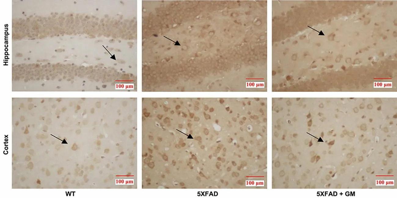
Figure 12 Images of CD11b immunohistochemical staining reactions in the hippocampus and cortex (×40)

Figure 13 Expression of GFAP in the hippocampus and cortex (immunohistochemical staining)

Figure 14 Expression of CD11b in the hippocampus and cortex (immunohistochemical staining)
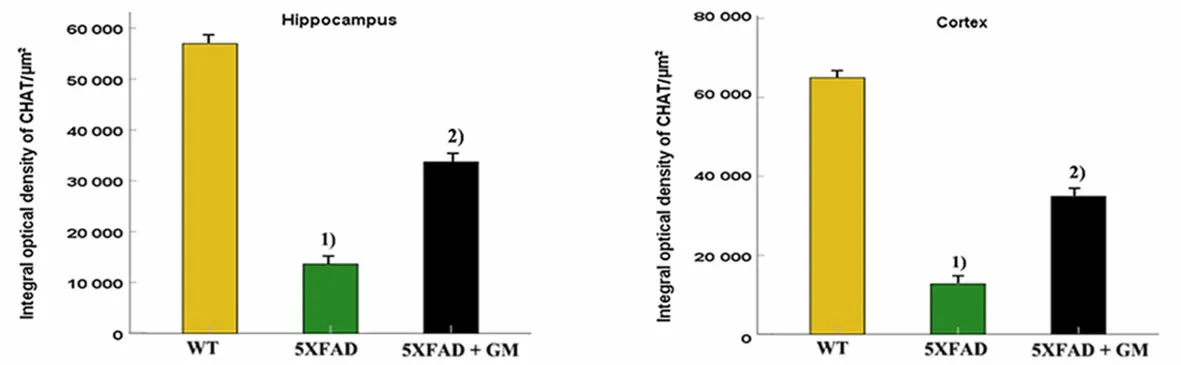
Figure 16 Expression of ChAT in the hippocampus and cortex (immunohistochemical staining)
BDNF is a key molecule in synaptic plasticity and plays an important role in the pathogenesis of AD. To investigate the effect of grain-sized moxibustion on BDNF, immunohistochemical staining was used in the present study. BDNF-positive cells were brown and distributed mainly in the cerebral cortex and the hippocampus cytoplasm (Figure 17).
Immunohistochemical staining results showed that the expression levels of BDNF in the hippocampus and cortex of the 5XFAD group were significantly lower compared with the WT group (P<0.05). After grain-sized moxibustion treatment, the expression levels of BDNF in the hippocampus and cortex in the 5XFAD + GM group were significantly increased compared with the 5XFAD group (P<0.05). These results demonstrate that grain-sized moxibustion at Xinshu (BL15) and Shenshu(BL23) may exert neuroprotective effects via the up-regulation of BDNF levels in the hippocampus and cortex of 5XFAD mice. See Figure 18.

Figure 17 Images of BDNF immunohistochemical staining reactions of the hippocampus and cortex from each group (×40)
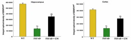
Figure 18 Expression of BDNF in the hippocampus and cortex (immunohistochemical staining)
3 Discussion
According to the theory of TCM, the main causes of AD lie in the deficiency of kidney essence, deficiency of marrow sea, and apraxia[12]. Xinshu (BL15) has the function of nourishing the heart and calming the mind,regulating Qi and blood, and adjusting the spirit[13].Shenshu (BL23) has the function of tonifying the kidney and nourishing the bone marrow. Experimental studies have shown that Shenshu (BL23) plays an important role in delaying aging through the regulation of free radical metabolism; moreover, moxibustion at Shenshu(BL23) is beneficial to the rehabilitation of dementia patients. Moxibustion is often used in TCM for disease prevention and health care and is highly regarded by physicians in China. According to TCM theory,moxibustion has the function of warming meridians and collaterals, and reinforcing and replenishing Yang Qi. It can improve the stagnation of meridians and mitigate Yang Qi deficiency that occurs during the process of aging. Thus, moxibustion is important for disease prevention and health as it improves blood circulation,regulates metabolism, and delays aging.
AD is a chronic neurodegenerative disease first described by Alois ALZHEIMER in 1907[14-15]. Ever since the first case report on AD, senile plaques and neurofibrillary tangles have been recognized as the neuropathological hallmarks of AD[16]. The amyloid cascade hypothesis was proposed by HARDY J A and other scholars in 1992 and is currently recognized as the mechanism of AD pathogenesis[17]. According to the amyloid cascade hypothesis, the deposition of Aβ is portrayed as an upstream event in the evolution of AD,leading to the formation of senile plaques, activation of oxidative stress and inflammatory cascades, synaptic,and neuritic injury, hyperphosphorylation of tau protein,widespread neuronal dysfunction and cell death with transmitter deficits, and ultimately clinical dementia[18-19].
The 5XFAD mice used in our study can simulate the pathological process and behavioral characteristics of AD comprehensively and rapidly. The 5XFAD mice generate and accumulate Aβ42almost exclusively and rapidly in the brain starting at the age of 1.5 months and exhibit cognitive deficits at the age of 4-5 months.Previous studies demonstrated that the levels of the ChAT and BDNF decreased significantly at the age of 6 months in 5XFAD mice[20-21]. Therefore, in the present study, grain-sized moxibustion was performed on mice starting at 1.5 months, which can be considered the early stage of AD pathogenesis.
Aβ peptides, as the initial pathological event in AD,are generated from APP[22]. APP undergoes subsequent cleavages by β-secretases and γ-secretases that ultimately generate Aβ peptides[23]. APP cleavage follows two pathways. One pathway is the nonamyloidogenic, and the other is the amyloidogenic pathway. In the amyloidogenic pathway, APP cleavage is initiated by β-secretase, generating a secreted form of APP and a 99-amino acid C-terminal fragment which is further cleaved sequentially by γ-secretase to generate 37-42-amino acid-long extracellular Aβ peptides, and most of the peptides are represented by deleterious Aβ42and Aβ40species[24]. Aβ42is the most neurotoxic of these species and is the primary component of the core of the senile plaque. It is shown that Aβ42can induce oxidative stress and neurotoxicity in the AD brain.Moreover, Aβ42leads to mitochondrial dysfunction and destruction of intracellular Ca2+homeostasis, ultimately leading to cell death[25]. In addition, studies suggest that the accumulation of Aβ42plays an important role in neurodegenerative processes in AD[26]. Studies suggest that preformed fibrils of Aβ40and Aβ42can promote each other’s aggregation[27]. Thus, reducing Aβ deposition, especially that of Aβ42, is a promising strategy for the treatment of AD[28]. According to our research, grain-sized moxibustion is a feasible and effective way for reducing Aβ40, Aβ42, and Aβ deposits in the brain of AD mice.
Neuroinflammation is another important hallmark of AD. The activated astrocytes and microglia surrounding amyloid plaques are the main characteristics of AD neuroinflammation. In the pathogenesis of AD, specific Aβ fragments such as Aβ42can further enhance proinflammation. Studies suggest that Aβ deposition is associated with increased neuronal toxicity and generation of reactive oxygen species, leading to a series of neuroinflammatory processes[29]. In an AD brain, Aβ can trigger the activation of microglia and astrocytes. It is shown that the activated microglia can enhance the level of inducible nitric oxide synthetase,promoting neurodegeneration[30]. In addition, apart from a direct neurotoxic effect, activated microglia and astrocytes can also produce Aβ deposition[31]. Many findings suggest that Aβ may induce toxicity through the aggravation of neuroinflammation in AD. The activated microglia can cause direct Aβ-induced neurotoxicity and supply an increased concentration of toll-like receptor ligands that fuels neuroinflammation[32]. The activated astrocytes could overproduce many proinflammatory cytokines and chemokines, which contribute to neuroinflammatory processes of AD[33]. The astrocyte-secreted proinflammatory factors can promote the conversion of APP into neurotoxic insoluble fibrillary Aβ[34].Neurotoxic Aβ can promote astrocytes to induce more inflammation-like glial responses, thus maintaining neurodegenerative injuries, such as progressive neuronal loss, enhanced astrocytosis, and amyloid plaque formation[35]. Aβ, astrocytes, microglia, and cytokines form a vicious cycle in the pathological process of AD that eventually leads to neuronal death.GFAP is recognized as the marker of astrocytes, and CD11b is recognized as the marker of microglia[36-37]. In our study, grain-sized moxibustion was a feasible and effective way to reduce the expression of GFAP and CD11b in transgenic AD mice. These results demonstrate that grain-sized moxibustion can restrain the activation of astrocytes and microglia and then slow down the neuroinflammatory process in 5XFAD mice.
The cholinergic system plays an important role in learning and memory and is adversely affected in AD[38].The degeneration of cholinergic transmission may affect all aspects of behavior and cognition[39]. Acetylcholine(ACh) is the first neurotransmitter identified, and published studies indicate that ACh plays a very important role in cognitive processes[40]. The concentration of ACh in the brain is dynamically regulated by the activities of enzyme ChAT and enzyme acetylcholinesterase[41-42]. ChAT is responsible for the synthesis of ACh, and enzyme acetylcholinesterase is responsible for hydrolyzing ACh. Reduced ChAT activity in the brain of patients with AD is correlated with an increased number of neuritic plaques[43]. The ChAT expression level is used as a marker for estimating ACh content in tissues[44]. In the brain of AD patients, a great number of cholinergic neurons are denatured and lost,which leads to decreased ChAT activity, resulting in decreased production, and the release and storage of ACh can lead to impairment of memory and cognitive function[45]. Therefore, increasing the ACh level can prevent neurodegeneration[46]. According to our study,grain-sized moxibustion increased the ChAT level in AD mice, accompanied by improved performance in the Morris water maze and Y-maze tests. These results suggest that grain-sized moxibustion can alleviate the deficits in memory and learning associated with AD.
BDNF is a neurotrophin that facilitates synaptic transmission and plasticity[47]. BDNF can protect neurons against neuroinflammation, repair damaged mitochondrial neurons, reduce the accumulation of free radicals, and promote neuronal regeneration[48]. It plays an important role in memory and learning[49]. BDNF expression in the brain decreases in patients with AD,which is found to be correlated with a decline in cognitive performance[50]. BDNF exerts neuroprotective effects against Aβ peptide toxicity and can prevent neuronal atrophy and neuronal death, thereby alleviating cognitive and behavioral deficits in AD[51].Our study showed that grain-sized moxibustion increased the BDNF level in AD model mice, further supporting the clinical applicability of this technique to the treatment of AD.
In the present study, grain-sized moxibustion treatment was performed on 5XFAD mice starting at the age of 1.5 months, which can be considered as the early stage of AD pathogenesis that Aβ42starts to generate and accumulate. The results showed that grain-sized moxibustion at Xinshu (BL15) and Shenshu (BL23)greatly improved learning and memory deficits in AD mice by decreasing Aβ deposition and neuroinflammation, as well as increasing ChAT and BDNF expression. This may indicate that the mechanism of grain-sized moxibustion in the treatment of AD may be associated with inhibiting the expression of Aβ42early,thereby interfering with the abnormal accumulation of Aβ to form a series of complex pathological processes with toxic neuronal products to further protect the learning and memory ability of AD mice. However, our current subject has not deeply studied the mechanism pathway of grain-sized moxibustion in the treatment of AD, which needs further research. Our findings require validation in clinical studies.
Results from this study demonstrate the efficacy of grain-sized moxibustion at Xinshu (BL15) and Shenshu(BL23) for 5XFAD transgenic mice. Grain-sized moxibustion can significantly ameliorate their spatial learning and memory ability. In addition, the results prove that grain-sized moxibustion can reduce the accumulation of Aβ42, Aβ40, and Aβ deposition, restrain the neuroinflammation process by reducing the expression of GFAP and CD11b, and increase the activity of ChAT and the expression of neurotrophin BDNF.These findings indicate that grain-sized moxibustion may provide significant therapeutic efficacy for the treatment of AD, and this study may also serve as a stepping stone to future exploration in the wide application of moxibustion.
Conflict of Interest
The authors declare that there is no potential conflict of interest in this article.
Acknowledgments
This work was supported by the Natural Science Foundation of Zhejiang Province (浙江省自然科学基金,No. LY13H270002).
Statement of Human and Animal Rights
The treatment of animals conformed to the ethical criteria in this experiment.
Received: 23 June 2021/Accepted: 4 January 2022
猜你喜欢
杂志排行

Journal of Acupuncture and Tuina Science的其它文章
- Effects of moxibustion on miRNA-133b, Pitx3/TH,and neurotransmitters in the midbrain of rats with diarrhea-predominant irritable bowel syndrome
- Effects of Tuina on serum creatine kinase and skeletal muscle mitochondria in delayed onset muscle soreness model rats
- Effects of herbal cake-partitioned moxibustion on the expression of thyroid autophagy-related factors LC3B and Beclin-1 in rats with autoimmune thyroiditis
- Clinical study of acupuncture combined with medication for the elderly with Alzheimer disease
- Clinical observation of acupuncture and moxibustion for functional dyspepsia due to Yang deficiency of the spleen and stomach
- Clinical observation of acupuncture combined with sitting-position knee-adjustment manipulations for patellofemoral arthritis