不同定位方法对早产儿脐静脉置管后异位的影响
2021-09-28应勤来刘珏张旭侯秋英朱雯
应勤来 刘珏 张旭 侯秋英 朱雯

[关键词] 早产儿;脐静脉置管;定位方法;超声
[中图分类号] R459.7 [文献标识码] B [文章编号] 1673-9701(2021)22-0062-04
Investigation on the effects of different positioning methods on ectopy after umbilical vein catheterization in premature infants
YING Qinlai LIU Jue ZHANG Xu HOU Qiuying ZHU Wen
Department of Neonatal,the Second Hospital of Jiaxing City in Zhejiang Province, Jiaxing 314000, China
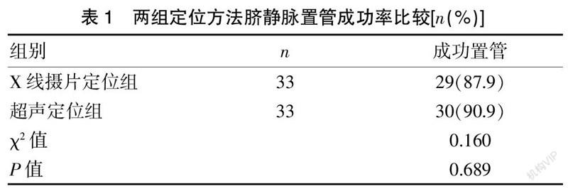
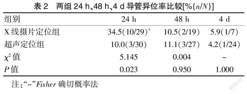
[Abstract] Objective To explore the effect of different positioning methods on ectopy after umbilical vein catheterization in premature infants. Methods From January 2018 to December 2019,a total of 66 cases of premature infants in the neonatal intensive care unit (NICU) of our hospital were selected and divided into two groups (X-ray radiography positioning group of 33 cases and ultrasound positioning group of 33 cases). The number of cases of successful catheterization was observed in both groups; the ectopic conditions were examined by ultrasound 24 h, 48 h, and 4 days after the catheterization. Results The success rates of X-ray radiography positioning and ultrasound positioning group after umbilical vein catheterization were 87.9% and 90.9% with no statistical significance(P=0.689); the position of the catheter was screened by ultrasound 24 hours after the catheterization. The ectopic rate of X-ray radiography positioning were 34.5% and 10.0%(P=0.023, with statistical significance); the ectopic composition ratio was compared between 48 h and 4 days,and the P value was not statistically significant(P>0.05). Conclusion Ultrasound positioning after umbilical vein catheterization is more accurate than X-ray radiography in positioning the catheter tip. The two positioning methods after catheter placement should be repeatedly screened with ultrasound technology on a regular basis. Regular inspections with ultrasound technology can ensure that the umbilical vein catheter is kept in place and prevent complications.
[Key words] Premature infants; Umbilical vein catheterization; Positioning methods; Ultrasound
早產儿是指胎龄<37周的新生儿,早产儿由于发育未成熟,尤其是出生后1周内往往因为喂养困难而需要静脉营养,因此,在临床实践中,选择合理的方式建立静脉通道十分重要。早产儿外周静脉腔径细,管壁薄,置管及护理的难度大。而出生后脐部结扎,可以暴露脐部的血管(1条脐静脉,2条脐动脉),脐静脉(Umbilical vein,UV)的管径相对外周静脉粗,上行可至下腔静脉,故可以作为早产儿出生后早期营养的优选静脉。脐静脉置管(Umbilical venous catheter,UVC)营养补液在早产儿特别是极低出生体重儿(VLBW)和超低出生体重儿(ELBW)中的应用也越来越广泛。置管后导管尖端的理想解剖位置是右心房外侧下腔静脉中或胸腔内下腔静脉中[1-2],尖端位置与置管后留置导管时间长短、并发症的发生密切相关,导管的异位与并发症的发生呈正相关[3]。导管异位指偏离理想解剖位置,本研究定义导管异位(图1):①过低:导管尖端处于膈肌平面以下或未入下腔静脉内;②过高:导管尖端处于上腔静脉中或进入心脏(右心房、右心室、左心房)。目前临床上可靠的定位方法是置管固定后行X线摄片定位和超声定位。故本研究选取66例早产婴儿,盲插置管后分别选用不同定位方法,并在使用过程中定期用超声技术反复检查,来探讨不同定位方法对早产儿脐静脉置管后异位的影响,现报道如下。