术中超声造影在肝肿瘤患者中的临床应用价值
2021-08-30贾孝艳袁宗成马栋孔海亮
贾孝艳 袁宗成 马栋 孔海亮
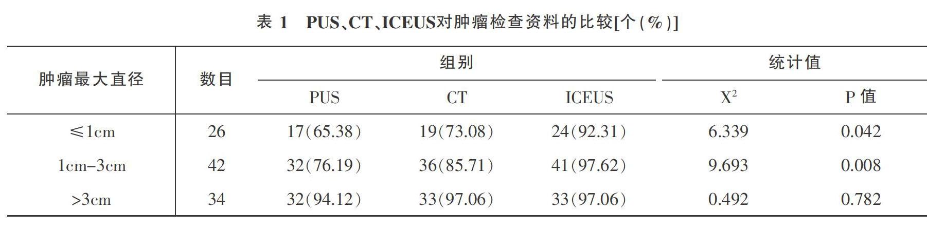

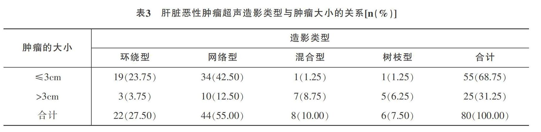
摘要:目的:探討术中超声造影在肝肿瘤患者中的临床应用价值。方法:回顾性分析我院2015年12月至2021年1月收治的86例肝肿瘤患者,依据术前彩超、增强CT或MRI、术中超声造影检查方法,分为术前超声(Preoperative ultrasonography,PUS)、CT、术中超声造影(Intraoperative contrast-enhanced ultrasonography,ICEUS)。结合术后病理检查结果,比较三种检查方法对不同直径肝肿瘤的敏感度,筛选出漏诊率低的检查方法;同时比较术中超声造影与术前常规检查对肝恶性肿瘤诊断的准确率;并观察肝脏恶性肿瘤超声造影类型与肿瘤大小、分化程度的关系。结果:ICEUS对于肝肿瘤数目的检出率为96.08%,显著高于PUS(79.41%)及CT(86.27%),差异有统计学意义(P<0.05),并发现这种差异主要体现在微小及小病灶肿瘤方面;但与术前检查比较,ICEUS对肝肿瘤性质的判断无明显优势,差异无统计学意义(P>0.05);80个恶性肿瘤病灶中,肿瘤超声造影类型与肿瘤大小存在明显相关性(X2=24.022,P=0.000),与肿瘤分化程度存在明显相关性(X2=23.236,P=0.001)。结论:术中超声造影能发现新的肿瘤病灶,有效补充术前检查不能发现的较小肿瘤病灶,能多角度显示肿瘤和管道的立体结构,保障手术精准操作,减少术后肿瘤残留及复发,而且发现超声造影类型为肿瘤分化程度提供有效依据,值得临床广泛应用。
关键词:肝肿瘤;超声造影检查;临床应用;术中
中图分类号:R735.7文献标识码:ADOI:10?郾3969/j.issn.1001-0270.2021.04.08
The Clinical Value of Intraoperative Contrast-enhanced Ultrasound in Patients with
Liver Tumors
JIA Xiao-yan, YUAN Zong-cheng, MA Dong, KONG Hai-liang(Corresponding author)
(Department of Ultrasound, the First Hospital of Hanbin District, Shaanxi 725000, China)
Abstract: Objective: To explore the clinical application value of intraoperative contrast-enhanced ultrasound in patients with liver tumors. Methods: A retrospective analysis of 86 patients with liver tumors admitted to our hospital was based on preoperative color Doppler ultrasound, enhanced CT or MRI, and intraoperative contrast-enhanced ultrasound. They were divided into preoperative ultrasonography (PUS), CT, and intraoperative contrast-enhanced ultrasound (ICEUS). Combined with postoperative pathological examination results, the sensitivity of the three examination methods to liver tumors of different diameters was compared, and the examination methods with low missed diagnosis rate were screened out. At the same time, the accuracy of intraoperative contrast-enhanced ultrasound and preoperative routine examinations in the diagnosis of liver malignant tumors were compared,also observed the relationship between the type of liver malignant tumor and the size and differentiation of the tumor.Results: The detection rate of ICEUS for the number of liver tumors (96.08%) was significantly higher than that of PUS (79.41%) and CT (86.27%). The difference was statistically significant (P<0.05), and it was found that this difference was mainly reflected in small tumor. Compared with intraoperative contrast-enhanced ultrasound examination and preoperative examination, ICEUS had no obvious advantage in judging the nature of liver tumors, and the difference was not statistically significant (P>0.05). Among 80 malignant tumor lesions, the contrast-enhanced type of tumor was significantly correlated with tumor size(X2=24.022, P=0.000), and significantly correlated with the degree of tumor differentiation(X2=23.236, P=0.001). Conclusion: Intraoperative contrast-enhanced ultrasound can find new tumor lesions, effectively supplement the smaller tumor lesions that cannot be detected by preoperative examination, and display the three-dimensional structure of tumors and pipelines from multiple angles, ensuring accurate surgical operations, reducing postoperative tumor residues and recurrence, and finding The type of contrast-enhanced ultrasound provides an effective basis for the degree of tumor differentiation, and is worthy of extensive clinical application.
Key Words: Liver tumor; Contrast-enhanced ultrasound examination; Clinical application; Intraoperative
我国肝癌患者占全世界病例的46.7%,肝癌在我国肿瘤死亡率中排名第2位[1-2],该疾病严重危害了人民的身心健康,对每位医务工作者提出巨大挑战。随着科学技术发展、仪器设备的换代及影像技术的更新,术前肝肿瘤的诊断率明显提高,但临床实际工作中仍有部分肝肿瘤在术前检查中未能很好定位及定性诊断[3]。……