A Mini Review on Polymer Dots: Synthesis, Properties and Optical Applications
2021-06-16LIHuijunGUIBojieZHIShiboWANGHuanHANSancanWANGDingWANGXianyingYANGJunhe
LI Hui-jun, GUI Bo-jie, ZHI Shi-bo, WANG Huan, HAN San-can, WANG Ding, WANG Xian-ying, YANG Jun-he
(School of Materials Science and Engineering, University of Shanghai for Science & Technology, Shanghai 200093, China)
Abstract: Polymer dots(PDs) have attracted intensive attention due to their advantages of tunable electrical and optical properties based on suitable manipulation of the structure and composition. As a new type of dots, the classification, synthetic methods and properties of PDs still lack systemic summarization. In this review, the polymer dots are divided into two kinds based on their structures: conjugated polymer dots(CPDs) and carbonized polymer dots(carbonized PDs). The definitions, synthetic methods and photoluminescence mechanisms of the two PDs will be discussed. Besides, their applications are demonstrated including bioimaging and fluorescent labelling, drug and gene delivery, sensing, photocatalysis and anti-counterfeiting.
Key words: polymer dots; conjugated polymer dots(CPDs); carbonized PDs; photoluminescence
1 Introduction
Polymer dots(PDs) are a new type of fluorescent carbon-based nanomaterial, which have drawn extensive attention due to their tunable structures and properties[1-2]. The phrase “polymer dots” was firstly used by Sano in 1997[3]. Sano reported a simple method of producing regular arrays polymer deposits on the surface of a silicon wafer. Yet the size of the reported polymer dots was different from that of the PDs defined nowadays. In 2007, Wuetal.[4]reported series of nanoparticles which consisted of π-conjugated polymers. They named the particles as “PDs” since the particle owned small size and exhibited high brightness. For a while, PDs were regarded as a subset of polymer nanoparticles(PNs) and the boundary between PDs and PNs was vague[4-7]. In some cases, researchers have even provided certain criteria to distinguish PDs and PNs[2,8],e.g.if the particles own semiconducting property, the size of the particle should be less than ~30 nm to satisfy the concept of the “dot” and the interior of the nanoparticle should be hydrophobic. On the other hand, as the properties and applications of PDs are similar to that of carbon dots(CDs), PDs were also defined as one kind of CDs before 2015 in many works[9-10].
Nowadays, two types of PDs have been proposed which are conjugated polymer dots(CPDs) and carbonized polymer dots(carbonized PDs), respectively. Wuetal.reported that if the precursors were conjugated polymers, the obtained PDs would be defined as CPDs[11]. CPDs can be obtained from the assembly of fluorescent conjugated polymers[2,9]. Otherwise, if the structure of the prepared PDs is not conjugated, the PDs are named as “non-conjugated polymer dots(NCPDs)”. Carbonized PDs are usually prepared from non-conjugated polymersviadehydration, condensation, carbonization or assembly routes[12-14]. Due to the differences in their precursors and preparation methods, CPDs and carbonized PDs exhibit quite different performance. In 2019, Xiaetal.[15]improved the classification and nomenclature of CDs, and the CDs are divided into four species, including graphene quantum dots(GQDs), carbon quantum dots(CQDs), carbon nanodots(CNDs) and carbonized PDs. According to their definition, carbonized PDs, which possess a hybrid structure of polymer and carbon dots, are a new form of CDs. Some reports have shown that the carbonized PDs are partially carbonized CDs with a core of carbonic structure and a shell of polymer structure[16]. Compared with CDs, PDs own similar characteristics of low toxicity, low cost, tunable electrical and optical properties, while PDs retain special mechanical properties and processing advantages of their polymer precursors.
Although the classification of PDs is more specific nowadays, it is still easy to confuse the usages of semiconducting polymer dots, carbonized polymer dots or polymer carbon dots, especially in the crossing field of carbon dots. It is also hard for some researchers to understand the relationships among these kinds of PDs. To better demonstrate the “PD” material for more potential applications, the classification, definition, property, preparation method and applications of different PDs are discussed in this work.
2 Classification of PDs
Herein, polymer dots(PDs) are divided into two categories according to their structures: conjugated polymer dots(CPDs) and carbonized polymer dots(carbonized PDs). The structure diagram of these PDs is shown in Fig.2.
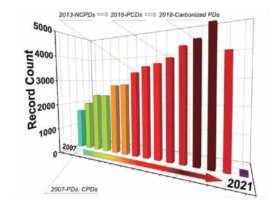
Fig.1 Graph of the record count about “polymer dots” on the web of science on January 16, 2021
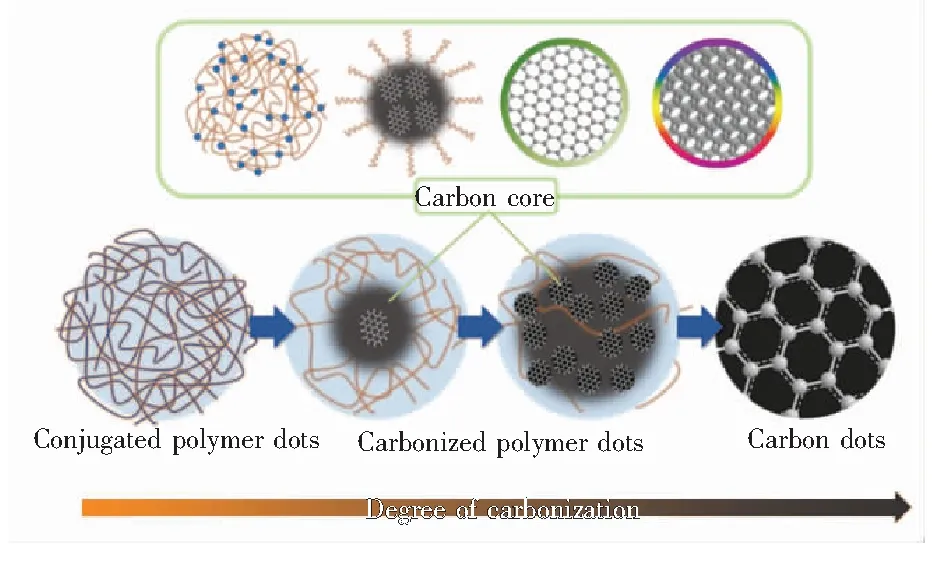
Fig.2 Structure diagram of PDs[9,16]
2.1 Conjugated Polymer Dots(CPDs)
CPDs exhibit excellent optical properties, such as high photostability, strong light-harvesting ability, low cytotoxicity, and high brightness. In general, CPDs are organic nanodots with extended π-conjugation along the molecular skeleton and delocalized electronic structures, which are favorable for two-photon absorption[17-18]. Similar with other common conjugated polymers, the fluorescence of CPDs is related to the π-conjugation in the molecular skeleton. And the extended π-conjugation skeletons make their optical and electronic properties unique, which can also be easily tuned through modifying the conjugated skeletons[19-22]. Compared to conjugated polymers, CPDs demonstrate better water solubility/dispersibility, which endows CPDs more possibility in biological-related applications[23].
Four backbone structures are frequently used in CPDs: poly(fluorene)(PF), poly(p-phenylenevinylene)(PPV), poly(p-phenyleneethynylene)(PPE) and poly(thiophene)(PT)(Fig.3(a)). The four basic structures can be achieved through Suzuki coupling, Heck coupling, Sonogashira coupling, oxidative polymerization and microwave-based polymerization. Most precursors of CPDs have the backbone structure like these four polymers.
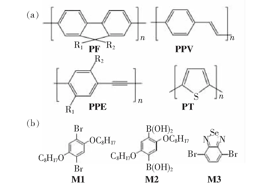
Fig.3 (a)Four basic backbone structures that conjugated polymers are based on: poly(fluorene)(PF), poly(p-phenylenevinylene)(PPV), poly(p-phenyleneethynylene)(PPE), and poly(thiophene)(PT). (b)Three kinds of conjugated polymers synthesized by Kim and co-workers[24].
On the other hand, when the conjugated structure contains repeatedly connected monomer, the band gap of the CPDs will be decreased due to the interaction of electron orbital domains with each other. Thus, the CPDs achieve the property of semiconductors or even conductors. At this situation, this kind of CPD is also defined as semiconducting polymer dots(SPDs), which are widely used in the field of light-emitting diodes, field-effect transistors, and photovoltaic devices[11,17,25-28].
Kimetal.[24]reported multi-color emitted CPDs through size manipulation. They firstly synthesized three kinds of conjugated polymers 1,4-Dibromo-2,5-bis(octyloxy)benzene, 2,5-bis(octyloxy) phenyldiboronic acid, and 4,7-dibromo-2,1,3-benzothiadiazole(M1, M2, and M3) which have same backbone structure but different compositions of repeating units(Fig.3(b)). These conjugated polymers emit different fluorescent emissions resulting from different intermolecular electron transfer paths between the conjugated polymer backbones. Then they fabricated CPDs of different sizes using the three conjugated polymers as the precursors by controlling the number of conjugated polymer chains within the final spherical polymer dots. Due to the size effect, the intermolecular electron transfer was then adjusted, which allowed diverse photoluminescent emission (Fig.4(a)-(b)). Wangetal.[18]have synthesized the CPDs which had an absorption peak at ~600 nm with a high PL quantum yield(QY) of (20.6±1.0)%(Fig.4(c)). The CPDs exhibit good biocompatibility, high photostability, and large two-photon absorption cross section. The CPDs excited by the second near-infrared(NIR-Ⅱ, 1 000-1 700 nm) light could emit NIR-Ⅰ(700-950 nm) emission, which is beneficial to realize deepinvivobrain imaging through intact skull.
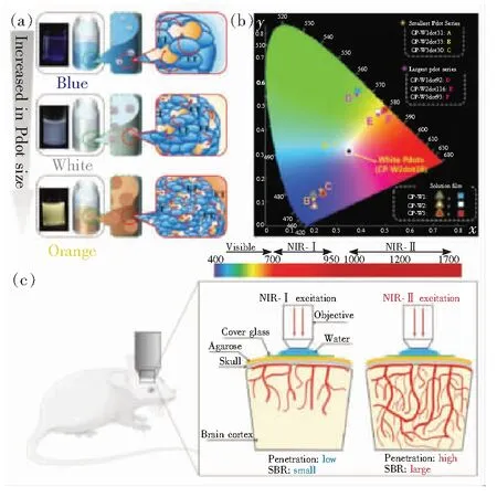
Fig.4 (a)Schematic illustration for the mechanism of various emission colors, depending on the sizes of PDs from a single conjugated polymer[24]. (b)Comparison of CIE coordinates of the largest and smallest PDs with those of CP solutions and the films of CPs[24]. (c)Schematic illustration of NIR-Ⅰ and NIR-Ⅱ excited in vivo 2PF imaging of mouse brain[18].
2.2 Carbonized Polymer Dots(Carbonized PDs)

Carbonized PDs possess a hybrid structure comprising of abundant polymer chains/functional groups on the surface and carbonic core. The core is derived from the dehydration and carbonization process of the connected polymer chain network structure[34-37]. Due to incomplete carbonization,short polymer chains or functional groups could be retained. Based on the degree of carbonization, the carbonic core can be divided into four subclasses: two kinds with complete carbonization cores similar to that of CQDs or CNDs[15,38-40], a para-crystalline carbonic core composed of tiny carbon clusters surrounded by polymer frames[41-42], and a highly dehydrated crosslinking and close-knit polymer frame structure[34-35].
Compared with CPDs, carbonized PDs show better water-solubility and chemical stability. The formation mechanisms of carbonized PDs are carbonization or dehydration of some commercial polymers, such as polyethyleneimine(PEI), poly(vinyl alcohol)(PVA), polyethylene glycol(PEG) and so on[43]. Chenetal.[44]reported a facile synthetic strategy for fluorescent carbonized PDsviautilizing PEI and L-ascorbic acid(AA) as the precursors. The carbonized PDs showed strong blue-green fluorescence emission, and the fluorescence could remain stable for over six months under ambient conditions(Fig.5(a)). The obtained carbonized PDs were then used for detecting tetracycline due to its fluorescence sensitivity. Vallanetal.[45]also reported a versatile room-temperature method to prepare customized fluorescent carbonized PDs(Fig.5(b)). They used four polycondensation pathways to prepare carbonized PDs from the same material, and the obtained carbonized PDs exhibited distinctly differences in fluorescence. They found that different PL properties(e.g. PL QY and excitation independence property) mainly originated from different degrees of conformational rigidity resulted from the intramolecular hydrogen bonding and electrostatic interactions within the four carbonized PDs.
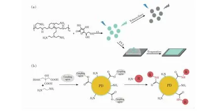
Fig.5 (a)Schematic illustration of the preparation and tetracycline detection of the PVA composite film with the PEI-AA carbonized PDs[44]. (b)Polymerization process of the carbonized PDs[45].

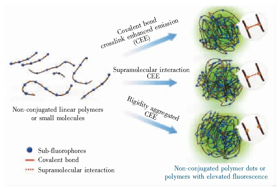
Fig.6 Representation of the covalent-bond, supramolecular- interaction, or/and rigidity-aggregated crosslink-enhanced emission(CEE) effect in carbonized PDs[32]. cases. Due to the unique hybrid structure, carbonized PDs demonstrate special properties such as tunable absorption, good biocompatibility, excellent photostability and high photoluminescence quantum yield[15,34-35,42].
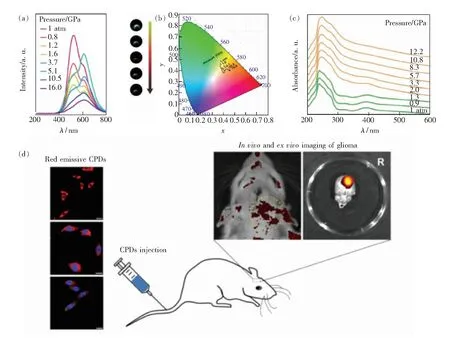
Fig.7 (a)PL spectra changes and the corresponding PL photographs of CPDs-1 under high pressure. (b)Pressure-dependent chromaticity coordinates(CIE) of liquid CPDs-1. (c)Ultraviolet-visible(UV-Vis) absorption against pressures of CPDs-1 in liquid[46]. (d)CLSM images of the cellular behaviors of CPDs[47].
3 Synthetic Methods
As shown above, polymer dots have different structural characteristics, and can be preparedviadifferent methods. The synthetic methods mainly include nano-precipitation method, mini-emulsion method, self-assembly method, hydro/solvo-thermal method, and some other methods.
3.1 Nano-precipitation Method
The nano-precipitation method is divided into three processes: particle nucleation, growth and aggregation.The precipitation of polymer is achieved by increasing solvent diffusion,viaadding more poor solvent or evaporating solvents[48](Fig.8(a)). Ideally, the polymer will be dissolved in the solvent, but not in the poor solvent. The technique is based on the interfacial deposition due to the displacement of solvent by poor solvent[49]. The nano-precipitation method has the advantages of short preparation time, less precursor consumption and energy saving. In details, the polymer precursors are firstly dissolved in water-miscible organic solvent, such as tetrahydrofuran (THF), dimethyl sulfoxide(DMSO), or methyl cyanide(MeCN). Under ultrasonic, excess water is quickly added into the polymer solution. Due to the abrupt change in solvent polarity, the conjugated polymer furtherly crosslinked and condensed, leading to the formation of CPDs. By adjusting the polymer concentration and the amount of amphiphilic polymers, the size of CPDs can then be controlled[50].
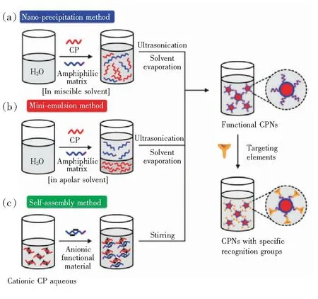
Fig.8 Scheme of nano-precipitation method(a), mini-emulsion method(b) and self-assembly method(c) to prepare PDs[22].
Wuetal.[51]have synthesized multi-color CPDs for biological fluorescence imaging by using nano-precipitation method. Polymers including poly(9,9-dioctylfluorenyl-2,7-diyl)(PFO), Poly(2,5-di(3′,7′-dimethyloctyl) phenylene-1,4-ethynylene(PPE), copolymer poly((9,9-dioctyl-2,7-divinylene- fluorenylene)-alt-co-(2-methoxy-5-(2-ethylhexyloxy)-1,4-phenylene))(PFPV) have been used as the precursors. They have found that the polymer precursors played a key role in tuning the light emissions of the obtained CPDsviaadjusting the size. In another work, Wuetal.[52]have successfully prepared CPDs by utilizing the mixture of hydrophobic polymer poly({9,9-dihexyl-2,7-bis(1- cyanovinylene)fluorene}-alt-co-{2,5-bis(N,N′-diphenylamino)-1,4-phenylene})(PDFDP) and amphiphilic polymer poly(styrene-co-maleic anhydride)(PSMA) to form uniform monodispersed nanoparticles. The PDs exhibit superior photostability that enables long-term stimulated emission depletion(STED) cellular imaging, which guarantees high spatiotemporal characterization of cellular structures and dynamics.
3.2 Mini-emulsion Method
Mini-emulsion method, a kind of heterogeneous polymerization method, is widely used for synthesis of various novel organic-inorganic hybrid materials. Many PDs have been prepared by mini-emulsion method. During the preparation, two incompatible solvents are mixed to form a homogeneous emulsion in the presence of proper surfactant.The reactions including the nucleation, growth, aggregation and agglomeration processes are confined in the emulsion droplet(Fig.8(b)). Due to the existence of independent droplets, spherical particles can be formed and agglomeration between particles is avoided. The mini-emulsion method requires an organic solvent that is insoluble in water, such as dichloromethane, to dissolve the conjugated polymer. Hashimetal.[53]synthesized luminescent quantum-dot-sized CPDs with the mean diameters ranging between 2 and 5 nmviathe mini-emulsion method. The CPDs exhibit excellent stability in solutions for several months. Cabezaetal.[54]have successfully loaded doxorubicin onto poly(butylcyanoacrylate) PDs by anionic polymerization used in mini-emulsion system, which can enhance antitumor activity of doxorubicin in breast cancer. However, the sizes of the PDs preparedviamini-emulsion method are usually larger than 40 nm, thus the method is not suitable for preparing small-sized PDs.
3.3 Self-assembly Method
Self-assembly has attracted attention recently since it provides a mild, bottom-up, and controllable method to assemble tiny atoms or molecules into large nanostructures. During the preparation process, oppositely charged conjugated polymers and co-assembling reagents are dissolved in water with a certain proportion, and stirred to obtain homogeneous solution[8](Fig.8(c)). Notably, the method can only be used to prepare water-soluble conjugated polymers. Without external intervention, the primary disordered systems gradually form into organized structureviathe interaction between individual components(e.g. attraction and repulsion, or spontaneous formation of chemical bonds).
The self-assembly method is unique attributed to the following characteristics: (1)Orderliness: the self-assembled structure would be more orderly than the individual components, which is different from traditional chemical reactions. (2)Interaction: the second feature is the weak interactions existed among the self-assembly like Van der Waals, capillary action, π-π interaction, and hydrogen bond. Compared with the traditional covalent bonding, these weak interactions play a more important role in the synthesis reaction and influencing the morphology and structure of the PDs, by influencing the physical properties of liquids, the solubility of solids, and the molecular assembly of biofilms. (3)Building blocks: the basic unit of the self-assembly includes nano-scale and micro-scale structures with different chemical compositions, structures, and functions[57-58]. The diverse choices of building blocks endow diverse compositions of PDs.
Zhaoetal.[55]have synthesized ultrasmall CPDs in the phosphate buffer solution(PBS)viathe self-assembly method, owing an amphiphilic structure with hydrophobic backbones and hydrophilic side chains(Fig.9(a)). The CPDs were prepared by assembling the phosphorescent platinum(Ⅱ) porphyrin (O2-sensitive) with the fluorene-based conjugated polyelectrolytes(O2-insensitive). Due to the dual sensitivities to oxygen of the two different compositions, the CPDs could exhibit excellent luminescence response to O2, with high reliability and full reversibility for measuring oxygen levels. The CPDs were then applied for luminescence imaging of tumor hypoxia in tumor-bearing mice. Zhangetal.[56]have synthesized rice-like NCPDs with a high fluorescence QY(Fig.9(b)). The NCPDs were self-assembledviaa mild thermal treatment method in aqueous solution, using PEI and CA as precursors. The obtained NCPDs have been successfully utilized for the detection of Cu2+and ClO-in environmental water samples. Liuetal.[59]have synthesized water-soluble nonconjugated PDs with strong fluorescence emission using hyperbranched polyethyleneimine(PEI) and D-glucose as the precursorsviaSchiff base reaction and self-assembly in aqueous phase.
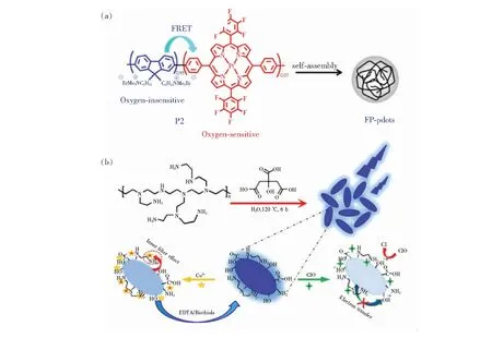
Fig.9 (a)Self-assembly behavior of the fluorescent/phosphorescent conjugated polyelectrolyte into the PDs[55]. (b)Synthesis of the carbonized PDs and the comprehensive sensing strategy[56].
3.4 Hydro/Solvo-thermal Method
Compared to the above methods, hydro/solvo- thermal methods allow for precise control over the size, shape distribution, composition and crystallinity of the polymer dots. Notably, for most PDs, the precursors utilized in the hydro/solvothermal method overlap with that for CDs. It has been found that shorter reaction time or lower reaction temperature leaded to the retaining of polymer structures. During the reaction process, the precursors firstly crosslink and agglomerate, and the degree of condensation and carbonization of the PDs will gradually increase with increasing reaction time[60].Viaproper manipulation of the reaction conditions, the final structure and property of the PDs can be precisely controlled.
Liuetal.[61]synthesized nitrogen-doped conjugated carbonized polymer dots with 31% efficient red emission forinvivoimaging by an one-step hydrothermal method, using o-phenylenediamine (OPD) with HNO3as the precursor and solvent(Fig.10(a)). Tan and co-workers[62]then reported red-emitting carbonized PDs obtained from p-phenylenediamine aqueous solution. They have utilized sulfuric acid(H2SO4), hydrochloric acid(HCl), and perchloric acid(HClO4) as the solvent, and H2SO4could greatly improve the PL QY of the red emission. It was found that ammonium salt precipitates were formed only in H2SO4-assisted system, which would release free reactants slowly and avoid the formation of large particle polymer precipitation, which promoted the formation of high-quality carbon dots.
Fig.10 (a)Possible formation mechanism of CPDs with conjugated aromatic benzene skeleton[61]. (b)Schematic representation of the formation of CDs from citric acid and ethylenediamine through the pyrolysis process[42].
3.5 Other Methods
In addition to the methods mentioned above, there are some common methods for preparing PDs, including microwave-assisted method, combustion/pyrolysis, and acid oxidation method. Similar to hydro/solvo-thermal method, microwave-assisted method exhibits short reaction time, rapid reaction rate, and controllable condition[63]. Zhaoetal.[64]have prepared a kind of PDs as a useful fluorescent probe for detecting Co2+and Mn2+. The PDs have been synthesized at 200 ℃ for 90 minviamicrowave-assisted method. The microwave-assisted method is convenient and time-saving. By comparison, PDs synthesizedviacombustion/pyrolysis and acid oxidation show lower product yields and PLQYs, as a result of plentiful group losses in the form of NH3/CO2/H2O and formation of water-insoluble carbon-sheet impurity[65].
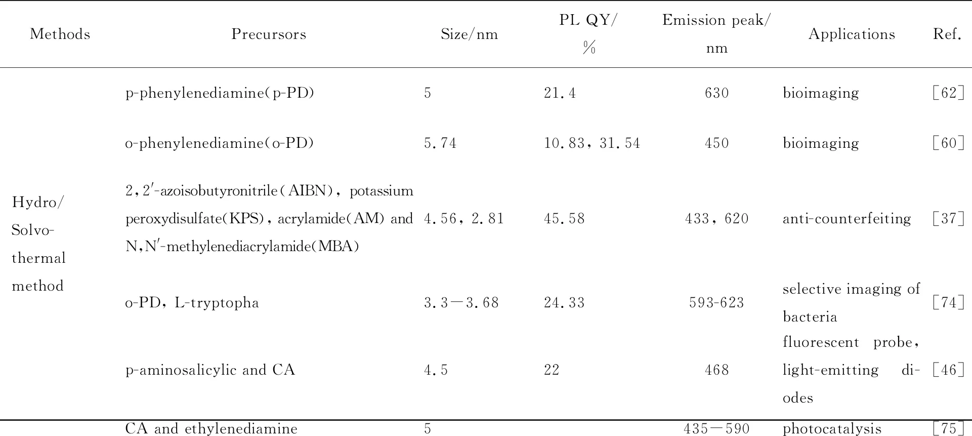
The PDs obtained from various precursors and preparation methods are summarized in Tab.1.
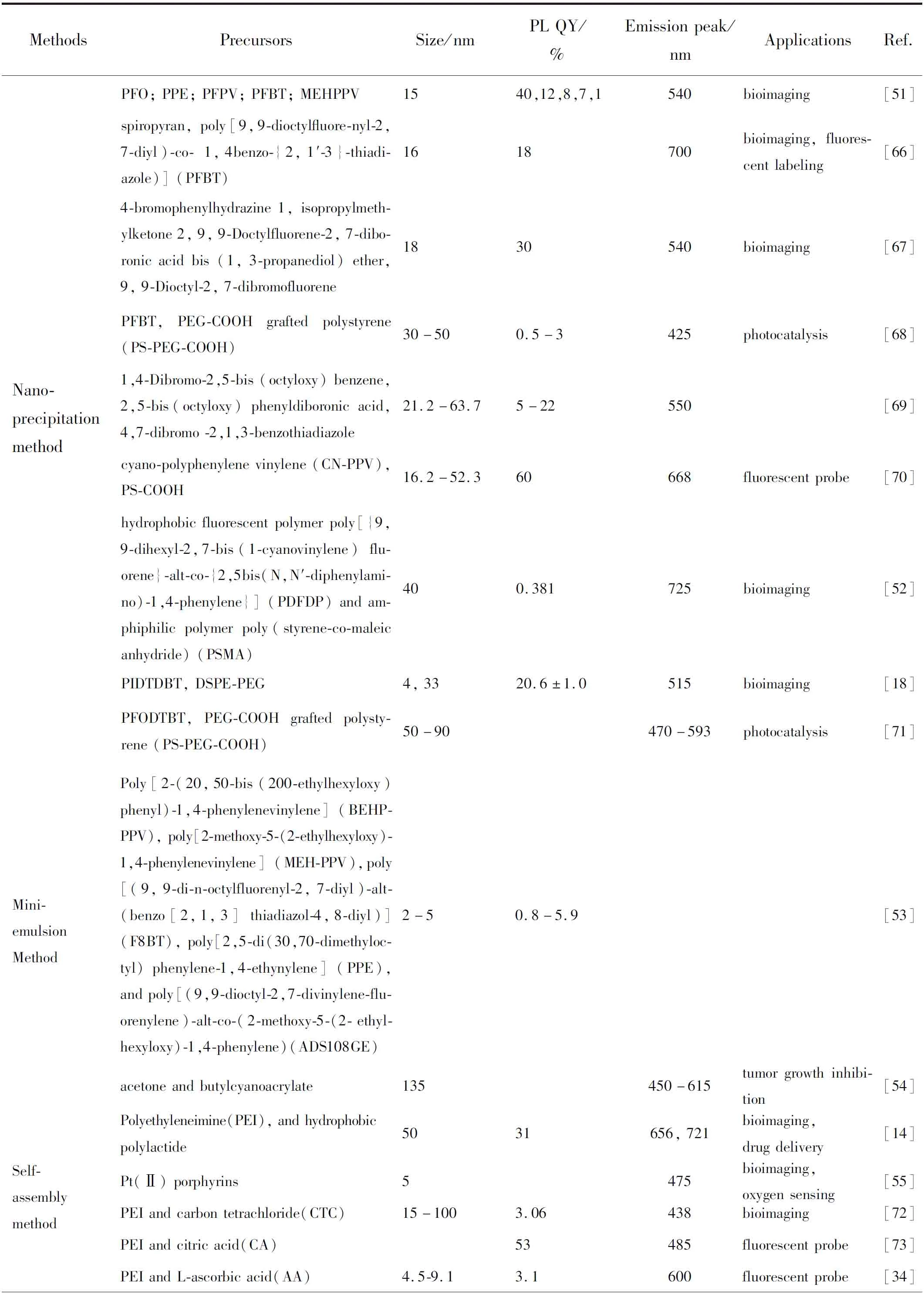
Tab.1 Summarization of the optical characters and applications of the PDs obtained from various precursors and preparation methods
Tab.1(continue)
4 Applications
PDs have been widely used in the fields of fluorescent labeling, drug delivery, bioimaging, catalysis, and anti-counterfeiting, due to their good biocompatibility, fluorescent properties and selectivity[2,32,51,71,76-77].
4.1 Bioimaging and Fluorescent Labeling
Most PDs, especially CPDs, could emit red or even near-infrared(NIR) fluorescence, which is a key factor ininvivobioimaging since long-wavelength light own strong penetrability through deep tissue. Besides, the light emission of long wavelength effectively avoids the influence of self-fluorescence of biomolecules or other materials which are usually blue or green. The NIR emission of PDs are owing to the special backbone structure of their precursors, which are usually semiconducting polymers(SPs). SPs consist of single or multiple π-conjugated segments. Electrons would travel along the backbones through tunneling and hopping by overlapping the electron clouds. When the electrons migrate from the donors to the acceptors within the backbones, energy transfer is amplified. These donor-acceptor units are connected directly or bound by the alkyne or alkene units which improve escalating energy transfer, thus leading to NIR emissions. The chemical structures of commonly used SPs are listed in Fig.11, and some typical structures exhibiting NIR-Ⅱ emission are included.
Fig.11 Chemical structures of semiconducting polymers[78]
Wuetal.[79]have synthesized CN-PPV PDs streptavidin probes via functionalization and bio-combination. The emitted orange PL from CN-PPV PDs exhibited high brightness, and excellent ability for highly targeting subcellular structures, showing great potential in label cell surface markers for microtubule structures in mammalian cells. Tanetal.[62]reported a facile method using acid-assisted hydrothermal route to prepare CPDs. The CPDs showed the two-photon photoluminescence properties by emitting 602 nm light under the excitation of 850 nm femtosecond pulse laser. The CPDs were then used in bioimaging for HeLa cells. Fangetal.[80]then reported hydrophobic PDs of small size prepared by nanoprecipitation, which can be used as an imaging probe for hypoxia(Fig.12(a)-(b)). The PDs exhibited high sensitivity, full reversibility and excellent hypoxia sensing in solution. Alifuetal.[81]synthesized a type of NIR-IIb light excitable deep-red emissive PDs, which has three-photon fluorescence and large three-photon absorption cross section(Fig.12(c)). With deep-red emission, the PDs can be utilized forinvivobioimaging of cerebral vasculature with and without the brain skull.
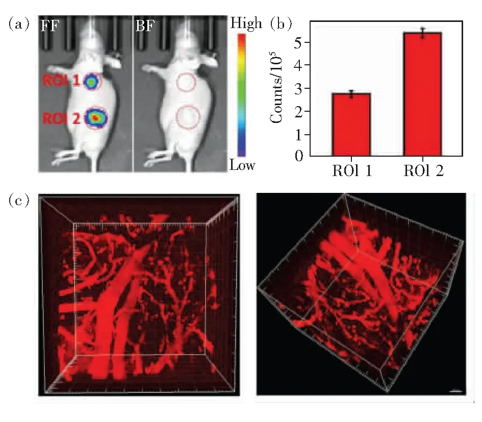
Fig.12 (a)Whole-body imaging of mice after subcutaneous (ROI 1) and intratumoral(ROI 2) injection of 1.6 mg·mL-1 CPDs solution. (b)Semiquantitative analysis of brightness in ROI 1 and ROI 2 area[80]. (c)3D reconstructed 3PFM three-photon fluorescence microscopic images of the brain blood vessels at depth from 0-350 μm[81].
4.2 Drug and Gene Delivery
Various well-organized drug and gene carriers have been designed and fabricated, including nanofibers, microparticles, nanoparticles, and nano dots[32]. These carriers could release the drugs or genes in specific part of organs, or cellular by an instant or batched drug-dosing type. PDs have been developed as smart replacements to the traditional metal quantum dots or polymer-based nanostructures, attributed to their high stability and bioavailability for stimuli-responsive drug dosing[2,82]. Various reports have shown that the PDs based carriers could improve preferential accumulation of drugs in tumor sites and extension of circulation time, and also reduce systemic side effects[32]. Compare to CDs, PDs are more suitable to encapsulate drugs or other therapeutic agents due to their flexible polymer matrix[2].
Lvetal.[83]have designed partially carbonized PDs of small size, which could function as vaccine carriers to deliver model antigen protein ovalbumin. Thus, the PDs could be utilized for potential subcutaneous vaccine delivery to induce efficient cancer immunotherapy. Weietal.[84]reported a type of positively charged fluorescent CPDs for gene delivery andin-situintracellular fluorescence imaging. Most of the cells were successfully loaded with CPDs after co-incubation for 2 h. Maetal.[85]have prepared theranostic liposomes containing CPDs. Theinvivostudies of the tumor-bearing mouse models revealed that the distribution of liposomes in tumor could be indicated accurately. The achievement has broadened the use of CPDs-based liposome by encapsulating various drugs into the theranostic liposomes.
4.3 Sensing
The unique structure and optical property of PDs bestow them potential applications as detector or sensor for various stimulus sources,e.g.gases, ions, biomolecules, pH or light[2,8,56,86].
Tongetal.[45]have reported carbonized PDs for Fe3+sensing. They attributed the quenching of the CEE fluorescence to surrounding Fe3+ions. The keto-enol tautomerism in amides is the key to capture Fe3+ions. Shown in the calculation results, the Gibbs free energy of the complex of Fe3+ion and the carbonized PDs is smaller than that of carbonized PDs. Thus, the electron cloud distribution of the CEE unit has been changed, which caused fluorescence quenching. Chengetal.[87]synthesized a sensitive PDs for determination of α-L-fucosidase, which is active in human serum. The PDs fluorescent sensor exhibits high sensitivity and selectivity, extending the application of PDs in biosensing field. Dongetal.[88]synthesized thermosensitive CPDsviasolvothermal method. The solid-state CPDs exhibited a reversible temperature-responsive photoluminescence enhancement behavior. They attributed this behavior to competitively restriction intramolecular vibrations(RIVs) and π-π interactions.
4.4 Light-emitting Devices
Fluorescent PDs have also attracted attention in the field of photoelectric devices. Some PDs of high PL QYs or long wavelength emission have been used in constructing LEDs. Zhaoetal.[74]have reported the carbonized PDs with dual-emission fluorescence of inherent blue and red emission. It was found that the quantitative structure and emission centers could be controlled by regulating the reaction conditions. They have successfully developed series of LED devices with yellow, red and blue emissions, by combining the PDs of dual-emission with commercially available GaN LED chips which emit UV light. Wangetal.[89]developed a low-cost, fast processable, environmentally friendly and one-step synthetic approach for preparing gram-scale single-component white-light-emissive carbonized PDs, which exhibited white light emission with the QY of ~41% and phosphorescence with the QY of ~23% under ambient conditions.
4.5 Photocatalysis
As introduced above, the PDs own semiconductor properties in some cases by properly adjusting the size and doping elements. This property can be utilized in the field of catalysisviapreparing their heterojunctions with other semiconductors or nanocomposites to improve the catalysis performance.Compared to traditional inorganic photocatalysts, organic photocatalytic PDs have gained enormous attention due to their several key features[90], including: (1)effective and facile preparation methods; (2)tunable size and surface hydrophilicity; (3)tunable optical gaps and relatively long excited state lifetimes; (4)electrons could move freely along the polymer backbone owing to the large π-conjugated structures. Due to these characters, many organic semiconducting PDs have been investigated as potentially photocatalysts. Wangetal.[75]have synthesized carbonized PDs/PbBiO2Br heterogeneous composite photocatalysts in the presence of self-sacrificing ionic liquid glueviaa facile solvothermal method. The heterojunction photocatalysts were proven to exhibit enhanced photocatalytic CO2reduction activities under light irradiation compared to the pure materials, which can be owing to the increased carrier separation and transfer rate caused by appropriate band structure in the heterojunction. Zhangetal.[91]have demonstrated an effective strategy to compartmentalize PDs into a liposome and successfully utilized the obtained nanoreactor as a photocatalyst forin-situhydrogen generation. The hydrogen is furtherly used forin-situhydrogen therapy, which is convenient and safe. This therapy method is also called photodynamic therapy, which has broad research prospects[92].
4.6 Anti-counterfeiting
Fluorescent anti-counterfeiting usually requires fluorescent materials with unique optical properties. The fluorescent materials could represent two or three kinds of information when exposed to different environments. The modulation factors could be light, pH, temperature, moisture or solvents. The fluorescence could be dual-emission, phosphorescence or other stimuli-responsive PL shift, which are also important characters of PDs. PDs have been proven to be suitable anti-counterfeiting ink materials based on their unique and tunable fluorescence and phosphorescence properties. Linetal.[93]have prepared PDs emitting photo-switchable ultrahigh-brightness red fluorescence. The special PL property was derived from the fluorescence resonance energy transfer(FRET) effect by introducing photochromic molecules into the PD structure. The red PL could then be reversibly quenched by UV irradiation, which is an ideal property for anti-counterfeiting. Abdollahietal.[94]also synthesized photo-switchable PDs as anti-counterfeiting materials. And the anti-counterfeiting ink was prepared and can be used to print clear QR code pictures, which is invisible under visible light, but not under UV irradiation.
5 Challenge and Outlook
As a new type of fluorescent nanomaterials, polymer dots own the benefits of tunable light emission, stability, biocompatibility and stimuli-sensitivity. Although tremendous efforts have been contributed to develop new precursors, synthetic methods and photoluminescence mechanism for PDs, some problems still exist and further researches are needed to improve their performance and applications.
(1)Precise definition. Since there are vague regions where carbonized PDs and polymerized CDs are usually regarded as the same family, more reasonable and unified classifications should be established and developed to elucidate their similarity and differences including precursors and polymeric characteristics.
(2)Purification treatment. The bottom-up pathways to PDs usually involve molecular precursors, inevitably introducing fluorescent byproducts of small molecular weight or oligomers. The existence of these byproducts becomes an obstacle to obtain reliable results while proper treatment is ignored in quite a few studies. To pave the way toward cellular imaging, lighting, catalysis and sensing, uniformly implemented purification steps for PDs, especially for carbonized PDs, are highly demanded. In addition to the common methods including time-consuming purification with dialysis membranes or high-cost HPLC technique, we could consider some novel methods developed for purifying carbon dots, for example, a new pH-controlled cloud point extraction(CPE) technique reported by Asadollah Beiraghietal.[95].
(3)Photoluminescence. The relationships between the structure and the fluorescence mechanism of PDs have not yet been studied clearly, due to the complexity of the structures of some PDs. Meanwhile, to realize betterinvivobioimaging and drug delivery for clinical examination, the PL emission of PDs with high QYs in the long wavelength region and their biocompatibility with improved stability require furtherly improvement. Thus, novel biocompatible precursors and solvents could be developed. Besides, it also requires precise purification treatment.
(4)NIR fluorescence imaging. Recently, PDs have been researched for bioimaging in the NIR-Ⅰ and NIR-Ⅱ region to introduce new opportunities in biomedical imaging. The NIR fluorescence imaging can improved the penetration depth to visualize deep tissues for treatment of cancer, bloodow, or brain tumors at an early stage. The research on the NIR fluorescence imaging of PDs is very meaningful and promising, including how to reduce costs, ensure long-term biocompatibility, and reduce the influence of water on fluorescence.