胃肠间质瘤的MRI影像学征象研究及在其病理侵袭危险度评估中的应用探讨
2021-05-10姜书山
姜书山
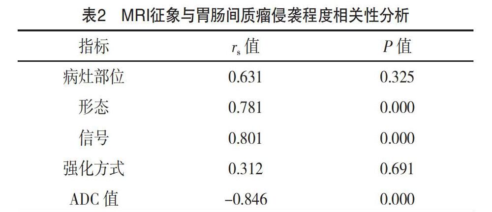
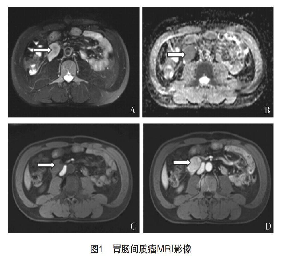
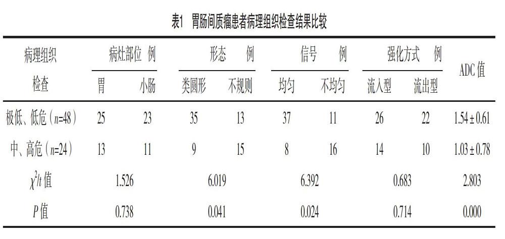
【摘要】 目的:探讨胃肠间质瘤的MRI影像学征象及在其病理侵袭危险度评估中的应用。方法:选取2017年5月-2020年1月本院72例胃肠间质瘤患者,所有患者均经病理組织及免疫组化确诊(金标准),确诊前均行MRI影像学检查,将MRI检查结果与金标准进行比较,根据检查结果比较极低、低危与中、高危患者不同侵袭程度MRI影像学征象,将MRI征象与胃肠间质瘤侵袭程度进行相关性分析。结果:72例胃肠间质瘤患者中极低、低危患者48例,中、高危患者24例,极低、低危患者与中、高危患者的形态、信号、ADC值比较,差异均有统计学意义(P<0.05)。MRI检查最终确诊70例,诊断符合率为97.22%。Spearman相关分析结果显示,胃肠间质瘤侵袭性与MRI征象中形态、信号呈正相关性(P<0.05),与ADC值呈负相关性(P<0.05)。结论:MRI用于胃肠间质瘤患者中影像学征象明显,能实现良、恶性的鉴别,且评估、预测病理侵袭危险程度,为临床诊疗提供影像学依据,值得推广应用。
【关键词】 胃肠间质瘤 MRI影像学征象 病理侵袭
[Abstract] Objective: To investigate the MRI imaging signs of gastrointestinal stromal tumors and its application in the assessment of the risk of pathological invasion. Method: A total of 72 cases of gastrointestinal stromal tumor patients were selected from May 2017 to January 2020 in our hospital, all patients were confirmed by histopathologic and immunohistochemical (the gold standard), MRI imaging examination were performed before diagnose, the MRI results comparing with the gold standard, according to the result of inspection is very low, low and medium, high risk patients with different degree of invasion MRI imaging signs, the MRI signs and gastrointestinal stromal tumor degree of invasive correlation analysis. Result: Among the 72 patients with gastrointestinal stromal tumor, there were 48 patients with very low/low level and 24 patients with medium/high risk. The differences in morphology, signal and ADC value between the two groups were statistically significant (P<0.05). MRI examination finally confirmed the diagnosis of 70 cases, the diagnostic coincidence rate was 97.22%. Spearman correlation analysis showed that the aggressiveness of gastrointestinal stromal tumor was positively correlated with the morphology and signal in MRI (P<0.05), negatively correlated with the ADC value (P<0.05). Conclusion: MRI has obvious imaging signs in patients with gastrointestinal stromal tumors, can distinguish between benign and malignant, evaluate and predict the risk of pathological invasion, provide imaging evidence for clinical diagnosis and treatment, it is worthy of popularization and application.
[Key words] Gastrointestinal stromal tumor MRI imaging signs Pathological invasion
胃肠间质瘤是一种起源于胃肠道间叶组织的肿瘤,占消化道间叶肿瘤的大部分[1]。既往研究表明:胃肠间质瘤与胃肠道肌间神经丛周围的Cajal间质细胞相似,具有c-kit基因、CD117表达阳性[2]。胃肠间质瘤好发于胃、小肠、食道及结直肠,临床多表现为包块、腹痛、胃肠道梗阻等,影响患者健康、生活。既往研究表明:胃肠间质瘤是一种具有潜在的恶性肿瘤,具有较高的复发率、转移率[3],患者预后与病理危险程度分级有关,即肿瘤的危险度分级越低,患者治疗预后越好。手术组织检查是胃肠间质瘤患者常用方法,并视为“金标准”,但是该方法检查前不宜使用激素治疗,再加上生物学形态上具有侵袭性等,难以动态了解患者病情变化情况[4]。MRI是临床上常用的影像学检查方法,能实现多平面、多参数成像,且患者诊断时能结合扩散加权成像(diffusion-weighted imaging,DWI)等功能成像,能清晰地显示病灶部位、病灶大小及病灶的内部结构等[5-6]。……