肝硬化患者的视网膜血管特征及其可能的诊断价值
2021-04-25黄茜钟跃思蔡满航
黄茜?钟跃思?蔡满航
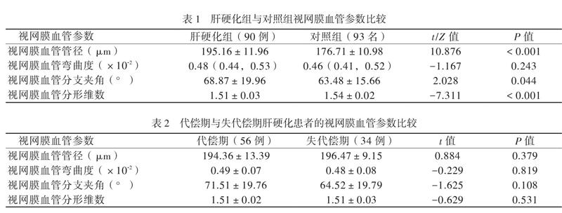

【摘要】目的 分析肝硬化患者的視网膜血管特征,并探讨视网膜血管参数在肝硬化中的诊断价值和眼底检查作为肝硬化筛查手段的可行性。方法 将90例确诊为肝硬化的患者和93名同期体检健康人员分别设为肝硬化组与对照组。使用眼底照相机和自动化图像分析系统对入组个体进行眼底图像采集和视网膜血管参数测量,包括视网膜血管管径、血管弯曲度、血管分支夹角和血管分形维数。比较2组各视网膜血管参数,采用受试者工作特征(ROC)曲线分析各视网膜参数单独和联合诊断肝硬化的效能。结果 与对照组相比,肝硬化组具有更宽的视网膜血管管径、更大的血管分支夹角和更小的血管分形维数(P均< 0.05)。视网膜血管管径、视网膜血管分形维数及两者联合诊断肝硬化的ROC曲线下面积分别为0.869(95%CI 0.820 ~ 0.919)、0.780(95%CI 0.713 ~ 0.847)和0.901(95%CI 0.860 ~ 0.942)。结论 肝硬化患者的视网膜血管系统发生了明显改变。视网膜血管管径和血管分形维数对肝硬化具有一定的诊断价值,诊断效能较佳,两者联合具有更高的诊断价值。眼底检查或可为肝硬化的便捷筛查和无创诊断带来更多的可能性。
【关键词】肝硬化;诊断;视网膜血管管径;视网膜血管分形维数;眼底检查
Retinal vascular features and diagnostic values in patients with liver cirrhosis Huang Xi, Zhong
Yuesi, Cai Manhang. Department of Hepatobiliary Surgery, the Third Affiliated Hospital of Sun Yat-sen University,
Guangzhou 510630, China
Corresponding author, Zhong Yuesi, E-mail: zhyues@ mail. sysu. edu. cn
【Abstract】Objective To analyze the retinal vascular features of patients with liver cirrhosis, investigate the diagnostic value of retinal vascular parameters for liver cirrhosis, and to evaluate the possibility of utilizing fundus examination as a screening method for liver cirrhosis. Methods Ninety liver cirrhosis patients (liver cirrhosis group) and 93 healthy subjects (control group) were enrolled. The funsus images and retinal vascular parameters including retinal vascular caliber, retinal vascular tortuosity, retinal vascular branching angle and retinal vascular fractal dimension were measured by fundus examination and automated image analysis system. The retinal vascular parameters were statistically compared between two groups. The diagnostic efficiency of single and combined retinal vascular parameters for liver cirrhosis was evaluated by the ROC curve. Results Compared with the control group, the retinal vascular caliber and the retinal vascular branching angle were significantly higher in the cirrhosis group (both P < 0.05). The area under ROC curve for retinal vascular caliber, retinal vascular fractal dimension and the two combined in the diagnosis of liver cirrhosis was 0.869 (95%CI 0.820-0.919), 0.780 (95%CI 0.713-0.847) and 0.901 (95%CI 0.860-0.942), respectively. Conclusions Significant changes in retinal vasculature were found in patients with liver cirrhosis. The retinal vascular caliber and the retinal vascular fractal dimension are of certain value in the diagnosis of liver cirrhosis with relatively high diagnostic efficiency, and the combination of these two retinal vascular parameters yields better diagnostic value. The fundus examination is considered as a potential convenient and non-invasive screening tool for liver cirrhosis.
【Key words】Liver cirrhosis;Diagnosis;Retinal vascular caliber;
Retinal vascular fractal dimension;Fundus examination
肝硬化是一种以肝组织弥漫性纤维化、假小叶和再生结节形成为特征的慢性肝病,常伴有高动力循环综合征和微循环障碍等血流动力学改变,威胁着中国约700万人的健康[1-4]。肝活组织检查(活检)是诊断肝硬化的金标准,但由于其存在有创性和采样误差等缺点而没能成为临床常规检查,目前多以胃镜、超声或CT等影像学检查辅助诊断肝硬化[1, 5]。胃镜检查为侵入性操作,检查过程中患者较为痛苦;超声检查与操作者经验关系较大,易受操作者主观判断影响而漏诊或误诊;CT则存在辐射,且费用高昂,上述原因造成部分患者接受检查的耐受性和依从性低。……