螺旋CT定性诊断肺磨玻璃结节的临床价值分析
2020-08-20林建华
林建华
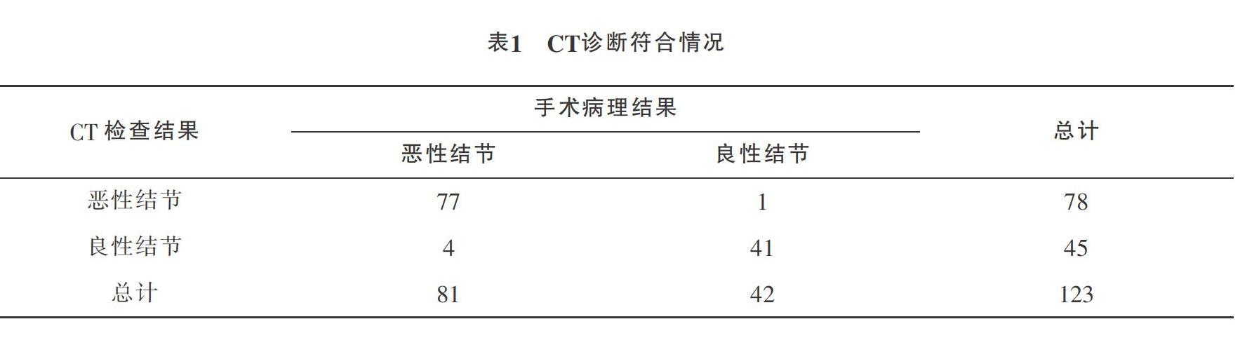

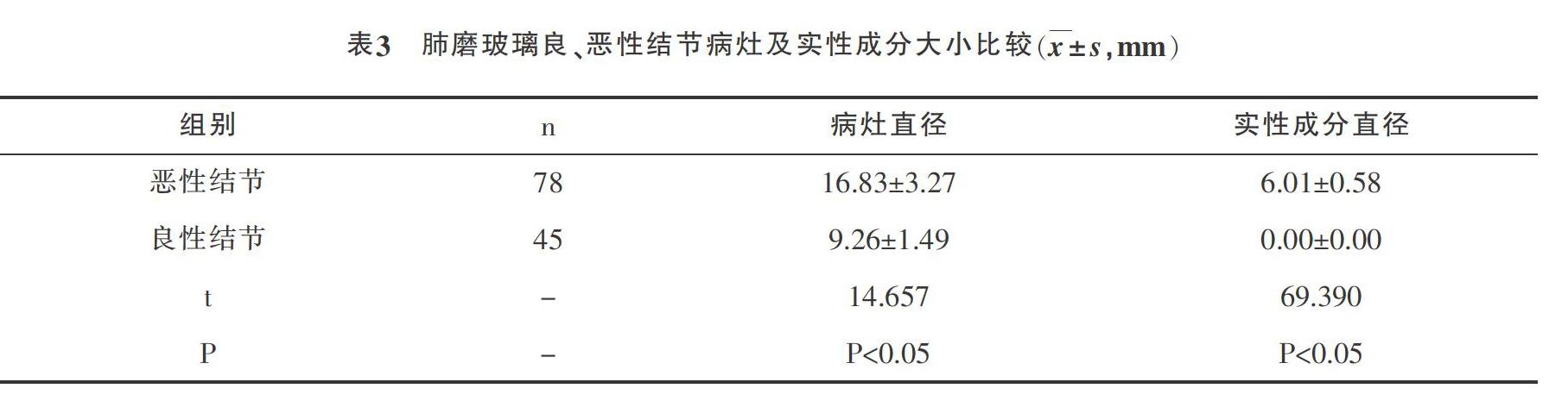
摘要:目的:研究在肺磨玻璃结节定性诊断中运用螺旋CT的意义。方法:纳入123例肺磨玻璃结节患者在我院开展研究。所有病人均行螺旋CT检查,经手术病理证实良性结节42个,恶性结节81个,统计诊断符合情况。结果:CT诊断与手术病理符合率为95.93%,其中良性结节符合率为97.62%,恶性结节符合率为95.06%;肺良性结节病灶及实性成分直径均显著小于恶性结节;肺恶性结节的分叶征、胸膜凹陷、毛刺征出现率高于良性结节,边缘清楚低于良性结节(P<0.05)。结论:在肺磨玻璃结节定性诊断中运用螺旋CT具有重要意义。
关键词:螺旋CT;肺磨玻璃结节;定性诊断;价值
中图分类号:R445.3;R734.2文献标识码:BDOI:10?郾3969/j.issn.1001-0270.2020.04.14
Clinical Value of Spiral CT in the Qualitative Diagnosis of Pulmonary Ground-glass Nodule
LIN Jian-hua
(Radiology Department, Peoples Hospital of Laibin, Guangxi 546100, China)
Abstract: Objective: To study the clinical significance of spiral CT in the qualitative diagnosis of pulmonary ground-glass nodule. Methods: 123 pulmonary ground-glass nodule patients underwent the spiral CT in our hospital were selected. According to surgical and pathological results, 42 cases confirmed with benign nodules and 81 cases confirmed with malignant nodules,the statistical diagnosis is consistent with the situation. Results: The diagnostic accordance rate of spiral CTand Surgical pathology was 95.93%, including 97.62% for benign nodules and 95.06% for malignant nodules; the diameters of benign nodules and solid components were significantly smaller than malignant nodules; the occurrence of lobular sign, pleural indentation and spicule sign in malignant nodules was higher than benign nodules; the marginal sharpness in benign nodules was better than malignant nodules (P<0.05). Conclusion: The spiral CT is great importance to qualitative diagnosis of pulmonary ground-glass nodule.
Key Words: Spiral CT; Pulmonary Ground-glass Nodule; Qualitative Diagnosis; Value
肺磨玻璃结节作为临床肺部常见占位性病变疾病之一,可分为恶性结节与良性结节[1]。恶性结节需早期通过手术治疗,以达到延长病人生存时间的目的。但是临床针对良恶性结节的诊断方法较少,且部分方法无法广泛使用。故采取科学合理的检查方法对肺磨玻璃结节进行早期定性诊断是改善病人疗效与疾病预后的重要前提,我院对此进行研究,阐述如下。
1 资料与方案
1.1 基线资料
纳入123例肺磨玻璃结节患者于2017年2月-2019年2月在我院开展研究,所有病人均知情同意,本次研究经伦理委员会批准。其中男女比例为70:53,年龄为35-74(54.47±4.26)岁。
1.2 方案
采用64层螺旋CT进行扫描,检查前指导病人正确屏气,屏气开始扫描。扫描范围:胸廓入口到肺底;扫描参数:螺距1.375,电流120mA,电压120kV,FOV为30cm;运用高分辨率骨算法进行MPR重建,层厚为1mm;由2名经验丰富的影像学医师观察图像并作出定性诊断(双盲法)。……