Sarcomatoid intrahepatic cholangiocarcinoma mimicking liver abscess:A case report
2020-04-22YanWangJiaLeiMingXingYuRenLuQiuLiJuanZhouShuDongYangXiangMingFang
Yan Wang,Jia-Lei Ming,Xing-Yu Ren,Lu Qiu,Li-Juan Zhou,Shu-Dong Yang,Xiang-Ming Fang
Yan Wang,Jia-Lei Ming,Xing-Yu Ren,Lu Qiu,Li-Juan Zhou,Xiang-Ming Fang,Department of Radiology,The Affiliated Wuxi People's Hospital of Nanjing Medical University,Wuxi 214023,Jiangsu Province,China
Shu-Dong Yang,Department of Pathology,The Affiliated Wuxi People's Hospital of Nanjing Medical University,Wuxi 214023,Jiangsu Province,China
Abstract
Key words: Sarcomatoid intrahepatic cholangiocarcinoma;Liver abscess;Radiological features;Case report
INTRODUCTION
Sarcomatous cholangiocarcinoma is a rare intrahepatic malignant tumor,accounting for 4.5% of intrahepatic cholangiocarcinomas[1].Spindle-shaped and pleomorphic cells and adenoid structures are observed in sarcomatous cholangiocarcinoma[1].In the relevant literature,it is also known as cholangiocarcinoma with sarcomatous changes[2].The tumor pathogenesis has not been sufficiently clarified.The prognosis of sarcomatous cholangiocarcinoma is worse than that of conventional cholangiocarcinoma[1].Pathological diagnosis is the gold standard for diagnosing sarcomatous cholangiocarcinoma.However,a preoperative biopsy can lead to intratumor bleeding and dissemination[2].It is imperative to improve the preoperative diagnosis using imaging examination.To our knowledge,the imaging findings of sarcomatous cholangiocarcinoma have rarely been reported,nor have its radiological features mimicking liver abscess.We report a patient with intrahepatic sarcomatoid cholangiocarcinoma (SICC) and discuss the imaging and clinical features of this unusual disease through a careful review of the literature.
CASE PRESENTATION
Chief complaints
The patient was a 43-year-old male suffering from repeated upper right abdominal discomfort and intermittent distension over a period of one month.There was no report of radiation of the pain to his shoulder and back.
History of present illness
There was neither a history nor symptoms of fever,yellowish eyes,weight loss,or vomiting.
History of past illness
He had a 25-year history of hepatitis B.He did not take any routine medication.
Personal and family history
His medical and family history did not reveal any history of serious or terminal illnesses or any other relevant information.
Physical examination upon admission
His temperature was 37°C,resting heart rate was 80 bpm,respiratory rate was 15 breaths/min,and blood pressure was 120/80 mmHg.
Laboratory examinations
The results of blood analysis were as follows:Red blood cells,5.08 × 1012/L [normal range:(3.5-5.5) × 1012/L];white blood cells,7.7 × 109/L [normal range:(4-10) × 1012/L],and platelet count,149 × 109/L [normal range:(80-300) × 109/L].The results of liver function tests were as follows:Albumin,39.3 g/L [normal range:(35.0-53.0) g/L];globulin,25.5 g/L [normal range:(17.0-33.5) g/L];lactic dehydrogenase,181.0 U/L[normal range:(109.0-245.0) U/L].The results of C-reactive protein and procalcitonin were 4 mg/L [normal range:(0-8) mg/L] and 0.04 ng/mL [normal range:(0-0.5)ng/mL],respectively.Other measurements were as follows:Serum α-fetoprotein(AFP) 66.91 ng/mL (normal range < 10 ng/mL),carcinoembryonic antigen 1.02 ng/mL (normal range < 5 ng/mL),serum carbohydrate antigen (CA) 19-9 and CA 125 levels were 19.9 U/mL and 26.3 U/mL (normal range:< 35 U/mL),respectively.
Imaging examinations
An abnormal mass of hypointensity on T1-weighted images (T1WI) and hyperintensity on T2-weighted images (T2WI) was observed in the right liver lobe(Figure 1).On post-contrast T1WI,honeycomb-like continuous enhancement,with slight transient hepatic parenchymal enhancement (THPE) around it,and adjacent proximal bile duct dilatation with enhancement of the wall were observed.In the arterial phase,blood vessels could be seen entering the lesion.Initially,a hepatic abscess could not be excluded.Further computed tomography (CT) examination(Figure 2) showed that the right lobe of the liver had a patchy low-density lesion of approximately 7.0 cm × 5.6 cm with heterogeneous enhancement.Mild dilation of the intrahepatic bile duct and enlarged lymph nodes of the retroperitoneal region were observed.
The diagnosing physician considered it to be a malignant occupation;thus,surgical resection was performed.During the operation,no ascitic fluid was found in the abdominal cavity.The liver was reddish-brown and nodular hyperplasia of the liver could be seen.A mass in the right lobe of the liver was observed,which had a hard texture.In addition,no satellite lesions were found around this mass.The surgeon diagnosed it as liver cancer and performed radical hepatectomy and lymphadenectomy.
The gross cross-section examination revealed a yellowish-white mass.On microscopic examination,the tumor consisted of an adenocarcinoma component and a sarcomatous component,and the background liver showed nodular hyperplasia.Immunohistochemical examination of the mass revealed positive staining for CD34,CK19,and pancytokeratin AE1/AE3,and negative staining for CA19,hepatocytes,AFP,HMBE-1,G3,TG,TTF1,and CK5/6.
FINAL DIAGNOSIS
Based on the histological and immunohistochemical findings,the tumor was diagnosed as an intrahepatic less differentiated sarcomatoid cholangiocarcinoma.
TREATMENT
The patient underwent radical hepatectomy.After surgery,he was treated with antiinfection agents,rehydration,and symptomatic treatment.
OUTCOME AND FOLLOW-UP
The overall duration of follow-up was only 2.5 mo as the patient passed away.
DISCUSSION
Sarcomatous changes that occur in cholangiocarcinoma are considered rare and the World Health Organization[3]classification of tumors defines sarcomatous intrahepatic cholangiocarcinoma (SICC) as a cholangiocarcinoma with spindle cell areas resembling spindle cell sarcoma or fibrosarcoma or with features of malignant fibrous histiocytoma.At present,the specific pathological mechanism of cholangiocarcinoma with sarcomatosis has not been clarified.Clinical manifestations as well as radiological imaging of this tumor are still limited.
An internet research was carried out using the search engines PubMed and Google with the keywords:“liver”,“sarcomatous”,“sarcomatoid”,“sarcomatosis” and“cholangiocarcinoma”.References of related literature were also reviewed to identify any other potentially relevant publications.After detailed search and analysis,43 nonrepeated cases of SICC were identified in 20 published studies[1,2,4-20].
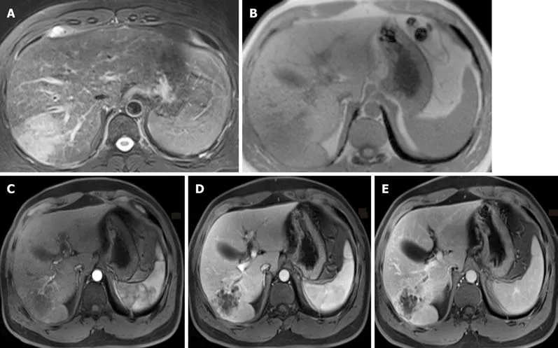
Figure1 Magnetic resonance imaging results.
The following tables (Tables 1-3) present a summary of the previous Englishlanguage literature with the addition of the present case.The clinical features of SICC were non-specific.Abdominal pain and fever were the most common complaints in patients.The age of the patients ranged from 37 to 87 years (median age,61.5 years)with SICC being more commonly observed in men.This is consistent with the age and sex of the patient in the current study.Similar to typical cholangiocarcinoma,SICC usually occurs in the left lobe of the liver.
Radiologic imaging of SICC is still limited.It usually shows a clear low or mixed echo mass on ultrasound[21].CT examination revealed low density lesions,clear or unclear,and sometimes with intra-tumor hemorrhage[15].Most lesions had enhanced peripheral areas in the arterial phase and gradually filled in the central region.In our case,on CT analysis,the lesion was seen as a patchy low-density mass with an unclear boundary.Heterogeneous enhancement was observed in peripheral areas,with no obvious enhancement in the inner necrosis area.In addition,there was slight THPE around the mass.As a result,it is difficult to distinguish SICC from an atypical liver abscess.It was reported that satellite foci may appear in SICC;and there is a certain relationship between satellite nodules and SICC[14].In the case reported herein,no daughter nodules were found.On magnetic resonance imaging,the lesion had low signal on T1WI and high signal on T2WI.After enhancement,a honeycomb-like structure and persistent enhancement with slight THPE and adjacent proximal bile duct dilatation with enhancement of the wall were observed.In addition,blood vessels were observed entering the lesion,which was also considered a distinguishing feature.The SICC involved multiple cystic changes accompanied by fibrous septum and was inhomogeneous and hyperintense in the center on DWI mapping,which was similar to atypical liver abscess on DWI mapping[18].Unfortunately,our patient had no evidence of diffusion restriction.Abdominal angiography showed that the tumor was supplied by the hepatic artery and was considered anemic[7],which was consistent with the results after enhancement.
The degree of SICC malignancy is higher than that of traditional cholangiocarcinoma.The general prognosis for this malignancy is 3 mo[1].It may be associated with an intrinsic sarcoma-like component,with high invasiveness.Most of these tumors are removed by surgery,and there is no unified comprehensive treatment for them.A previous study reported that surgery and postoperative treatment of a combination of gemcitabine and cisplatin adjuvant therapy can improve the prognosis of patients,and survival can be as long as 29 mo[13].However,the effectiveness of chemotherapy remains unclear.According to the survival curve(Figure 3),using the Kaplan-Meier method,in 44 patients with SICC reported in the literature and our case,the survival rate of patients who underwent surgery was significantly higher than those without surgery.The present patient did not undergo chemotherapy or radiotherapy after surgery,only supportive treatment.This was likely a contributing factor in his postoperative survival time of only 2.5 mo.
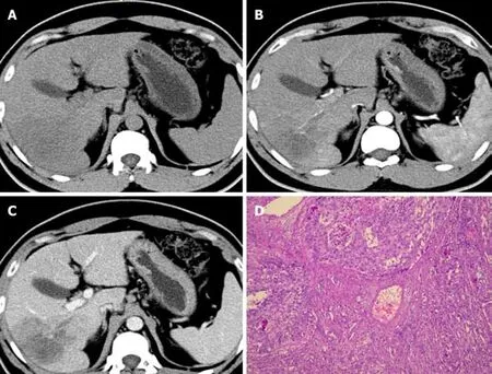
Figure2 Computed tomography images and histopathologic findings (100 ×).
CONCLUSION
In summary,the clinical and radiological features of SICC are non-specific.The usual symptoms are abdominal pain and fever and the imaging features are hypovascularity and progressive enhancement.SICC can present as a multilocular cyst on radiological images and it is necessary to distinguish it from an atypical abscess.It is very difficult to diagnose SICC accurately prior to surgery.The last necessary preoperative biopsy is used for identification.Pathological diagnosis is the gold standard for diagnosing SICC.We report a case of SICC mimicking liver abscess and previous reports were reviewed to increase our understanding of the clinical and imaging evidence of SICC in order to improve the preoperative diagnostic ability for radiologists as well as surgeons.
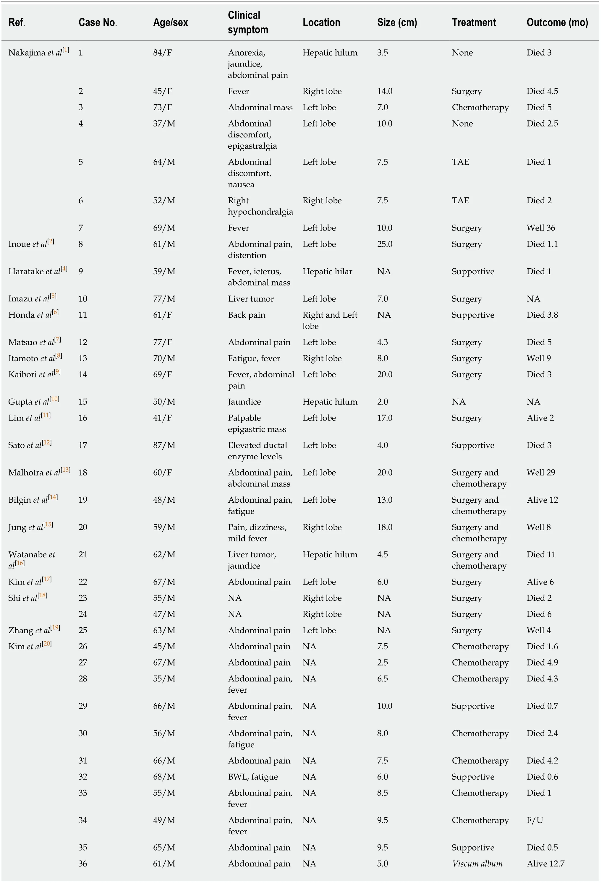
Table1 Summary of sarcomatous intrahepatic cholangiocarcinoma reported in English-language literature from 1992 to 2019
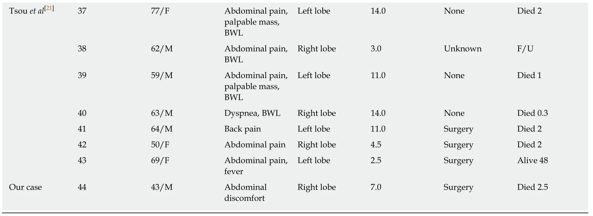
F:Female;M:Male;BWL:Body weight loss;TAE:Transarterial embolization;NA:Not available;F/U:Lost to follow-up.
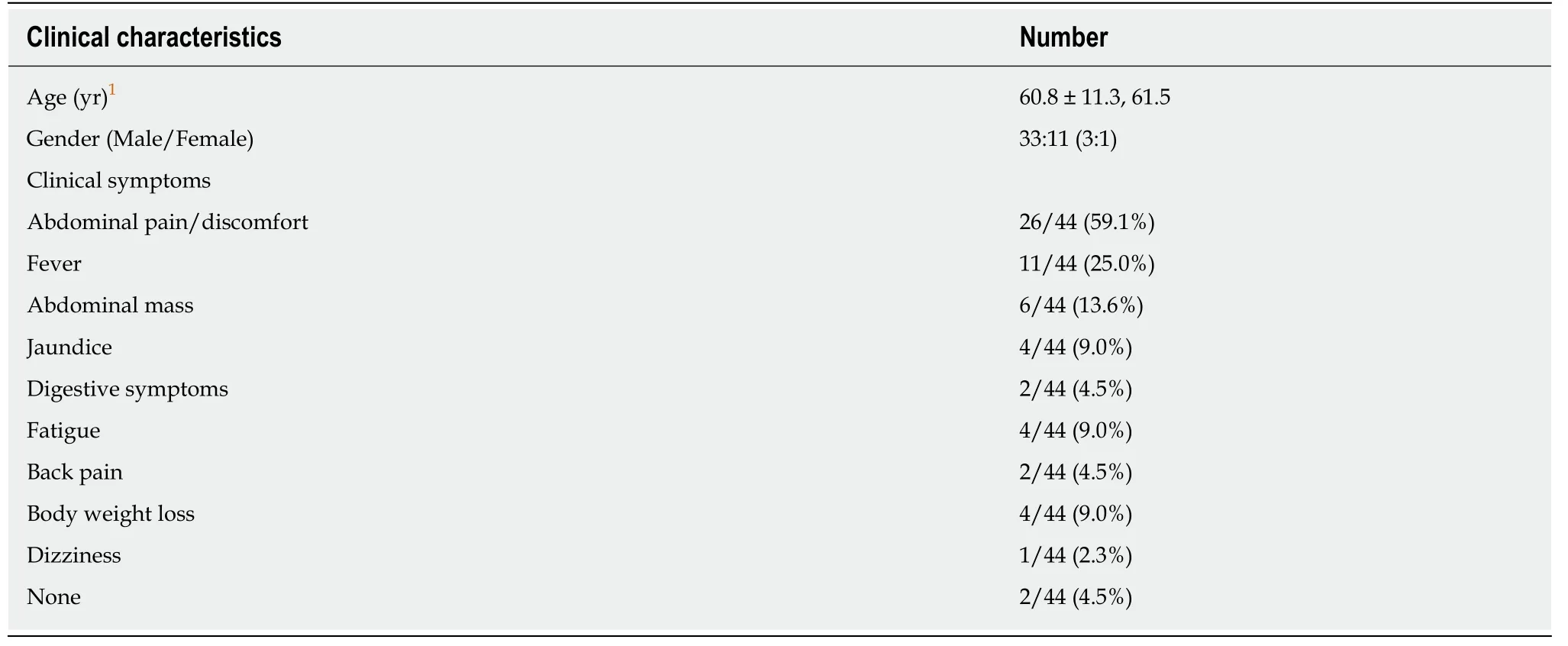
Table2 Clinical characteristics of sarcomatous intrahepatic cholangiocarcinoma reported in the English-language reports and our case
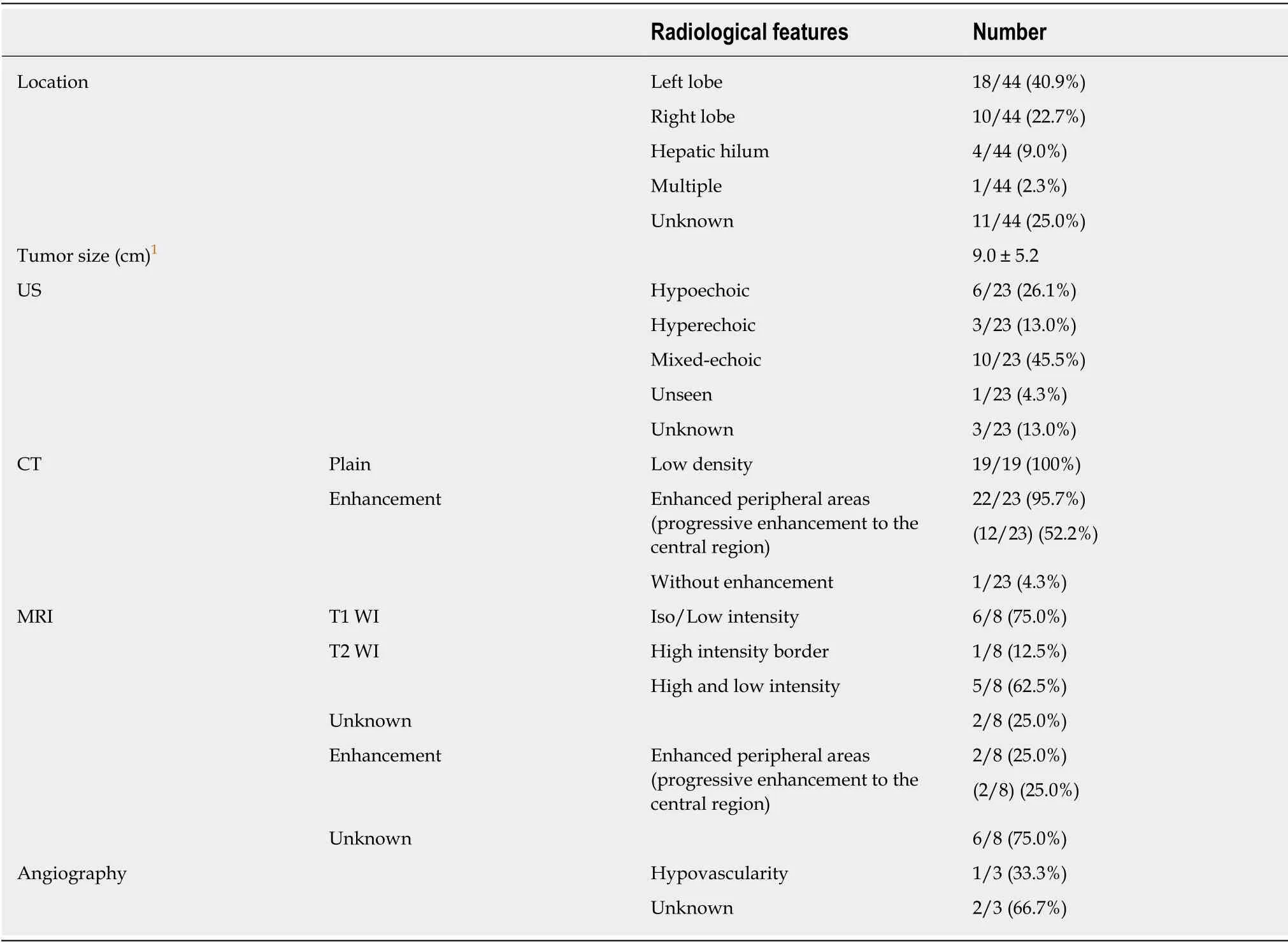
Table3 Summary of radiological features of 44 patients

Figure3 Survival rates of operated sarcomatous intrahepatic cholangiocarcinoma and non-operative sarcomatous intrahepatic cholangiocarcinoma.
ACKNOWLEDGEMENTS
We are grateful to our foreign teacher,a Canadian,for his contribution to language polishing and thank the patients and their family members who agreed to publish this case.
杂志排行
World Journal of Clinical Cases的其它文章
- Role of oxysterol-binding protein-related proteins in malignant human tumours
- Oncogenic role of Tc17 cells in cervical cancer development
- Acute distal common bile duct angle is risk factor for postendoscopic retrograde cholangiopancreatography pancreatitis in beginner endoscopist
- Three-dimensional computed tomography mapping of posterior malleolar fractures
- Application of a modified surgical position in anterior approach for total cervical artificial disc replacement
- Potential role of the compound Eucommia bone tonic granules in patients with osteoarthritis and osteonecrosis:A retrospective study