Postpartum pubic symphysis diastasis-conservative and surgical treatment methods,incidence of complications:Two case reports and a review of the literature
2020-04-22KristinaNorvilaiteMonikaKezeviciuteDianaRamasauskaiteAudroneArlauskieneDaivaBartkevicieneValentinasUvarovas
Kristina Norvilaite,Monika Kezeviciute,Diana Ramasauskaite,Audrone Arlauskiene,Daiva Bartkeviciene,Valentinas Uvarovas
Kristina Norvilaite,Diana Ramasauskaite,Audrone Arlauskiene,Daiva Bartkeviciene,Clinic of Obstetrics and Gynaecology,Institute of Clinical Medicine,Faculty of Medicine,Vilnius University,Vilnius LT-08661,Lithuania
Monika Kezeviciute,Vilnius University,Institute of Clinical Medicine,Faculty of Medicine,Vilnius LT-08661,Lithuania
Valentinas Uvarovas,Department of Orthopedics and Traumatology,Institute of Clinical Medicine,Faculty of Medicine,Vilnius University,Vilnius LT-08661,Lithuania
Abstract
Key words: Pubic symphysis diastasis;Conservative treatment;Internal pubic synthesis;Pregnancy;Vaginal delivery;Case report
INTRODUCTION
The pubic symphysis is a non-synovial joint that connects the right and left superior pubic rami with a normal radiographic separation width of 4 to 5 mm.Due to hormone-related changes and physiological alterations observed during pregnancy,the gap can increase by 2-3 mm and remain after delivery,such a separation is called physiological pubic symphysis diastasis.Infrequently vaginal delivery might lead to joint widening of > 10 mm which is diagnostic and defined as pathological pubic symphysis diastasis[1].A physiological widening of the symphysis joint is based on normal endocrine changes during pregnancy;therefore,it does not depend on the mode of delivery,and the pathological diastasis is considered a complication of the vaginal method of childbirth and can be prevented by Cesarean delivery[2].A separation larger than 25 mm involves damage of ligaments linked to the pubic bones;thus,pubic rupture can be determined[3].This is a rare pathology found in postpartum women with an estimated prevalence ranging from 1 in 300 to 1 in 30000 pregnancies[4].Possible predisposing factors involve the number of previous pregnancies,fetal macrosomia,narrow pelvic outlet or cephalo-pelvic disproportion,rapid and dense contractions during labour,epidural route of anaesthesia,previous trauma in the pelvic region,osteomalacia,chondromalacia,and infections[5,6].Pubic symphysis diastasis can be symptomatic or asymptomatic.Although the exact number of women with an asymptomatic condition is unclear,literature reports show that postpartum patients with asymptomatic separation are at a higher risk of developing symptoms after secondary trauma or weight stress[7].The leading symptom of symphyseal separation is pubic joint pain and inflammation.Pain can radiate to the abdominal or inguinal area,to lower extremities or to the back.The symptoms tend to worsen while moving,bearing a load,or raising a leg.Sometimes it may contribute to symphyseal or lower back discomfort or be followed by complicated locomotion leading to instability and incapacity while walking or standing[1].Less common manifestations are urinary dysfunction and increased and/or uneven movement of the pelvic joints[8].There are various non-operative and operative treatment options,but no gold standard treatment has been defined.The aim of this study was to review currently available approaches underlining pubic symphysis diastasis treatment indications,their effectiveness and complications.Two clinical cases of postpartum pubic symphysis diastasis are presented,and their postoperative conservative treatment,complications and outcomes are described.
CASE PRESENTATION
Chief complaints
Case 1:A 28-year-old patient (gravida 1,para 1) presented to the clinic 3 mo postpartum after an uneventful vaginal delivery with the complaint of a sharp,extremely intense pain in the pubic region.
Case 2:A 32-year-old woman (gravida 2,para 2) developed acute-onset anterior pubic pain during delivery.This anterior pubic pain radiated to the left buttock and thigh.The pain persisted postpartum and was exacerbated by any movement.Moreover,pain in the sacroiliac region contributed to the condition.
History of present illness
Case 1:The patient had a normal pregnancy until the last trimester when she started to complain of pain in the pubic region.She had an uneventful vaginal delivery of healthy twins.The pain increased after labour and became unbearable.Clinical and imaging examinations confirmed pubic symphysis diastasis with secondary pubic osteitis.Conservative treatment was administered for persistent pubic pain.The condition improved.Unfortunately,the pain recurred 3 mo later.The patient started to complain of severe pain accompanied by complicated locomotion.Progression of symphysial separation with expanded signs of osteitis and displacement of pubic bones were detected radiographically.Taking into account that the symptoms recurred after conservative treatment,surgical treatment was selected.Internal pubic synthesis under spinal anesthesia was performed.The pubic symphysis was reached layer by layer through a Pfannenstiel incision.Surgery revealed vertical and horizontal instability of the pelvis and the presence of 0.5 mm inflammatory fluid in the pubic symphysis.Debridement and repositioning were performed and a 6-hole plate and 6 screws were used for fixation of the superior symphysis diastasis.After the operation,the patient was allowed partial weight-bearing with the assistance of crutches for 3 mo.The postoperative period was without complications.However,several months later,a fistula appeared in the scar location.Diagnostic examination showed no pelvic instability or disruption of the inserted plate.Antibacterial therapy was administered and the fistula healed.However,the positive outcome was only temporary as the fistula recurred several times,causing severe aches in the region of the pubic joint.X-ray showed four loose screws,thus removal of the fixation plate was performed.The patient recovered well and one year later had no complaints.Furthermore,no radiological signs of skeletal instability or infection were detected.
Case 2:The patient with an uncomplicated prenatal course developed acute-onset anterior pubic pain during her first and otherwise normal delivery.This anterior pubic pain radiated to the left buttock and thigh.The pain persisted postpartum and was exacerbated by any movement.Radiographs confirmed pubic symphysis diastasis and conservative treatment was administered.The effect was only shortterm and there were several periods of exacerbations of symptoms.When the patient conceived for the second time,acute-onset anterior pubic pain recurred and was accompanied by pain in the sacroiliac region.Symphysis diastasis and secondary pubic osteitis were confirmed,but this time no benefit following conservative treatment was observed.Persistent pain and complicated locomotion led to scoliotic deformation of the lumbar part of the spine and leg length discrepancy.
History of past illness
Case 1:The patient had no previous history of any major illness or any surgical interventions in the past.
Case 2:The patient had no previous history of any major medical illness or any surgical interventions in the past.
Personal and family history
Case 1:No allergies,harmful habits or medicines taken were recorded.
Case 2:No allergies or harmful habits were recorded.The patient was taking vitamin D supplements.
Physical examination upon admission
Case 1:Clinical examination demonstrated painful palpation of the pubic region.Furthermore,pubic pain was provoked by pressure applied to the iliac crests in the antero-posterior and medial directions.There was no neurological pathology.
Case 2:Palpation of the anterior surface of the symphysis pubis elicited pain.Positive Trendelenburg's sign was observed.Active straight leg raising was limited because of yielding pain.Bilateral trochanteric compression also induced pain.
Laboratory examinations
Case 1:Leukocytosis (11 × 109/L) was detected during the first visit,which resolved during conservative treatment and was recorded again when the pain recurred.Neutrophilic leukocytosis (15 × 109/L) and elevation of C reactive protein (350 mg/L)were documented when the fistula occurred in the scar location.No other significant abnormalities in laboratory examinations were observed.
Case 2:No significant abnormalities in laboratory examinations were observed.
Imaging examinations
Case 1:On admission,pelvic X-ray showed a symphyseal gap of 1.5 cm with radiological characteristics of secondary pubic osteitis (Figure 1A).As the pain recurred 3 mo later,X-ray was repeated and a 2.5 cm symphysial separation with expanded signs of osteitis and displacement of pubic bones were detected (Figure 1B).After surgical treatment,the fistula in the scar location appeared several times;thus,X-ray was performed again and it showed four loose screws (Figure 1C).Removal of the fixation plate was performed and no radiological signs of skeletal instability or infection were detected (Figure 1D).
Case 2:On admission,radiographs confirmed pubic symphysis diastasis complicated by osteitis (Figure 2A).X-ray was repeated after repositioning and fixation of pubic symphysis diastasis with a plate and screws (Figure 2B).
FINAL DIAGNOSIS
Pubic symphysis diastasis and secondary pubic osteitis.
TREATMENT
Case 1
Oral non-steroidal antiinflammatory drugs (NSAIDs) and steroid injections into the symphysis pubis joint were administered for persistent pubic pain.Despite temporary improvement the symptoms recurred and surgical treatment was selected.Internal pubic synthesis under spinal anesthesia was performed.Pubic symphysis was reached layer by layer through a Pfannenstiel incision.Surgery revealed vertical and horizontal instability of the pelvis and the presence of 0.5 mm of inflammatory fluid in the pubic symphysis.Debridement and repositioning were performed and a 6-hole plate and 6 screws were used for fixation of the superior symphysis diastasis.After the operation,the patient was allowed partial weight-bearing with the assistance of crutches for 3 mo.The postoperative period was without complications.Several months later,a fistula appeared in the scar location and was successfully managed with antibacterial therapy.However,the positive outcome was only temporary as the fistula recurred several times and four loose screws were detected radiographically;thus,removal of the fixation plate was performed.
Case 2
Following the occurrence of the first episode of pain,NSAIDs and acetaminophen were administered for pain management.However,analgesia was insufficient;thus,intrasymphyseal steroid injections in combination with local anaesthetics were administered.The effect was only short-term and there were several periods of exacerbations of symptoms.During the second pregnancy,the same medications were administered,but this time no benefit following conservative treatment was observed.Taking into account the complications (scoliotic deformation of the lumbar part of the spine and leg length discrepancy),surgery was indicated.The operation was performed in two stages,firstly,the left sacroiliac joint was fixed with a plate and screws,followed by the second stage of repositioning and fixation of pubic symphysis diastasis with a plate and screws.
OUTCOME AND FOLLOW-UP
Case 1
The patient recovered well after surgery and one year later had no complaints.Furthermore,no radiological signs of skeletal instability or infection were detected.
Case 2
Unfortunately,the symptoms remained after surgery.As the operative treatment was not effective,the pain was managed by analgesics,antidepressants and neuroleptics in combination with physiotherapy and pelvic binders.To correct leg length discrepancy,an insole was adjusted for the shorter leg.
DISCUSSION
Postpartum pubic symphysis separation is a clinical diagnosis based on clinical signs,of which the most common is pain in the symphyseal and sacroiliac joints,in some cases complicated locomotion or waddling gait is observed[1].The literature mentions three clinical tests,highly ranked for their specificity and sensitivity,and used to evaluate symphysiolysis:tenderness in the particular joint area on palpation,positive Patrick's (Faber) test,and positive Trendelenburg sign that showed the highest sensitivity[9].Even though clinical assessment is sufficient to identify pubic symphysis diastasis[10],diagnostic imaging methods such as radiography,ultrasound,computed tomography and MRI are used for confirmation[11].
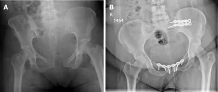
Figure2 X-ray images of Case 2.
Prompt detection of the symptoms and establishment of the diagnosis are essential for early management strategies and may decelerate progression of the condition[1].Although there are many treatment options varying from conservative to surgical methods,it is a rare pathology with insufficient evidence-based indications for each of them as well as a lack of studies highlighting the complications.A minimal widening of the symphyseal joint frequently has no clinical manifestations and requires no treatment at all[12].Most symptomatic cases involve mild complaints and,as recommended in the literature,conservative treatment as an initial treatment option is sufficient.The selection between surgical or non-surgical strategies is significant as early operative treatment may not only have a faster beneficial effect on general health and pain reduction,but can also help to avoid incomplete healing or subsequent treatment difficulties[13].
In general practice,uncomplicated and mild cases are dealt with by conservative treatment.If conservative treatment fails or severe and/or complicated cases develop,surgery is performed.In general,uncomplicated cases and those with mild symptoms are more common.Many conservative methods are applied.Their efficiency has been discussed by many authors who are working on new non-interventional methods.
Conservative treatment is carried out to relieve pain and is usually combined with other methods to provide adequate analgesia.NSAIDs and acetaminophen as firstline analgesics for postpartum pain management are considered to be appropriate for pregnant and postpartum women and during lactation.Nevertheless,controversial results were observed in a meta-analysis,which reported similar effectiveness of Paracetamol and placebo in non-pregnant women[14,15].
NSAIDs therapy usually starts with ibuprofen ranging from 400 to 600 mg four times a day.Another option is naproxen varying from 250 to 500 mg twice a day.The doses can be adjusted per requirement and be reduced as tolerated[16].
In cases with a vicious cycle of pain accompanied by muscle spasms,lumbar epidural analgesia may be administered for 24 to 72 h,as several cases have reported good outcomes using epidural morphine,bupivacaine or fentanyl.A suggested dosage for lumbar epidural analgesia is bupivacaine 0.1% combined with 2 μg/mL of fentanyl for intermittent top-ups within 72 h[17].
There is research-based data of successful pain management with intrasymphyseal steroid injections in combination with local anaesthetics,particularly hydrocortisone,chymotrypsin and lidocaine[18].Bonninet al[19]suggested a protocol for local infiltration that consisted of 5 mL of lidocaine 1% and 40 mg of methylprednisolone.These authors presented a case report where infiltration with lidocaine alone was not as effective as lidocaine combined with methylprednisolone.An intracutaneous injection is inserted perpendicularly toward the pubic symphysis.When the needle reaches the fibroelastic cartilage it must be slightly withdrawn and the injection should be completed without resistance.Local infiltration is an easy and quick method with reported long-term effectiveness.Although this procedure demands the skills of an anaesthesiologist,the risk of developing an iatrogenic infection or an allergy to medications remains.Thus,these injections are contraindicated for patients with hypersensitivity to steroids or local anaesthetics[19].In addition,external heat,ice or massage may aid in diminishing the symptoms.The administration of corresponding analgesia should be supported by bed rest,where the keystone is lateral decubitus positioning,or lying in a hammock is advised[1].As an additional therapy,transcutaneous electrical nerve stimulation has also shown positive clinical outcomes[20].There are several articles supporting the benefits of physiotherapy and acupuncture,as stabilising exercises show a significant improvement in functional status[21].Physiotherapy focuses on strengthening muscles of the trunk and pelvis,and patients should learn how to avoid strain on the pelvis.These techniques combined with acupuncture are superior to traditional treatment alone and are recommended as an adjunct to standard treatment[22].Nevertheless,effective pain management is necessary,as pain has a negative impact on patients' psychological state.Taking into account that postpartum patients have a higher demand for emotional and social support,self-help group meetings are advantageous where helpful written information is available and practical solutions can be discussed between patients affected by the same problems[23].
In line with adequate analgesia,other techniques of conservative treatment are used to ensure effective healing.For example,pelvic ring integrity should be maintained and circular compression is necessary.To achieve this goal,supports or braces such as pelvic binders,belt braces or supportive pelvic/symphyseal belts are used.A brace or a girdle provides compression and stability to the sacroiliac joints and improves the disbursement of weight-bearing forces in the pelvis,back,hips and legs.Maintenance braces are beneficial for healing as they provide pelvic support in locomotion and reduce pain.A recent study on the effectiveness of pelvic binders applied multi-detector computed tomography and compared the treatment outcomes of diverse pelvic instability grades.The findings suggested that in globally unstable cases over-reduction of a binder may lead to overriding impacted symphysis[11,24].Mulchandani and colleagues confirmed the efficiency of the pelvic binder in a review of four cases of conservative treatment with diastasis varying from 4 to 9.6 cm.A surgical waiver in those cases resulted in fast discharge postpartum and pain-free follow-up[8].A novelty in this area is an elastic band device made from neoprene straps.It limits the contractility of the internal rotation muscles,movements of the pelvis and has proved to reduce the pathologic widening of the symphysial joint and minimise the pain syndrome.The elastic band is a European Conformity-certified medical device[25].
Conservative treatment consists of several different components and should be based on a multidisciplinary team approach.
Surgical treatment is rarely obligatory.Undoubtedly,an indication for operation is diastasis complicated by nerve compression,urogenital tract trauma or massive bleeding.Another indication is inefficient conservative treatment lasting from 1 to 1.5 mo;therefore,patients should be carefully followed-up after conservative treatment.Another indication is a large widening of the joint.A previous indication for surgery was a widening exceeding 2.5 cm[26],while recent studies suggest that conservative management has good outcomes and can be efficient in cases with wider separations.Therefore,surgery is now indicated only in cases where the diastasis is more than 4 cm[27].On the other hand,anterior separation of the pubic symphysis of more than 2.5 cm causes progressive injury to the posterior pelvic ring,including disruption of the sacroiliac joint or sacral fracture,thus pain in the sacroiliac region might be indicative of further impairment[28].
An orthopaedic surgical correction in patients with a symphyseal gap over 4 cm was supported by the reduced duration of hospitalisation,a faster return to nonaffected daily life,necessity for infant care,a shorter number of days in pain and no side effects on defecation[29].The main surgical treatment methods are anterior cerclage wiring,anterior plating and external fixation[12].The internal fixation procedure using a plate is reported to have fewer complications compared to fixations using only a wire or a screw alone.Therefore,this method is most commonly used in general practice.Some studies have compared the outcomes of using different types of plates[30],and in reduction of the diastasis,a two-hole plate technique is described as superior to a four-hole plate[31].Beneficial outcomes of a complete symphysis disruption following internal fixation have been observed in acute,subacute and chronic cases[32].
Internal fixation is questionable in cases where organs of the reproductive system are damaged as it may increase the risk of infection in bones or soft tissues;therefore,in these cases external fixation should be considered as the method of choice[33].
On the other hand,surgical treatment with plate fixation is associated with frequently observed complications,the most common being contamination of the inserted pin or other infections,irritation of soft tissues,failure to fixate,loosening or replacement of screws,and recurrent widening that may require revision surgery[11,30].One study retrospectively reviewed 148 patients treated with plate fixation and found that hardware breakage occurred in 43% of patients,although most were asymptomatic;therefore,the authors suggest that a high incidence of late fixation failure is clinically unimportant[34].In this context,infectious complications are of primary concern,because any infection is more likely to have a severe course or lead to complications as immune insufficiency is observed during pregnancy and it was found that the Th1 axis and natural killer cytotoxicity suppression are also retained in the early postpartum.Complete immune recovery may take from 3 to 4 mo after delivery[35].If posterior pelvic-arch instability is involved,open reduction and internal anterior-plate fixation of the pubic symphysis with posterior percutaneous screw fixation of the sacroiliac joints is a treatment option for simultaneous correction of symphyseal and sacroiliac joint instability[28].
Over the last 20 years,in many surgical specialties,the use of minimally invasive surgery has expanded widely as it is considered to be a safer and more effective technique to meet surgical needs than open surgery;therefore,laparoscopic techniques are increasingly used for this pathology.Considering that the main drawback of open surgery for a symphyseal diastasis is a high risk of infection,laparoscopic techniques may be beneficial due to smaller wounds and no need to remove the inserted plate.Moreover,a study demonstrating the repair of symphysis separation by Anchor and Suture Tape stabilisation also emphasises that such treatment has a reduced risk of hernia,decreased postoperative pain is observed and the absence of inserted rigid constructions such as plates or screws allows motion which is more physiological and more beneficial for healing.In the postoperative period,mobilisation with limitations of several activities for two weeks is promoted.However,despite the lack of studies on this method,several disadvantages have been demonstrated.Firstly,it demands a technically skilled general surgeon,and secondly,the operation might be unsuccessful and lead to sustained pain if the anchors are placed incorrectly or the suture is under-tensed[36].Another minimally invasive technique is a pelvic bridge,a percutaneous method of subcutaneous fixation for the anterior pelvic ring,made through two incisions over each anterior iliac crest and one incision over the symphysis.A reconstruction plate or a spinal rod is placed through a subcutaneous tunnel overlying the external oblique fascia in the subcutaneous tissue,and fixation to the iliac crest and the pubis is achieved to ensure stability.This method is advantageous and includes fewer wound complications and less pain in the surgical site[37].
A rapid improvement in conservative and minimally invasive therapies suggests that surgical treatment is only necessary in very few cases.Surgery requiring open techniques is associated with a high rate of complications,especially infections;therefore,conservative therapy appears to be a better option in most pelvic symphysis diastasis cases.The possibility of persistent pain after surgery must be taken into account.Our clinical cases also support the opinion that the necessity of surgery should be strongly considered.In terms of surgical complications and improving the outcomes of non-interventional methods,some criticism may be referred to the indication for surgical treatment in cases of separation of less than 4 cm with the possibility of conservative treatment in even wider separations[8,27].
CONCLUSION
Pubic symphysis diastasis is a rare pathology mostly affecting postpartum women.A clear aetiology has not been defined,and only the predisposing factors are described in the literature.It can be symptomatic or asymptomatic.The main symptom is mild or severe pain which is managed by symptomatic or pathogenetic treatment.Taking into account that standardisation of conservative and surgical treatment or studies of possible complications were not found in the literature,we overviewed recent experiences and practical approaches as well as several new methods.After presenting our clinical cases of surgical treatment resulting from insufficient conservative treatment,we revealed a high risk of postoperative infections that are likely to trigger complications in treating postpartum symphysis pubis diastasis.
杂志排行

World Journal of Clinical Cases的其它文章
- Role of oxysterol-binding protein-related proteins in malignant human tumours
- Oncogenic role of Tc17 cells in cervical cancer development
- Acute distal common bile duct angle is risk factor for postendoscopic retrograde cholangiopancreatography pancreatitis in beginner endoscopist
- Three-dimensional computed tomography mapping of posterior malleolar fractures
- Application of a modified surgical position in anterior approach for total cervical artificial disc replacement
- Potential role of the compound Eucommia bone tonic granules in patients with osteoarthritis and osteonecrosis:A retrospective study