超声在膝骨关节炎患者软骨厚度检查中的有效性及其安全性分析
2019-11-22孙莹汪洋吴冬平
孙莹 汪洋 吴冬平
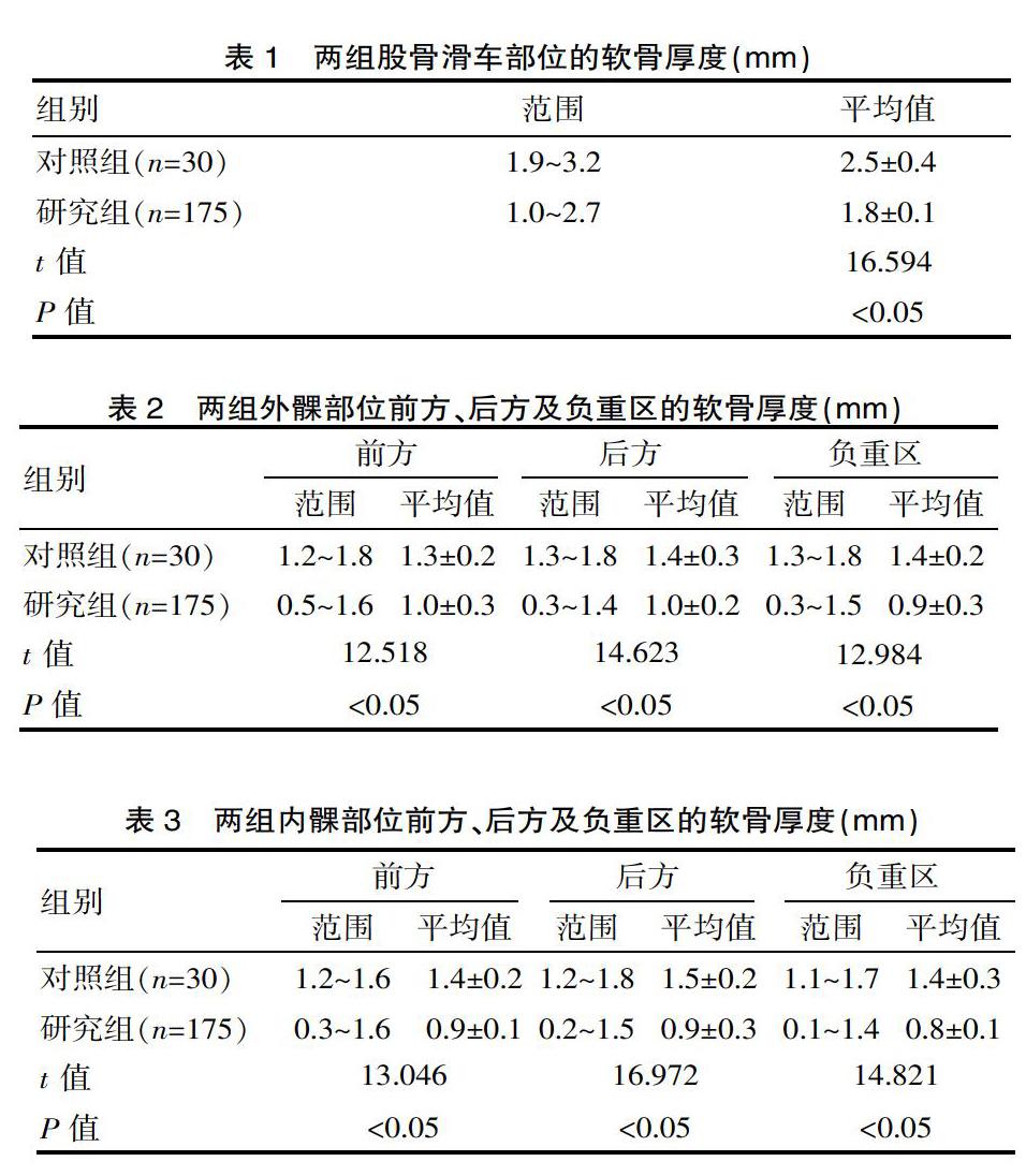
[摘要] 目的 探討分析超声在膝骨关节炎患者软骨厚度检查中的有效性及其安全性。方法 便利选取2016年1月—2017年1月该院收治的膝骨关节炎患者100例,175个膝关节作为研究组;同时选取15名健康体检人群,30个膝关节作为对照组。两组均使用多普勒彩色超声对膝部关节不同部位的软骨厚度进行检查,主要包括:股骨滑车、外髁及内髁的前方、后方及负重区等部位。结果 研究组股骨滑车的软骨厚度(1.8±0.1)mm,显著小于对照组股骨滑车的软骨厚度(2.5±0.4)mm(t=16.594,P<0.05);研究组外髁前方、后方及负重区的软骨厚度分别为(1.0±0.3)mm、(1.0±0.2)mm及(0.9±0.3)mm,均显著小于对照组的外髁前方、后方及负重区的软骨厚度(1.3±0.2)mm、(1.4±0.3)mm及(1.4±0.2)mm(t=12.518、14.623、12.984,P<0.05);研究组内髁前方、后方及负重区的软骨厚度分别为(0.9±0.1)mm、(0.9±0.3)mm及(0.8±0.1)mm,均显著小于对照组内髁前方、后方及负重区的软骨厚度(1.4±0.2)mm、(1.5±0.2)mm及(1.4±0.3)mm(t=13.046、16.972、14.821,P<0.05)。结论 超声可以较为准确的对骨关节炎患者的软骨厚度变化情况进行评估;且超声检查更加简便价廉,可以将其作为临床诊断膝骨关节炎患者软骨病变的辅助检查手段。
[关键词] 超声;膝骨关节炎;软骨厚度;有效性
[中图分类号] R684.3 [文献标识码] A [文章编号] 1674-0742(2019)08(c)-0179-04
[Abstract] Objective To investigate the efficacy and safety of ultrasound in cartilage thickness examination in patients with knee osteoarthritis. Methods Convenient selected from January 2016 to January 2017, 100 patients with knee osteoarthritis admitted to our hospital and 175 knee joints were selected as the study group. 15 healthy subjects and 30 knee joints were selected as the control group. Both groups used Doppler color ultrasound to examine the thickness of cartilage in different parts of the knee joint, including: the front, the rear and the weight-bearing area of the femoral block, the external malleolus and the medial malleolus. Results The cartilage thickness of the study group was (1.8±0.1)mm, which was significantly lower than that of the femoral block of the control group (2.5±0.4) mm (t=16.594, P<0.05). The anterior, posterior and weight-bearing areas of the external sac cartilage thickness of the study group was (1.0±0.3) mm, (1.0±0.2) mm and (0.9±0.3) mm, respectively, which were significantly smaller than the cartilage thickness of the anterior, posterior and weight-bearing areas of the control group (1.3±0.2) mm, (1.4±0.3) mm and (1.4±0.2) mm (t=12.518, 14.623, 12.984, P<0.05); the cartilage thickness of the anterior, posterior, and weight-bearing caries in the study group was (0.9±0.1) mm, (0.9±0.3)mm and (0.8±0.1)mm, both were significantly smaller than the cartilage thickness (1.4±0.2)mm, (1.5±0.2)mm and (1.4±0.3)mm in front of, behind and under weight in the control group (t=13.046, 16.972, 14.821, P<0.05). Conclusion Ultrasound can accurately evaluate the changes of cartilage thickness in patients with osteoarthritis. Ultrasound examination is more convenient and cheaper. It can be used as an auxiliary examination for cartilage lesions in patients with knee osteoarthritis.
[Key words] Ultrasound; Knee osteoarthritis; Cartilage thickness; Effectiveness
随着科学及医学技术的不断进步,核磁共振成像(简称MRI)技术开始在临床骨骼检查系统当中广泛应用,有效提高临床早期诊断膝关节病变的准确率[1]。但是MRI检查的价格十分昂贵,且在检查时对患者的要求较高(需要长时间保持静止),很难被患者及患者家属所接受。因此,目前临床中在对膝关节类疾病检查时,仍未将MRI检查替代X光线作为一种常规检查方式[2]。超声检查膝关节的优点在于其检查迅速、操作简便、无创、重复性高,且费用相对较低,更易于患者接受。除此之外,超声检查的最大优点就是可以对患者进行动态性的监测,且能够行随访检查,确认疗效[3]。该次便利选取2016年1月—2017年1月该院收治的膝骨关节炎患者100例,175个膝关节与健康体检人群15名,30个膝关节,对比两组膝关节软骨的超声检查结果,以探讨超声在检查膝骨关节炎患者软骨厚度中的有效性。……