超声图像引导的膀胱超声自动扫描系统研究
2019-10-21朱海煜白景峰杨聪
朱海煜 白景峰 杨聪
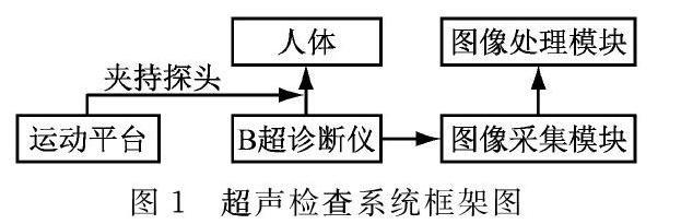
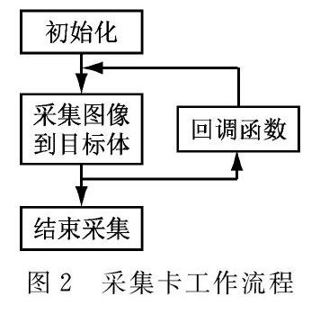
摘 要: 为解决超声检查中的医师职业病、过于依赖医生经验、对传染疾病的隔离等问题,提出一种超声图像引导的膀胱超声自动扫描系统,该技术可根据超声图像判断超声探头位置,从而实现自动扫描。本研究搭建了各自动扫描系统各个模块,并针对超声图像引导技术进行实验。系统通过OK-MC10A图像采集卡从B超诊断仪Mylab 90上取得实时医学图像,经过滤波、二值化等预处理后,使用连通域标记、K均值聚类等算法对其进行图像分割,实时确定膀胱区域质心坐标,并以此判断超声探头位置。人体实验表明,该系统可以在超声扫描过程中实时识别膀胱图像,并测量出准确的膀胱直径。
关键词: 超声扫描;图像采集;图像分割
中图分类号: TP393
文献标志码: A
文章编号:1007-757X(2019)06-0014-03
Abstract: In order to solve the problems of thatdoctors occupational diseases, the doctor over-reliance relies on doctors' his/her experience and isolation ofes infectious diseases in ultrasonic examination, an ultrasonic automatic scanning system of bladder guided by ultrasonic image was is proposed. This technology system can judge the position of ultrasonic probe according to the ultrasonic image and realize automatic scanning. In this study, the automatic scanning system modules were are built and the experiment was is conducted on the ultrasonic image guidance technology. The system obtains real-time medical images from Mylab 90, a b-mode ultrasonic diagnostic instrument, through OK-MC10A image acquisition card. After preprocessing such as filtering and binarization, the system USEuseSs connected domain markers, k-means clustering and other algorithms to segment the images and determine the centroid coordinates of the bladder region in real time, so as to determine the position of the ultrasonic probe. Human experiments show that the system can recognize the bladder image in real time and measure the bladder diameter accurately.
Key words: Ultrasonic scanning; Image acquisition; Image segmentation
0 引言
由于超聲扫描所具有的安全、无创、低成本等优点,医生们常常使用该技术获取体内器官的实时影像,并进行诊断。通常来说,每位医生同时只能对一名患者进行超声检查操作,由于医师数目有限且检查需求量大,因此,超声医师往往面临着较高的工作压力。扫描过程中皮肤接触不可避免,医师容易暴露于未知的病菌环境中。同时,传统的B超扫描方法,对医生的个人经验依赖性过大,往往需要较多时间来积累,导致检查效果参差不齐。因此,需要寻找一种机器人辅助超声扫描的超声检查手段,减少超声检查中医师的比重。
1999年,Pieroot等首次将工业机器人应用于超声自动扫描,开启了机器人辅助超声扫描手段的先河[1]。……