Clinical evaluation of endoscopic resection for treatment of large gastric stromal tumors
2019-04-16YuanYuanXiangYuanYuanLiLingYeYinZhuXiaoJiangZhouYouXiangChenGuoHuaLi
Yuan-Yuan Xiang,Yuan-Yuan Li,Ling Ye,Yin Zhu,Xiao-Jiang Zhou,You-Xiang Chen,Guo-Hua Li
Abstract
Key words: Endoscopic resection;Surgery;Gastric stromal tumor;Clinical evaluation
INTRODUCTION
Gastric stromal tumor is a digestive tract mesenchymal tumor with malignant potential.Studies[1]have reported that this will increase the psychological burden of patients and the possibility of deterioration of gastric stromal tumor,because some researchers[2]believe that small gastric stromal tumor may also be malignant,and the size of gastric stromal tumor may increase during long-term follow-up.Although the NCCN[3]guidelines do not recommend immediate surgical treatment for gastric stromal tumors,researchers such as Yeginet al[4]believe that once gastrointestinal stromal tumors are suspected,they should be treated by surgical operation or endoscopic resection.In the past,surgical resection is considered to be the standard treatment for gastrointestinal stromal tumors (GISTs),but open surgery is more traumatic,the intraoperative complications are greater,the surgical field is not obvious,and the amount of bleeding is increased.With the development of endoscopic techniques,the use of endoscopy to completely remove gastric stromal tumors has become possible,and the long-term efficacy of endoscopic treatment of gastric stromal tumors < 3 cm in diameter has been determined[5-7].However,the safety and efficacy of endoscopic treatment of large gastric stromal tumors (≥ 3 cm)remains controversial and few studies focusing on the comparison between endoscopic and surgical methods have been published[8-11].Therefore,the aim of this retrospective study was to evaluate the clinical long-term efficacy and safety of endoscopic resection for large (≥ 3 cm) GISTs in the stomach.
MATERIALS AND METHODS
Patients
All patients who underwent endoscopic resection or surgery at our hospital from 2012 to 2017 for pathologically confirmed gastric stromal tumor with a maximum diameter of ≥ 3 cm were collected.The clinical data,intraoperative conditions,pathology,oral chemotherapy,and follow-up data were analysed.
Pathological evaluation
For the evaluation of postoperative GIST biological behavior,the improved NIH classification system[12]was adopted for risk classification.
Follow-up methods
Tumor recurrence was defined as local gastric muscular lesions found on abdominal computed tomography (CT) or endoscopic ultrasonography,or gastric stromal tumors identified at the same location in surgery.
Statistical analysis
All statistical analyses were performed using SPSS 17.0 software.Continuous variables are expressed as the mean ± SD,and categorical data are displayed as numbers (n) and percentages (%),and compared using theX2test.The statistical methods of this study were reviewed by Gui-Lian Lan.
RESULTS
A total of 261 patients were included,including 37 patients in the endoscopy group and 224 patients in the surgical group.In the endoscopy group,the maximum tumor diameter was 3-8 cm;34 cases had low-risk tumors,3 had intermediate-risk,and 0 had high-risk;the mean follow-up time was 30.29 ± 19.67 mo (range,7-76),no one was lost to follow-up,and no patient had chemotherapy after operation;two patients had recurrence,both of whom had low-risk stromal tumors and did not undergo complete endoscopic resection.In the surgical group,the largest tumor diameter was 3-22 cm;103 patients had low-risk tumors,75 had intermediate-risk,and 46 had high-risk;the average follow-up time was 38.83 ± 21.50 mo (range:7-78 mo);a total of 53 patients were lost to follow-up,79 patients received oral chemotherapeutic drugs,and 8 patients had recurrence after operation (6 cases had high-risk tumors,1 had intermediate-risk,and 1 had low-risk) (Table 1).
We found that the maximum tumor diameter was significantly larger in the surgical group than in the endoscopy group,and the number of patients with highrisk stromal tumors was far greater in the surgical group than in the endoscopy group(P< 0.05).The number of patients taking oral chemotherapy drugs was far greater in the surgical group than in the endoscopy group (P< 0.01).The recurrence rate was similar between the endoscopy group and the surgical group (5.4%vs4.7%,P> 0.05),but the endoscopy group had no high-risk stromal tumors,while patients with the same size tumors were considered high risk in the surgical group.We hypothesized that this finding may be related to the tumor volume.Therefore,we performed an analysis based on the volume of tumor.
According to the tumor volume analysis,we found that the average tumor volume of the endoscopy group was 26.67 ± 26.22 cm3(range,3.75-120),all of which were less than 125 cm3.The average volume of the surgical group was 273.03 ± 609.74 cm3(range,7-4114),and 78 cases had a tumor volume ≥ 125 cm3,with the intermediaterisk and high-risk stromal tumors each accounting for 50% (39/78) of these tumors.Seven patients with a tumor volume < 125 cm3also had high-risk stromal tumors.The tumor volumes of the endoscopy group were significantly smaller than those of the surgical group (P< 0.05).Among all patients with a tumor volume < 125 cm3,only seven patients in the surgical group had high-risk stromal tumors (37.625 cm3to 115.2 cm3),accounting for 3.8% (7/183);of tumors with a volume < 125 cm3,high-risk tumors accounted for 50% (39/78) (Table 2).
Therefore,we found that both patients with a tumor volume ≥ 125 cm3and < 125 cm3have high-risk gastric stromal tumors.The pathological grade of tumor cannot be determined according to the volume of tumor.Therefore,how should we assess the pathological grade of gastric stromal tumors before surgery?
We analysed whether preoperative endoscopic findings of different pathological grades of gastric stromal tumors showed ulcer haemorrhage on the surface of the tumor and whether there was tumor liquefaction on abdominal CT or ultrasound.We found that the rates of patients who were positive for both were 57.1% in the high-risk group (12/22),12.7% (6/47) in the intermediate-risk group,and 2.1% (2/97) in the low-risk stromal tumor group;there were statistically significant differences among the three and between any two groups (P< 0.01).The percentage of patients with simple surface ulcer bleeding was 33.3% (7/21) in the high-risk group,46% (23/50) in the intermediate-risk group,and 29.8% (29/97) in the low-risk group;there was no statistically significant difference among the three groups (P= 0.15 > 0.05).There were no high-risk patients with liquefaction alone,while 20% (10/50) and 11.3% (11/97) of intermediate-risk and low-risk patients had liquefaction alone;there was no statistically significant difference among the three groups (P= 0.62 > 0.05).A total of 22 patients had tumors with a volume of less than 125 cm3without liquefaction or surface ulcer bleeding,including 1 high-risk (4.5%,1-22) and 21 (95.5%,21/22) low or medium-risk stromal tumors.Of seven patients with high-risk stromal tumors < 125 cm3,60% (3/5) had endoscopic surface ulcer bleeding and tumor liquefaction on abdominal CT or ultrasound (Table 3).
Two patients with recurrence in the endoscopic group had low-risk stromal tumors(one patient had a maximum diameter of 3 cm and a volume of 18.75 cm3and was followed for 6 mo;the other patient had a maximum diameter of 5 cm and a volume of 37.5 cm3and was followed for 6 mo);neither was treated with oral chemotherapy and was completely removed under endoscopy.In the surgical group,8 patients had recurrence (one was metastatic),including 6 cases of high-risk stromal tumors,1 case of intermediate-risk stromal tumor,and 1 case of low-risk stromal tumor.Among them,six patients with high-risk stromal tumors had a maximum tumor size of 7-20 cm and a volume of 40-3840 cm3,five patients received oral chemotherapy (6-21 mo),and the follow-up period was 6-35 mo.One patient with an intermediate-risk stromal tumor had a maximum diameter of 4 cm and a volume of 40 cm3and was followed for 17 mo without oral chemotherapy.One patient with a low-risk stromal tumor had a maximum diameter of 5 cm and a volume of 56.25 cm3and was followed for 34 mo without oral chemotherapy (Table 4).

Table 1 Data on gastric stromal tumors of different sizes
DISCUSSION
GISTs account for 1% to 2% of gastrointestinal tumors[13].They can occur in any part of the gastrointestinal tract,and the most common part is the stomach (50%-60%)[14].With the development of endoscopic techniques in recent years,several studies have demonstrated the safety and efficacy of different endoscopic methods for treating small GISTs[15-20],However,only a few data are available for the application of endoscopic methods to large-size tumors[21-25]and their follow-up periods were relatively short (range,3-73 mo),so the long-term efficacy and safety of endoscopic treatment of large gastric stromal tumors remain unclear.This study mainly analysed the clinical data of endoscopic resection or surgical treatment of large (≥ 3 cm) gastric stromal tumors from January 2012 to December 2017 at our hospital,as well as the long-term efficacy of endoscopic resection for large gastric stromal tumors.
正常情况下,附着龈呈浅粉色,而牙槽黏膜则呈亮红色。Müller等[22]认为,与牙槽黏膜相比,附着龈表面的角化层更厚,下方固有层内的胶原纤维更加致密,且潜在血管数量更少,加之其表面不规则的点彩分布,均可能会增加其对光的散射。除此之外,附着龈的厚角质层还会阻止光线进入更深的血管层,这也可能会降低潜在血管的显色。Kerdvongbundit等[23]曾利用激光多普勒测速仪对牙龈组织的微循环进行了研究,结果表明:在牙龈无炎症的状态下,牙槽黏膜的血流量明显高于附着龈。
Of the 261 included patients,37 were in the endoscopy group and 224 were in the surgical group.In the endoscopy group,the maximum diameter was 3-8 cm;34 patients had low-risk tumors,3 had intermediate-risk,and no patient had high-risk;the average follow-up time was 30.29 ± 19.67 mo (range,7-76 mo),none of the patients were lost to follow-up,and no patient received oral chemotherapeutic drugs;two patients had recurrence,both of whom had low-risk stromal tumors and did not undergo complete endoscopic resection.In the surgical group,the largest tumor diameter was 3-22 cm,and 31 tumors were ≥ 10 cm;103 patients had low-risk tumors,75 had intermediate-risk,and 46 had high-risk;the average follow-up time was 38.83± 21.50 mo (7-78 mo),53 patients were lost to follow-up,and 8 patients had recurrence after operation (6 cases were high-risk tumors,1 was intermediate-risk,and 1 was low-risk).
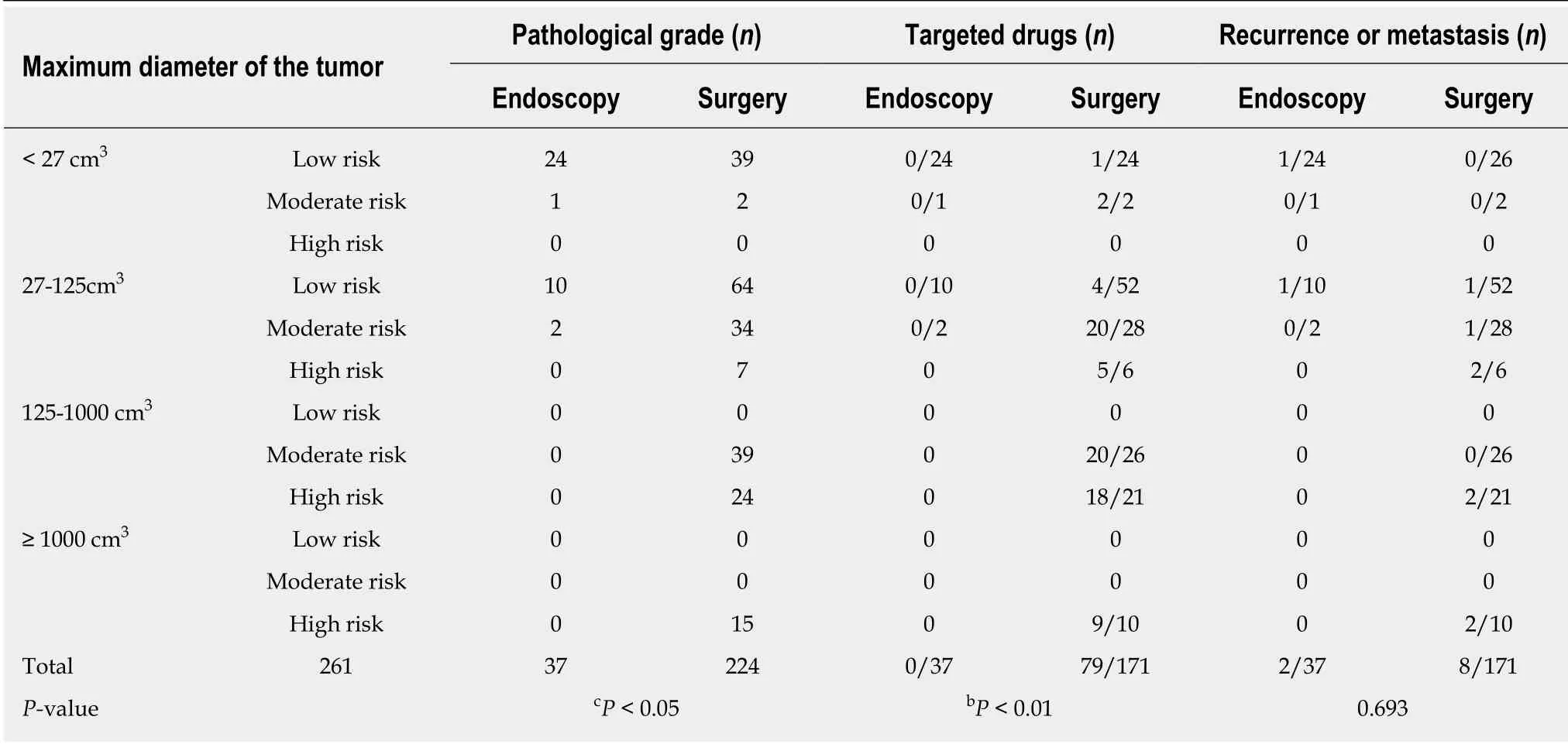
Table 2 Data for gastric stromal tumors of different volumes
We found that the maximum tumor diameter in the surgical group was much larger than that in the endoscopy group,and there were far more high-risk patients than in the endoscopy group,but the endoscopy group had no high-risk patients,while patients with the same size tumors were considered high-risk patients in the surgical group.We hypothesized that this finding was likely related to the volume of the tumor,and thus we performed further analysis based on the volume of tumor.
The average tumor volume in the endoscopy group was 26.67 ± 26.22 cm3(3.75-120),all less than 125 cm3,and no one was at high risk.The average tumor volume in the surgical group was 273.03 ± 609.74 cm3(7-4114).The tumor volumes of the endoscopy group were smaller than those of the surgical group.Among patients with a tumor volume < 125 cm3,7 had high-risk tumors (37.625 cm3-115.2 cm3),while among 78 patients with a tumor volume > 125 cm3,intermediate- and high-risk stromal tumors each accounted for 50% (39/78).Both patients with a tumor volume ≥125 cm3and < 125 cm3had high-risk gastric stromal tumors.There was no high-risk stromal tumor with a volume < 125 cm3in the endoscopy group.The pathological grade of the tumour cannot be determined according to the volume of tumor.We then asked,how should we determine the pathological grade of the gastric stromal tumor before surgery?
Previous studies have reported that endoscopic tumor ulcer haemorrhage and tumor liquefaction observed by ultrasound or abdominal CT may be related to the malignant degree of gastric stromal tumors[26,27].Thus,we analysed the presence or absence of tumour surface ulcer bleeding and whether there was tumor liquefaction on ultrasound or abdominal CT in patients with different pathological grades of gastric stromal tumors.We found that the positive rate for both was 57.1% (12/21)with high-risk tumors,and intermediate-risk and low-risk tumors accounted for 12.7% (6/47) and 2% (2/97),respectively;there were statistically significant differences among the three and between any two of groups (P< 0.01,P< 0.01,P<0.05,andP< 0.01).The proportion of high-risk patients with simple surface ulcer bleeding was 33.3% (7/21),and the intermediate-risk and low-risk patients accounted for 46% (23/50) and 29.8% (29/97),respectively;there was no significant difference among the three groups (P= 0.15 > 0.05).No high-risk patients had liquefaction alone,while the proportion of intermediate-risk and low-risk patients with simple liquefaction was 20% (10/50) and 11.3% (11/97),respectively;there was no statistically significant difference among the three groups (P= 0.62 > 0.05).Therefore,patients with endoscopic tumor surface ulcer bleeding and tumor liquefaction on ultrasound or abdominal CT are considered to have a high possibility of malignant stromal tumor,which should not be excluded from endoscopic treatment.

Table 3 Characteristics of preoperative examination of gastric stromal tumors with different pathological grades
In our study,of 22 patients with a tumor volume < 125 cm3and no surface ulcer bleeding or liquefaction,1 (4.5%,1/22) had a high-risk stromal tumor,and 21 (95.5%,21/22) had low-risk and intermediate-risk stromal tumors.Of 7 patients with highrisk stromal tumors < 125 cm3,3 (60%,3/5) had endoscopic surface ulcer bleeding and tumor liquefaction on abdominal CT or ultrasound.Two patients in the endoscopic group recurred due to incomplete tumor removal;thus,we believe that endoscopic complete resection is safe for 95.5% of patients with a tumor volume < 125 cm3without surface ulcer haemorrhage or tumor liquefaction.Of all patients with a tumor volume ≥ 125 cm3,the high-risk and intermediate-risk groups accounted for 50%(39/78).Therefore,we found that patients with a tumor volume ≥ 125 cm3had a high probability of high risk stromal tumor (50%) and were not suitable for endoscopic treatment.All patients with a maximum diameter of ≥ 10 cm accounted for 96.7%(30/31) of the high-risk group;therefore,they are not suitable for endoscopic resection.However,in the endoscopic group,there was no tumor with a maximum diameter ≥ 10 cm,a volume of ≥ 125 cm3,and a classification of high risk,thus,the safety of endoscopic treatment remains unclear.Therefore,a larger multi-center study is needed to confirm these findings.
Research reports[28]indicate that the use of chemotherapy before surgery can reduce the tumor volume and the scope of surgery to improve the overall cure rate.For GISTs of important organs,it can retain the structure and function of organs to the utmost extent and reduce the risk of tumor rupture and bleeding.It is currently recommended that the target drug treatment time should be more than 1 year for mid-risk patients,and more than 3 years for high-risk patients;patients at a high risk of tumor rupture should extend the medication time[29].In our study,78 (29.8%)patients were in the intermediate-risk group,including 3 patients in the endoscopy group and 75 patients in the surgical group.Among them,3 intermediate-risk patients in the endoscopic group were not treated with oral chemotherapy,and no recurrence or metastasis was observed during the follow-up period.There were 14 intermediaterisk patients who did not take oral chemotherapeutic drugs after surgery,but only 1 patient had recurrence (7.1%,1/14).Therefore,we believe that oral chemotherapy drugs may not be needed for intermediate-risk patients,but larger sample studies are still needed for further confirmation.
In summary,this study provided several important findings.First,prior to endoscopic treatment of gastric stromal tumors with a maximum diameter ≥ 3 cm,tumor volume should be evaluated preoperatively by ultrasound.Second,endoscopic treatment was found safe for 95.5% of patients with gastric stromal tumors having a tumor diameter ≥ 3 cm and a tumor volume < 125 cm3without endoscopic surface ulcer bleeding or CT liquefaction.Third,endoscopic treatment should not be performed for patients with gastric stromal tumors with a maximum diameter ≥ 10 cm and a volume ≥ 125 cm3.Lastly,oral chemotherapeutics may not be needed for patients with intermediate-risk stromal tumors.

Table 4 Clinical data for 10 patients with recurrence
ARTICLE HIGHLIGHTS
Research background
Gastrointestinal stromal tumors (GISTs),first proposed by Mazuret alin 1983,are a group of gastrointestinal stromal tumors with malignant differentiation potential,which are not sensitive to radiotherapy and chemotherapy.The pathological examination technique is difficult to popularize widely,so it is recommended to follow the diameter < 2 cm at present,while the diameter > 2 cm is treated surgically,but the malignant change and metastasis may occur during the long-term follow-up period.Therefore,surgical resection is the only way to treat GISTs,mainly including traditional open surgery,laparoscopic surgery,and endoscopic digestive surgery.Past transmission Open laparotomy is the first choice for the treatment of gastrointestinal stromal tumors,but for the patients with smaller diameter of the tumor,the surgical trauma is greater.In addition,the perioperative mortality of elderly patients can be as high as 1% or more.At the same time,the traditional open operation time is long,intraoperative bleeding is much,and the operation cost is expensive.Laparoscopic surgery has been proved to be a safe and effective method for the treatment of gastric stromal tumors with a diameter ≤ 5 cm,and it is also effective in the treatment of gastric stromal tumors with a tumor diameter > 5 cm.However,there are some limitations to the microstromal tumors whose diameter is smaller than 1 cm and the gastric stromal tumors with special location.If it is difficult to remove the large tumor through the orifice,it is necessary to cut open the abdominal wall or prolong the surgical incision.Moreover,it is difficult to expose and operate the tumors near the gastric cardia and the great curvature of the stomach body near the fundus of the stomach.In recent years,with the continuous development of endoscopic technology and endoscopic instruments,endoscopic resection of gastric GISTs is possible.Many studies have shown that endoscopic resection is safe and effective in treating gastric GISTs.It provides an effective minimally invasive method for the treatment of gastric stromal tumors.However,endoscopic resection is prone to major complications,such as bleeding,perforation,and positive margin.There are still doubts about the safety and efficacy of endoscopic treatment for gastric stromal tumors.In recent years,there have been many reports about the clinical evaluation of endoscopic treatment of gastric stromal tumors,which confirmed the efficacy and safety of endoscopic treatment of < 3 cm gastric stromal tumors.However,there are few reports about endoscopic treatment of large gastric stromal tumors (≥ 3 cm).The efficacy and safety of endoscopic treatment for < 5 cm gastric stromal tumors were confirmed,but the follow-up time was short and the sample size was small.More literature reports are needed to further confirm the long-term efficacy of endoscopic treatment for large stromal tumors of the stomach.
Research motivation
The purpose of this study was to collect the clinical and pathological data of all patients diagnosed with gastric stromal tumors with the largest diameter ≥ 3 cm at our hospital during the last six years from 2012 to 2017,who were treated by endoscopy or surgery.The long-term curative effect was evaluated by follow-up to evaluate the long-term efficacy of endoscopic treatment of large gastric stromal tumors,and to provide a direction for the choice of surgical methods for large gastric stromal tumors in the future.
Research objectives
The main purpose of this study was to observe the long-term efficacy of endoscopic treatment of gastric macrostromal tumors,and to reduce the pain associated with surgical treatment in patients with large gastric stromal tumors.More patients with large stromal tumors of the stomach can enjoy the benefits of minimally invasive surgery and improve the quality of life.
Research methods
From 2012 to 2017,the clinical data of all patients with large stromal tumor of the stomach treated by endoscopy or surgery were analyzed and a long-term follow-up was carried out.In this study,the chi-square test was used to test the statistical differences between groups,which clearly confirmed the differences between the two groups.
Research results
In this study,we found that endoscopic therapy can be used to treat large gastric stromal tumors,but it needs to meet certain conditions,and should be evaluated according to the results of preoperative ultrasound.There is controversy about whether endoscopic treatment of large gastric stromal tumors can be performed in previous studies.This study provides a good answer to the circumstances in which patients with large stromal tumors of the stomach can be treated by endoscopy.It provides a direction for the treatment of large stromal tumors of the stomach in the future.
Research conclusions
In this study,we found that the volume of gastric stromal tumor treated by endoscopy is smaller than that by surgical treatment.For patients with moderate-risk stromal tumors,oral chemotherapeutic drugs may not be needed,and endoscopy can be used to treat large gastric stromal tumors.However,certain conditions need to be met.Endoscopic treatment is safe for 95.5% of patients with gastric stromal tumors with a diameter ≥ 3 cm and a volume < 125 cm3 without endoscopic surface ulcer bleeding or CT liquefaction.This study will provide a direction for the choice of treatment methods for patients with large stromal tumors of the stomach in clinical work.
Research perspectives
There are many other aspects to judge the pathological grade of gastric stromal tumors before operation.We can continue to study more factors affecting the pathological grade of gastric stromal tumors on the basis of this study in the future.This study is a retrospective single-center study.There were no high-risk stromal tumors in the endoscopy group and no patients with a maximum tumor diameter ≥ 10 cm or volume ≥ 125 cm3.In the future,a multicenter prospective study of larger stromal tumors of the stomach could be attempted,to further explore the longterm safety and efficacy of endoscopic treatment for large stromal tumors of the stomach.
ACKNOWLEDGEMENTS
I show my sincere gratitude to my supervisor,professor Guo-Hua Li,for proposing this novel and clinically significant topic and guiding me to collect and analyze the data.At the same time,I would like to thank professor You-Xiang Chen,Xiao-Jiang Zhou,and other professors for their valuable comments on my article.In addition,I would like to thank The First Affiliated Hospital of Nanchang University for providing me with an excellent data collection platform.
猜你喜欢
杂志排行

World Journal of Clinical Cases的其它文章
- Ultrasound imaging of abdominal sarcoidosis:State of the art
- Porphyromonas gingivalis and digestive system cancers
- Value of superb micro-vascular imaging in predicting ischemic stroke in patients with carotid atherosclerotic plaques
- Open anterior glenohumeral dislocation with associated supraspinatus avulsion:A case report
- Vein of Galen aneurismal malformations - clinical characteristics,treatment and presentation:Three cases report
- Non-lnvasive management of invasive cervical resorption associated with periodontal pocket:A case report