64排CTA、MRA和彩超在颅内动脉硬化狭窄的临床价值
2018-04-13李学应孙成晖利建吴扬燕
李学应 孙成晖 利建 吴扬燕
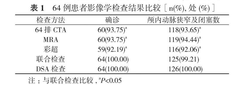
【摘要】 目的 探讨64排螺旋CT血管成像(CTA)、磁共振血管成像(MRA)和彩超在颅内动脉硬化狭窄患者中的诊断效果及价值。方法 64例颅内动脉硬化狭窄患者, 均采用经数字减影血管造影技术(DSA)确诊, 且患者均行64排CTA、MRA和彩超检查, 将64排CTA、MRA和彩超检查结果与DSA结果进行对照。结果 64例患者均完成DSA检查, 患者均得到确诊, 临床确诊率为100.00%, 且64例患者中共检查出126处颅内动脉狭窄及闭塞。64排CTA、MRA和彩超确诊率、颅内动脉狭窄及闭塞发生率两两比较差异无统计学意义(P>0.05);联合检查确诊率、颅内动脉狭窄及闭塞发生率均高于64排CTA、MRA和彩超检查, 差异具有统计学意义(P<0.05)。结论 颅内动脉硬化狭窄患者采用64排CTA、MRA和彩超联合检查有助于提高临床确诊率, 能为临床治疗提供影像学依据和参考, 值得推廣应用。
【关键词】 颅内动脉硬化狭窄;64排螺旋CT血管成像;磁共振血管成像;彩超;联合检查;影像学依据
DOI:10.14163/j.cnki.11-5547/r.2018.10.015
【Abstract】 Objective To discuss the diagnostic effect and value of 64 row computed tomography angiography (CTA), magnetic resonance angiography (MRA) and color Doppler ultrasound in patients with intracranial arteriosclerosis stenosis. Methods A total of 64 patients with intracranial arteriosclerosis stenosis confirmed by digital subtraction angiography (DSA) all examined by 64 row CTA, MRA and color Doppler ultrasound, and the results of 64 row CTA, MRA and color Doppler ultrasound were compared with those of DSA. Results All 64 patients completed the DSA examination. All patients were confirmed with clinical diagnosis rate as 100.00%. A total of 126 intracranial arterial stenosis and occlusion were detected in 64 patients. 64 row CTA, MRA and color Doppler ultrasound had no statistically significant difference in diagnosis rate and discovery rate of intracranial artery stenosis and occlusion (P>0.05). Combined examination had higher diagnosis rate and discovery rate of intracranial artery stenosis and occlusion than single 64 row CTA, MRA or color Doppler ultrasound, and the difference was statistically significant (P<0.05). Conclusion The combined examination of
64 row CTA, MRA and color Doppler ultrasound is helpful to improve the clinical diagnosis rate and provide imaging evidence and reference for clinical treatment, and it is worthy of popularization and application.
【Key words】 Intracranial arteriosclerosis stenosis; 64 row computed tomography angiography; Magnetic resonance angiography; Color Doppler ultrasound; Combined examination; Imaging basis
颅内动脉硬化狭窄是临床上常见的疾病, 是缺血性脑卒中最重要的病因, 加强患者早期诊断、早期干预对改善患者预后具有重要的意义。目前, 临床上对于颅内动脉硬化狭窄以DSA检查为主, 并将其视为“金标准”[1]。但是, DSA检查时具有一定的创伤性, 诊断可重复性较差, 导致临床诊断依从性较低, 难以达到预期的诊断效果[2]。研究表明[3], 将64排CTA、MRA和彩超用于颅内动脉硬化狭窄患者中能提高临床确诊率, 能为临床治疗提供影像学依据, 但是不同学者试验结果存在争议。因此, 本文以2015年6月~2017年7月科室收治的颅内动脉硬化狭窄患者64例作为研究对象, 探讨64排CTA、MRA和彩超在颅内动脉硬化狭窄患者中的诊断效果及价值, 报告如下。