TAZ与胰腺癌细胞增殖和抗凋亡能力的关系研究
2017-08-29罗和生黄晓东
占 婷,罗和生,田 霞,黄晓东
1.武汉大学附属同仁医院(武汉市第三医院)消化内科,湖北 武汉430060; 2.武汉大学人民医院消化内科
TAZ与胰腺癌细胞增殖和抗凋亡能力的关系研究
占 婷1,2,罗和生2,田 霞1,黄晓东1
1.武汉大学附属同仁医院(武汉市第三医院)消化内科,湖北 武汉430060; 2.武汉大学人民医院消化内科
目的 探讨转录共激活因子TAZ对胰腺癌细胞增殖与抗凋亡能力的影响及其作用机制,为胰腺癌寻找新的诊断与治疗靶点提供思路。方法 用空载病毒(对照组)、TAZ过表达病毒(Lv-TAZ)和TAZ干扰病毒(siTAZ)转染胰腺癌细胞后用CCK8法检测各组细胞的增殖情况;流式细胞仪检测各组细胞的凋亡率;Western blotting检测各组细胞的PTEN、Akt、p-Akt的表达情况。结果 与对照组相比,Lv-TAZ组细胞增殖率升高,siTAZ组细胞增殖率下降。与对照组相比,Lv-TAZ组细胞凋亡率下降,siTAZ组细胞凋亡率升高。Western blotting结果显示,Lv-TAZ组细胞PTEN表达水平下调,p-Akt上调;siTAZ组细胞PTEN表达水平上调,p-Akt下调。结论 TAZ在胰腺癌细胞中可能通过调控PTEN/Akt信号通路发挥其促增殖和抗凋亡作用。
胰腺癌;TAZ;PTEN/Akt信号通路
近年来胰腺癌的发病率和死亡率不断升高,使其成为消化系统恶性程度最高的肿瘤之一[1]。目前,手术切除是胰腺癌唯一根治的方法,但是由于侵袭性强,大部分胰腺癌患者就诊时已经发生局部或远处转移,失去根治性手术的机会[2]。因此,我们迫切需要对胰腺癌发病机制进行深入研究,以寻找新的治疗策略。
转录共激活因子TAZ (transcriptional co-activator with PDZ-binding motif) 于2000年作为14-3-3蛋白的结合蛋白而被发现,参与哺乳动物多种组织的生长[3]。作为一种转录共激活因子,TAZ通过与多种转录因子如TEA结构域家族转录因子、Runt功能域转录因子等结合而发挥不同的作用[4-5]。越来越多研究已经证实,TAZ参与肿瘤形成、细胞增殖与迁移、上皮细胞间质转化(epithelial mesenchymal transition,EMT)等一系列过程[6-8]。研究[9]发现,TAZ在胰腺癌组织和细胞中高表达,且能够促进胰腺癌细胞的迁移、侵袭、EMT。然而TAZ对胰腺癌细胞增殖与抗凋亡能力的影响仍未见报道。因此,本研究着重讨论TAZ对胰腺癌细胞的增殖与凋亡能力的影响及其可能机制。
1 材料与方法
1.1 细胞系及细胞培养 人胰腺癌细胞PANC1购自中国科学院上海细胞库。细胞在37 ℃、体积分数为5%的CO2培养箱中培养,培养细胞的完全培养基为含1%双抗(Thermo Fisher Scientific,美国)、100 g/L胎牛血清(NQBB, USA)的高糖培养基DMEM (Gibco, Carlsbad,美国)。
1.2 细胞转染 TAZ的靶向siRNA慢病毒(siTAZ)以及过表达病毒(Lv-TAZ)由上海吉凯基因化学技术有限公司合成。将细胞接种于6孔板,于第2天按照操作说明书加入适量慢病毒、Ploybrene和Enhance培养12 h后换成完全培养基后继续培养。
1.3 Western blotting检测 转染后的细胞用RIPA裂解液(北京普利莱基因技术有限公司)裂解提蛋白后,用BCA蛋白浓度测定盒(碧云天生物技术研究所)测蛋白浓度。将蛋白与loading buffer混合后100 ℃变性10 min,再用100 g/L SDS-PAGE胶分离蛋白,然后转到NC膜上,随后用兔抗人TAZ(CST,美国)、PTEN(CST,美国)、Akt(CST,美国)、p-Akt(CST,美国)、GAPDH(Santa Cruz,美国)多克隆抗体于4 ℃中孵育过夜。最后用羊抗兔二抗(Lincoln,美国)在室温下孵育1 h,蛋白表达水平用ECL化学发光显色试剂盒检测。
1.4 CCK-8检测 将转染后的细胞接种于96孔板中,然后用CCK8试剂(日本同仁化学研究所)分别检测各种细胞在24 h、48 h、72 h、96 h时450 nm的OD值以反映细胞的增殖情况。
1.5 流式细胞检测 将转染后的细胞于6孔中培养3 d后,根据Annexin V-PE凋亡试剂盒(BD Bioscience,美国)的说明书处理细胞后用流式细胞仪检测细胞的凋亡情况。
1.6 统计学处理 采用SPSS 19.0软件进行统计学处理,说明数据的图用GraphPad Prism 5制作,采用t检验和单因素方差分析进行统计学分析,P<0.05为差异有统计学意义。
2 结果
2.1 TAZ对胰腺癌细胞增殖与抗凋亡能力的影响 首先用空载对照病毒(对照组)、TAZ干扰病毒(siTAZ组)和过表达病毒(Lv-TAZ 组)转染胰腺癌细胞PANC1,转染效率见图1。CCK-8检测结果显示,与对照组比较,Lv-TAZ 组细胞增殖率升高,siTAZ 组细胞增殖率下降(见图2)。将转染后的胰腺癌细胞用流式细胞仪检测其凋亡情况,结果显示,与对照组相比,Lv-TAZ 组细胞凋亡率下降,siTAZ 组细胞凋亡率升高(见图3)。

图1 转染病毒后各组细胞TAZ蛋白的表达情况;图2 转染病毒后各组细胞的增殖情况
Fig 1 The expression of TAZ protein after transfection of the virus in each group; Fig 2 The proliferation of cells after transfection of the virus in each group
2.2 TAZ在胰腺癌细胞中对PTEN/Akt信号通路的作用 将PANC1细胞用空载病毒、TAZ干扰病毒和过表达病毒转染后提取蛋白,然后用Western blotting检测PTEN、Akt、p-Akt蛋白的表达情况。结果显示,与对照组相比,Lv-TAZ组蛋白PTEN表达下调,siTAZ组蛋白PTEN表达上调(见图4)。而与对照组相比,Lv-TAZ组蛋白p-Akt表达上调,siTAZ组蛋白p-Akt表达下调,而Akt表达量无明显改变(见图5)。
3 讨论
近几年来虽然胰腺癌的手术治疗已经有了很大的提升,但大多数患者在确诊的时候已经在中晚期,失去了切除机会[10],因此,我们迫切需要研究早期胰腺癌的检测和治疗靶点。最近研究[11-12]显示,胰腺癌本质上是一种由于基因和表观遗传学的改变而导致的基因性疾病。因此,研究能够促进胰腺癌发生、发展的关键基因能够为胰腺癌早期检测及治疗提供新的思路。
TAZ是一种含有WW结构域的转录共激活因子,是Hippo信号通路重要的下游效应因子。该信号通路在器官大小、干细胞功能维持、肿瘤发生调节上都发挥着重要作用[13]。当Hippo信号通路激活时,该通路中的丝/苏氨酸蛋白激酶MST1/2作用于肿瘤抑制基因LATS1/2激酶使其磷酸化,导致其下游的效应因子TAZ失活[14]。近年来,TAZ被发现在多种恶性肿瘤中表达升高,如肝癌[15]、胃癌[16]、乳腺癌[17]等,提示TAZ在肿瘤的发生、发展中发挥着重要作用。
研究[9]发现,TAZ在胰腺癌组织和细胞中高表达,并能够促进胰腺癌细胞的迁移、侵袭、EMT。

图3 转染病毒后各组细胞的凋亡情况Fig 3 Apoptosis after transfection of the virus in each group

注:与对照组比较,*P<0.05,**P<0.001。
图4 转染病毒后各组细胞PTEN蛋白的表达情况
Fig 4 The expression of PTEN protein after transfection of the virus in each group
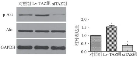
注:与对照组比较,*P<0.05。
图5 转染病毒后各组细胞p-Akt及Akt蛋白的表达情况
Fig 5 The expressions of p-Akt protein and Akt protein after transfection of the virus in each group
然而TAZ对胰腺癌细胞增殖与抗凋亡能力的影响及作用的可能机制仍未见报道。而在我们的实验中,通过过表达TAZ的表达和干扰TAZ的表达,发现TAZ的表达水平与胰腺癌细胞的增殖率呈正相关,与胰腺癌细胞的凋亡率呈负相关。这些结果证实了TAZ能够增强胰腺癌的增殖与抗凋亡能力。PTEN 是公认的肿瘤抑制因子,与多种肿瘤的发生、发展密切相关[18],而我们在实验中发现,TAZ的表达水平与PTEN蛋白的表达水平呈负相关。PTEN是PI3K-Akt通路上的一个重要的抑制因子,它可以使PIP3去磷酸化成为PIP2从而抑制 Akt的磷酸化使其失活[19]。P13K/Akt信号通路过度激活常见于多种肿瘤,与人类多种肿瘤的发生、发展密切相关[20]。活化的Akt是肿瘤发生和发展的一个重要因素,它能够使PIP2促进细胞增殖,减少细胞凋亡,降低细胞对药物的敏感性[21-22]。 为了探讨TAZ与活化的Akt之间的潜在关系,我们检测p-Akt与Akt在转染后的胰腺癌细胞中的表达水平,结果发现在TAZ过表达的胰腺癌细胞中p-Akt表达升高,在干扰了TAZ表达的胰腺癌细胞中,p-Akt表达下降,而与Akt表达水平基本一致。这些结果提示,TAZ在胰腺癌细胞中可能通过调控PTEN的表达进而激活Akt信号通路。
总之,我们的研究表明,TAZ在胰腺癌细胞中可能通过调控PTEN/Akt信号通路来发挥其促增殖和抗凋亡作用。因此,我们可以认为TAZ是胰腺癌早期诊断和治疗的潜在靶点。
[1]Chen W, Zheng R, Zhang S, et al. Report of cancer incidence and mortality in China, 2010 [J]. Ann Transl Med, 2014, 2(7): 61.
[2]Von Hoff DD, Ervin T, Arena FP, et al. Increased survival in pancreatic cancer with nab-paclitaxel plus gemcitabine [J]. N Engl J Med, 2013, 369(18): 1691-1703.
[3]Kanai F, Marignani PA, Sarbassova D, et al. TAZ: a novel transcriptional co-activator regulated by interactions with 14-3-3 and PDZ domain proteins [J]. EMBO J, 2000, 19(24): 6778-6791.
[4]Cui CB, Cooper LF, Yang X, et al. Transcriptional coactivation of bone-specific transcription factor Cbfa1 by TAZ [J]. Mol Cell Biol, 2003, 23(3): 1004-1013.
[5]Mahoney WM Jr, Hong JH, Yaffe MB, et al. The transcriptional co-activator TAZ interacts differentially with transcriptional enhancer factor-1 (TEF-1) family members [J]. Biochem J, 2005, 388(Pt 1): 217-225.
[6]Xiang L, Gilkes DM, Hu H, et al. Hypoxia-inducible factor 1 mediates TAZ expression and nuclear localization to induce the breast cancer stem cell phenotype [J]. Oncotarget, 2014, 5(24): 12509-12527.
[7]Bartucci M, Dattilo R, Moriconi C, et al. TAZ is required for metastatic activity and chemoresistance of breast cancer stem cells [J]. Oncogene, 2015, 34(6): 681-690.
[8]Huang W, Lv X, Liu C, et al. The N-terminal phosphodegron targets TAZ/WWTR1 protein for SCFbeta-TrCP-dependent degradation in response to phosphatidylinositol 3-kinase inhibition [J]. J Biol Chem, 2012, 287(31): 26245-26253.
[9]Xie D, Cui J, Xia T, et al. Hippo transducer TAZ promotes epithelial mesenchymal transition and supports pancreatic cancer progression [J]. Oncotarget, 2015, 6(34): 35949-35963.
[10]Xie D, Xie K. Pancreatic cancer stromal biology and therapy [J]. Genes Dis, 2015, 2(2): 133-143.
[11]Campbell PJ, Yachida S, Mudie LJ, et al. The patterns and dynamics of genomic instability in metastatic pancreatic cancer [J]. Nature, 2010, 467(7319): 1109-1113.
[12]Yachida S, Jones S, Bozic I, et al. Distant metastasis occurs late during the genetic evolution of pancreatic cancer [J]. Nature, 2010, 467(7319): 1114-1117.
[13]Pan D. The hippo signaling pathway in development and cancer [J]. Dev Cell, 2010, 19(4): 491-505.
[14]Harvey KF, Zhang X, Thomas DM. The Hippo pathway and human cancer [J]. Nat Rev Cancer, 2013, 13(4): 246-257.
[15]Xiao H, Jiang N, Zhou B, et al. TAZ regulates cell proliferation and epithelial-mesenchymal transition of human hepatocellular carcinoma [J]. Cancer Sci, 2015, 106(2): 151-159.
[16]Zhou GX, Li XY, Zhang Q, et al. Effects of the hippo signaling pathway in human gastric cancer [J]. Asian Pac J Cancer Prev, 2013, 14(9): 5199-5205.
[17]Rashidian J, Le Scolan E, Ji X, et al. Ski regulates Hippo and TAZ signaling to suppress breast cancer progression [J]. Sci Signal, 2015, 8(363): ra14.
[18]McCabe N, Kennedy RD, Prise KM. The role of PTEN as a cancer biomarker [J]. Oncoscience, 2016, 3(2): 54-55.
[19]Carnero A, Paramio JM. The PTEN/PI3K/AKT Pathway in vivo, cancer mouse models [J]. Front Oncol, 2014, 4: 252.
[20]Bartholomeusz C, Gonzalez-Angulo AM. Targeting the PI3K signaling pathway in cancer therapy [J]. Expert Opin Ther Targets, 2012, 16(1): 121-130.
[21]Hers I, Vincent EE, Tavaré JM. Akt signalling in health and disease [J]. Cell Signal, 2011, 23(10): 1515-1527.
[22]Steelman LS, Chappell WH, Abrams SL, et al. Roles of the Raf/MEK/ERK and PI3K/PTEN/Akt/mTOR pathways in controlling growth and sensitivity to therapy-implications for cancer and aging [J]. Aging (Albany NY), 2011, 3(3): 192-222.
(责任编辑:李 健)
Relationship of TAZ with proliferation and anti apoptotic ability of pancreatic cancer
ZHAN Ting1,2, LUO Hesheng2, TIAN Xia1, HUANG Xiaodong1
1.Department of Gastroenterology, Wuhan Third Hospital, Tongren Hospital of Wuhan University, Wuhan 430060; 2.Department of Gastroenterology, People’s Hospital of Wuhan University, China
Objective To investigate the effect and functionary mechanism of TAZ on the proliferation and anti apoptotic ability of pancreatic cancer cells, and to provide a new way for the diagnosis and treatment of pancreatic cancer.Methods Pancreatic cancer cells were transfected with lentiviral vector expressing TAZ (Lv-TAZ) and TAZ siRNA lentiviral vector (siTAZ), and then CCK8 was used to detect the proliferation. Flow cytometry was used to detect the apoptosis. Western blotting was used to detect the expressions of PTEN, Akt, p-Akt.Results Compared with the control group, the proliferation rate of cells transfected with Lv-TAZ was increased, and the cell proliferation rate of cells transfected with siTAZ was decreased. Compared with control group, the apoptosis rate of cells transfected with Lv-TAZ was decreased, and the apoptosis rate of cells transfected with siTAZ was increased. Western blotting results showed that the expression of PTEN in cells transfected with Lv-TAZ was downregulated, and the expression of p-Akt was upregulated. On the contrary, the expression of PTEN in cells transfected with siTAZ was upregulated, and the expression of p-Akt was downregulated.Conclusion TAZ may play a role in promoting the proliferation and anti apoptosis by regulating PTEN/Akt signaling pathway in pancreatic cancer cells.
Pancreatic cancer; TAZ; PTEN/Akt signaling pathway
湖北省科技支撑计划项目(2015BHE022)
占婷,在读硕士研究生,研究方向:胰腺癌。E-mail:472308046@qq.com
黄晓东,博士,主任医师,研究方向:消化道动力障碍性疾病。 E-mail:13886190549@163.com
10.3969/j.issn.1006-5709.2017.07.006
R735.9
A
1006-5709(2017)07-0741-03
2016-12-17
