重视老年髋部骨折患者术前骨密度与骨结构的影像学评价
2017-08-10程晓光王玲苏永彬杨明辉王满宜吴新宝
程晓光 王玲 苏永彬 杨明辉 王满宜 吴新宝
. 述评 Editorial .
重视老年髋部骨折患者术前骨密度与骨结构的影像学评价
程晓光 王玲 苏永彬 杨明辉 王满宜 吴新宝
髋部骨折;骨密度;定量 CT;术前评价
随着社会人口老龄化的发展,老年人群髋部骨质疏松性骨折的发生率明显增加[1-4],骨质疏松症的最严重临床后果是具有极高的致死率和致残率,其中,骨折后 1年的死亡率即高达 20%[5-7]。以往对老年人群髋部骨折以保守治疗为主,现在随着对老年髋部骨折认识的提高和围手术期综合保障能力的提高,越来越多的患者接受了手术治疗,明显降低了患者的死亡率和致残率。北京积水潭医院创伤骨科为探索老年髋部骨折治疗开展的绿色通道项目,可以在老年髋部骨折患者就诊时,快速进行全面检查,准确诊断,从而对患者情况进行全面评估,并据此决定是否手术治疗及采取何种治疗方案。2015年开展此项目以来,已经治 1500余例,取得了明显的效果[8-9]。
其中一项重要评价指标是对这些老年患者股骨上段骨密度与骨结构的评价,其对于决定手术方式和植入物的选择有很大帮助。例如,对于股骨粗隆间骨折患者,其股骨头的骨质情况对于内固定螺钉头钉部分的选择是至关重要的,如果其股骨头骨密度高、骨强度好,其头钉部分可以选用拉力螺钉类,这样可以保证获得足够的把持力;反之,则适合选用螺旋刀片类的头钉[10-11]。目前,临床常用的影像检查手段包括 X 线片、CT 等,但这些技术用来判断骨密度或者骨强度是非常不精确的。双能 X 线骨密度仪 ( dual energy X-ray absorptiometry,DXA ) 是使用比较广泛的骨密度测量方法之一,但 DXA 往往对患者的拍摄体位要求比较高,尤其是在髋部 DXA 测量时,被测下肢需内旋约 15°,但是髋部骨折患者疼痛剧烈,难以配合,这样就限制了DXA 在髋部骨折患者骨密度测量的应用[12]。
定量 CT 骨密度测量技术 ( quantitative computed tomography,QCT ) 克服了上述困难,通过采用 CT 图像结合 QCT 体模的方法,经过分析软件进行处理,从而得到骨密度和骨结构参数[13]。其在髋部的应用称为髋部 QCT,是利用临床髋部 CT 扫描得到的原始数据进行图像后处理,经由 1次 CT 扫描完成解剖显示与骨密度测量。因此,在不增加患者受辐射剂量的情况下,使得髋部骨折患者的手术前骨密度和骨结构测量成为可能[14-16]。北京积水潭医院放射科 2007年以来,一直开展老年髋部骨折的 QCT 研究和临床应用[17]。特别是在创伤骨科此次开展的扬帆项目中,髋部 QCT 得到了充分应用,体现了技术优势,即老年髋部骨折患者在急诊通过 1次 CT 扫描,既可以判断是否存在骨折以及评价骨折的详细解剖和分型,为手术方案的选择提供定性影像依据,也可以进行髋部骨密度测量和骨结构分析,从而为治疗方案的选择提供了定量影像依据。在近10年的临床应用中,累计测量 3000余例,业已成为髋部骨折手术前的常规检查。现将方法介绍如下,希望对国内同行有所借鉴。

图1 左侧股骨颈骨折,CT 扫描时髋部下面放置 QCT 校准体模,右侧股骨颈正常用于 QCT 骨密度测量,结果与 DXA 一致图2 QCT 分析软件从三个相互垂直的平面调整股骨颈角度,使股骨颈与水平面平行图3 髋部 QCT 骨密度分析结果,位置,感兴趣区 ROI 以及 BMD 结果与 DXA 一致Fig.1Left femoral neck fracture. QCT calibration model was placed under the hip in the CT scanning. QCT bone density measurement for the right femoral neck as normal. Results consistented with the DXAFig.2QCT analysis software adjusted the femoral neck angle from three mutually perpendicular plane, so that the femoral neck paralleled to the horizontal planeFig.3Location, ROI and BMD results in hip QCT consistented with the DXA
我科在急诊检查室配备了东芝 16排螺旋 CT 扫描机和美国 Mindways 公司的 QCT 分析系统。在我院,当骨科医师怀疑老年患者可能发生了髋部骨折时,在 X 线片检查基础上,髋部 CT 平扫是常规的检查项目。在做髋部 CT 扫描前,将 QCT 的校准体模置于患者髋部水平检查垫的下方,对患者体位无特殊要求,采用统一的扫描条件和重建参数,对髋部进行 CT 扫描。对常规髋部 CT 图像进行多平面重建和三维重组,观察是否存在骨折及骨折的解剖细节。同时,将带有 QCT 体模信息的 CT 原始数据传输到 QCT 分析工作站,进行髋部骨密度测量 ( computed tomography X-ray absorptiometry,CTXA ) 和骨结构分析 ( bone investigation toolkit,BIT )。QCT 分析软件可以对 CT 图像在冠状面、矢状面、横轴面上进行旋转调节,从而保证所有患者测量平面相一致,避免了 DXA 扫描所需要的患者体位配合问题。CXTA 是通过模拟 DXA 的成像方式,对髋部的二维投影图像进行骨密度测量 ( 图 1~3)。有研究证明 QCT 测量的骨密度与 DXA 测量的骨密度具有很好的一致性,二者可以互换使用[14]。更重要的是,此技术利用 3D 容积数据,可以进行股骨上段任何区域的骨密度测量。根据骨科医师的要求,影像科常规对股骨头,股骨颈与股骨粗隆间区域进行螺钉方向的骨密度测量( 图 4,5),为骨科制订手术方案和内固定物的选择提供了定量数据支持[18]。QCT 分析软件的高级图像处理模块 BIT 可以对股骨颈进行更详细的区域分析,包括股骨颈不同区域的皮质骨厚度、松质骨和皮质骨骨密度以及相关的力学参数测量[19-20],这些测量参数可以帮助临床医生更好地了解老年髋部骨折的发生机理。另外,髋部 QCT 还可以进行更复杂的图像处理,用于研究髋部骨折的发生机理或药物治疗的反应等[21-22]。
除上述骨质分析外,髋部 QCT 还可以用于髋部软组织的研究,通过应用 QCT 软件测量髋部不同水平的肌肉面积和密度,脂肪面积和含量等。这些测量参数可以用于研究髋部软组织在骨折发生中的作用[23]。
开展这项工作以来,髋部 QCT 检查已经成为我院髋部骨折患者术前的常规检查,到目前为止,已经完成了 3000余例髋部 QCT 测量,取得了良好的效果,充分显示出这种髋部骨折手术前的检查方式具有很好的临床应用前景,值得推广应用。
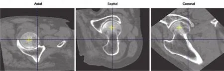
图4 髋部 QCT 分析软件可以对髋部图像进行旋转,使测量平面符合骨科要求Fig.4Hip QCT analysis software could rotate the hip image to make sure the measurement plane was in line with orthopedic requirements

图5 髋部 QCT 分析软件测量股骨头和粗隆的骨密度,为骨科手术方案选择提供依据Fig.5Hip QCT analysis software measured the BMD of the femoral head and trochanter, providing a reference for orthopedic surgical options
[1] Zhang L, Cheng AG, Bai ZQ, et al. Epidemiology of cervical and trochanteric fractures of the proximal femur in 1994in Tangshan, China[J]. J Bone Miner Metab, 2000, 18(2):84-88.
[2] Bow CH, Tsang SW, Loong CH, et al. Bone mineral density enhances use of clinical risk factors in predicting ten-year risk osteoporotic fractures in Chinese men: the Hong Kong Osteoporosis Study[J]. Osteoporos Int, 2011, 22(11):2799-2807.
[3] Cooper C, Cole ZA, Holroyd CR, et al. Secular trends in the incidence of hip and other osteoporotic fractures[J]. Osteoporos Int, 2011, 22(5):1277-1288.
[4] Dhanwal DK, Cooper C, Dennison EM. Geographic variation in osteoporotic hip fracture incidence: the growing importance of asian inf l uences in coming decades[J]. J Osteoporos, 2010.
[5] Randell AG, Nguyen TV, Bhalerao N, et al. Deterioration in quality of life following hip fracture: a prospective study[J]. Osteoporos Int, 2000, 11(5):460-466.
[6] von Friesendorff M, Besiakov J, Akesson K. Long-term survival and fracture risk after hip fracture: a 22-year follow-up in women[J]. J Bone Miner Res, 2008, 23(11):1832-1841.
[7] Baudoin C, Fardellone P, Bean K, et al. Clinical outcomes and mortality after hip fracture-A 2-year follow-up study[J]. Bone, 1996, 18(3Suppl):S149-157.
[8] 杨明辉, 吴新宝, 龚晓峰, 等. 骨科与老年科共管模式治疗老年髋部骨折及与英国的比较[J]. 中国骨与关节杂志, 2017, 6(3):169-173.
[9] 吴新宝. 杨明辉, 张萍, 等. 老年病科和骨科共管模式缩短老年髋部骨折患者术前等待时间和住院时间[J]. 骨科临床与研究杂志, 2017, 2(2):96-100.
[10] McCormack R, Panagiotopolous K, Buckley R, et al. A multicentre, prospective, randomized comparison of the sliding hip screw with the Mdoff sliding screw and side plate for unstable intertrochanteric hip fractures[J]. Injury, 2013, 44(12):1904-1909.
[11] Born C, Karich B, Bauer C, et al. Hip screw migration testing: fi rst results for hip screws and helical blades utilizing a new oscillating test method[J]. J Orthop Res, 2011, 29(5):760-766.
[12] Fan B, Lu Y, Genant H, et al. Does standardized BMD still remove differences between Hologic and GE-Lunar state-of-the-art DXA systems[J]? Osteoporos Int, 2010, 21(7):1227-1236.
[13] Genant HK, Boyd D. Quantitative bone mineral analysis using dual energy computed tomography[J]. Invest Radiol, 1977, 12(6):545-551.
[14] Cheng X, Wang L, Wang Q, et al. Validation of quantitative computed tomography-derived areal bone mineral density with dual energy X-ray absorptiometry in an elderly Chinese population[J]. Chin Med J (Engl), 2014, 127(8):1445-1449.
[15] Lang TF, Keyak JH, Heitz MW, et al. Volumetric quantitative computed tomography of the proximal femur: precision and relation to bone strength[J]. Bone, 1997, 21(1):101-108.
[16] Bousson VD, Adams J, Engelke K, et al. In vivo discrimination of hip fracture with quantitative computed tomography: results from the prospective European Femur Fracture Study (EFFECT)[J]. J Bone Miner Res, 2011, 26(4):881-893.
[17] Cheng X, Li J, Lu Y, et al. Proximal femoral density and geometry measurements by quantitative computed tomography-association with hip fracture[J]. Bone, 2007, 40(1):169-174.
[18] 危杰, 伊辰, 王满宜, 等. 股骨近端骨密度定量 CT 测量及其在骨折手术前评估的作用[J]. 中国骨与关节杂志, 2014, 3(11):830-834.
[19] 王玲, 张勇, 程晓光, 等. 定量 CT 评价股骨近段骨皮质厚度及骨密度[J]. 中国医学影像技术, 2015, 31(10):1461-1465.
[20] Wang L, Cheng XG, Su YB, et al. Sex-related variations in cortical and trabecular bone of the femoral neck in an elderly Chinese population[J]. Osteoporos Int, 2017.
[21] Carballido-Gamio J, Harnish R, Saeed Streeper T, et al. Structural patterns of the proximal femur in relation to age and hip fracture risk in women[J]. Bone, 2013, 57(1):290-299.
[22] Yu A, Carballido-Gamio J, Wang L, et al. Spatial differences in the distribution of bone between femoral neck and trochanteric fractures[J]. J Bone Miner Res, 2017, 13.
[23] Edmunds KJ, Gíslason MK, Arnadottir ID, et al. Quantitative computed tomography and image analysis for advanced muscle assessment[J]. Eur J Transl Myol, 2016, 26(2):93-100.
( 本文编辑:李贵存 )
. 会议 ●征文 ●消息 Conference / Call for Paper / News .
本刊被美国化学文摘数据库收录公告
本刊现为中国科技论文统计源期刊。2013年 1月,本刊经美国化学文摘 ( Chemical Abstracts,CA ) 数据库审理委员会审核通过,并从 2013年第 1期开始,正式被美国化学文摘数据库收录。特此公告!
CA 只收录本刊论著,其它文章不收录。
查询本刊请使用拼音:Zhongguo Gu Yu Guanjie Zazhi或本刊标准国际刊号 ( ISSN ):2095-252X,查询网址:http://cassi.cas.org/search.jsp。
《中国骨与关节杂志》编辑委员会
The pre-operation bone mineral density and bone structure assessment with quantitative computed tomography in osteoporotic hip fractures
CHENG Xiao-guang, WANG ling, SU Yong-bin, YANG Ming-hui, WANG Man-yi, WU Xin-bao.
Department of Radiology, Beijing Jishuitan Hospital, Beijing, 100035, China
Operation is one of the choices for the treatment of hip fractures in elders. However, how to assess the bone mineral density ( BMD ) and bone structure pre-operation are challenging. Dual-energy x-rays absorptiometry ( DXA ) is widely used for BMD measurement of the hip, but limited in elder hip fractures due to the pain leading to the diff i culty of positioning. Furthermore, DXA is unable to measure the BMD of the femoral head. In this commentary, we introduced “Beijing Jishuitan hospital sailing project” experience which is to combine the routine clinical hip CT scan with the quantitative computed tomography ( QCT ) of the hip for elder hip fractures. With a single hip CT scan, the details of the hip fracture can be shown, meanwhile the CT data are used for QCT analysis of the the hip, in which the DXA-equivalent BMD is measured. BMD of the femoral head, neck and trochanteric area can be measured as well. All measurements are very useful for the pre-operation planning. QCT is utilizing the clinical CT scan with QCT calibration phantom and software, further post-processing of CT data, with no additional radiation. We completed more than 3000cases of QCT and it is proved to be very successful, so we recommend to use QCT for hip fractures.
Hip fracture; BMD; QCT; Pre-operation assessment
10.3969/j.issn.2095-252X.2017.08.001
R683.3
首都临床特色应用研究项目 ( Z141107002514072)
100035北京积水潭医院放射科 ( 程晓光、王玲、苏永彬 ),创伤骨科 ( 杨明辉、王满宜、吴新宝 )
2017-07-01)