A case report of partial molar pregnancy associated with a normal appearing dizygotic fetus
2016-10-18RahamniMaryamParvizSara
Rahamni Maryam, Parviz Sara
Advanced Diagnostic and Interventional Radiology Research Center (ADIR), Imam Khomeini Hospital, Tehran University of Medical Science, Tehran, Iran
A case report of partial molar pregnancy associated with a normal appearing dizygotic fetus
Rahamni Maryam, Parviz Sara*
Advanced Diagnostic and Interventional Radiology Research Center (ADIR), Imam Khomeini Hospital, Tehran University of Medical Science, Tehran, Iran
ARTICLE INFO
Article history:
Partial mole
Normal fetus
Dizygotic karyotype
ABSTRACT
Partial molar pregnancy is a rare entity in which there is usually a triploid abnormal fetus associated with a large placenta with cystic changes .The incidence of a normal diploid fetus and a partial molar placenta is extremely rare . Here we report a case of partial molar pregnancy in which a normal appearing male fetus with diploid karyotype coexist . A focal placental abnormal region was detected at 11 week of gestation as enlargement associated with cystic changes . Fetus showed no obvious abnormality . The patient underwent amniocentesis and the result was compatible with a normal diploid male fetus. Regarding these findings the patient continued her pregnancy under close observation and advanced sonographic evaluations were made to rule out other differentials. There were no obstetric complications until the 26th gestational week when preterm rupture of the membranes occurred . The patient underwent induced vaginal delivery and the products were sent for pathologic evaluation which confirmed the partial molar changes.
Document heading doi: 10.1016/j.apjr.2016.01.015
1. Introduction
A partial molar pregnancy is a variation of a molar pregnancy in which an embryo either develops incompletely or with multiple structural anomalies[1].
In this kind of abnormal pregnancy, the egg usually receives two sets of chromosomes from the father, usually because two sperm have fertilized the egg. The egg now has 69 chromosomes, instead of the normal 46 [1]. Most pregnancies in which molar change has been reported in association with a normal fetus represent a dizygotic twin pregnancy with one complete hydatidiform mole and other normal twin with clearly distinguishable molar regions in the placenta [2]. We present a case of singelton pregnancy in which placental molar change was associated with normal appearing dizygotic fetus.
2. Case report
The patient was a 23 year old white woman with first trimester spotting.She was on her second pregnancy and the first was a normal live birth.No personal or familial history of genetic abnormalities was detected.
刺史吕某……致祭于苏升、陈寅、李宽、秦陈甫、鲁余之灵。尔等五人感余诚信,力输公税,争赴先期。溪山阻深,淫潦暴至,不忍欺我,忘其险艰。[6]6372
Ultrasound examination revealed a singelton pregnancy with no fetal structural abnormalities and fetal biometery consistent with gestational age (11w+4d). There was a fondal placenta with focal area of enlargement in its central part associated with numerous lucent cyst (central placental diameter was about 75 mm) (Figure 1). No other seprate normal placental tissue was detected. The umblical cord attached to the central placenta adjacent to thefocal abnormality. Maternal serum毬-HCG level was 64 750 mIU/ mL. The patient underwent amniocentesis at her 13w+4d and the karyotype was 46xy compatible with apparently normal male fetus.
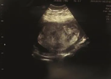
Figure 1. Ultrasound examination.
Even though no other significant abnormalities were noted during serial examinations, the risk of subsequent fetal and maternal complications was explained to the patient and her family. The patient elected to continue her pregnancy and received close observasion at outpatient clinic. During that period, evaluation revealed no obstetric complication. Maternal serum titres of 毬-HCG were found to be 37 098 mIU/mL and 36 000 mIU/mL at 18 and 22 weeks gestation respectively. The serial biometrics of the fetus were measured every 2 weeks from weeks 18 to 24 and showed a normal growth pattern corresponding to the gestational age. By ultrasound, the placenta showed mild enlargement up to 100 mm width in its central part in 21 weeks gestation.
No evidence of placental insufficiency such as growth retardation or oligohydramniosis was detected in the course of follow up. The biophysical profile and non-stress test was evaluated biweekly.
The clinical course was smooth until the 26th gestational week, when the patient was admitted to the labour room due to premature rupture of the membranes. Admission laboratory findings of the patient included haemoglobin 11.5 g/dL and platelet count 247×104/mm3. The serum titre of毬-HCG was 5258.1 mIU/mL. Ultrasonography showed macerated fetus with decreased amniotic fluid index. The patient went under a induced vaginal delivery and the pregnancy products were sent for pathologic evaluation.
Pathological study showed placental tissue measuring 20 cm x15 cm x 5 cm and weighting 1 100 mg. Umblical cord measures 20 cm in length and 1 cm in maximum diameter. Multiple vesicles with maximum diameter of 2 cm on the maternal surface of the placenta were seen.
The histopathological findings were compatible with a partial molar pregnancy. The serum titre of毬-HCG of the mother decreased to undetectable levels 1 month after delivery without any chemotherapy. She was doing well and in follow-up sonography no evidence of retained tissue or recurrence was detected after 6 months.
3. Discussion
Partial molar pregnancy with coexisting fetus is a rare complication with the incidence of 0.005 % to 0.01% of all the pregnancies[3]. It usually derive from dispermic fertilization of a haploid normal oocyte and produce a triploid set of chromosomes [4]. Ultrasonography has made it possible a diagnosis of a hydatidiform mole and co-existent fetus in the first trimester. Ultrosound finings include a greatly enlarged placenta relative to the size of the uterine cavity associated with cystic spaces (“molar placenta”) [5]. An amniotic cavity (gestational sac), either empty or containing amorphous inappropriately small fetus with multiple structural anomalies. However in some situations molar changes in placenta is associated with a normal diploid fetus .
In cases of a singleton normal fetus with partial molar placenta, the fetus must have normal karyotype to survive in utero , although its placenta can have some chromosomal variation, from diploidy of the amnion to triploidy of the chorionic villi [7]. From this clinical perspective, there are two different types of US findings in the placenta: the focal and diffuse molar changes[11]. The former shows a normal placenta with a focal area of hydropic changes as in the case of our patient. The difference between a focal partial molar degeneration and twin pregnancy with complete mole could be challenging by ultrasound per se because they both present with two distinct regions of the placenta. A practical method is to follow the fetus umblical cord. If it connects to the molar placenta one could exclude the twin pregnancy, but if the cord insertion is in normal placental site differentiatin of two entity is not possible by ultrosound.
Another consideration in the setting of focal vascular placental lesion associated with normal diploid fetus is placental tumor such as chorioangioma. However the differentiation is made by its sonographic feature which shows a well-circumscribed lesion with different echo pattern from the rest of the placenta, and can be located on the fetal placental surface or protruding into the amniotic cavity. The sonographic diagnosis of chorioangioma is based upon an increased vascularity or a large feeding vessel inside the tumor with the same pulsation rate as in the umbilical cord [8] the finding that is absent in molar placenta.
Another uncommon vascular placental lesion should be kept in mind as a rare differential diagnosis is placental mesenchymal dysplasia (PMD). It has been documented to be more in female fetuses with a F:M of ~3.5:1[12]. The sonographic appearance ofPMD is a thickened placenta with hypoechoic areas[9]. Although PMD may be associated with different patterns of blood flow with advancing gestation, the absent or low venous signals inside the placental lesion (at least during the first two trimesters) may be of value in differentiating PMD from chorioangioma or a molar pregnancy that are characterized by high velocity blood flow[10].
Management of molar changes associated with normal appearing fetus still remains challenging. The serum b-hCG level can be a helpful marker , when the serum b-hCG level remains greater than 106 mIU/mL, TOP (termination of pregnancy) should be considered. In contrast, in cases of successful pregnancy outcomes with viable fetuses, the serum b- hCG level usually starts to decline from the beginning of the second trimester, and sonography usually reveals a decrease in the size of the molar portion of the placenta [13].
Conflict of interest statement
We declare that we have no conflict of interest.
Refrences
[1] American Pregnancy Association. Molar pregnancy: Symptoms, risks and treatment[Online]. Available at: http://americanpregnancy.org/pregnancycomplications/molar-pregnancy/. [Accessed on 1/8/2015].
[2] Hsieh CC, Hsieh TT, Hsueh C, Kuo DM, Lo LM, hung TH. Delivery of a severely anaemic fetus after partial molar pregnancy: Clinical and ultrasonographic findings. Hum Reprod 1999;14:1122-1126.
[3] Suzuki M, Matsunobu A, Vakita K, Osanai K. Hydatidiform mole with surviving co-existent fetus. Ombt Gynecol 1980; 56: 384-388.
[4] Vaisbuch E, Ben-Arie A, Dgani R, Perlman S, Sokolovsky N, Hagay Z. Twin pregnancy consisting of a complete hydatidiform mole and coexistent fetus: report of two cases and review of literature. Gynecol Oncol 2005;98:19–23.
[5] Rezaee A, Weerakkody Y. Partial hydatidiform mole [Online]. Available at: http://radiopaedia.org/articles/partial-hydatidiform-mole.
[6] Changchien CC, Eng HL, Chen WJ. Twin pregnancy with hydatidiform mole (46,XX) and a coexistent fetus (46,XY): report of a case. J Formos Med Assoc 1994; 93: 337–339.
[7] Sarno AP, Moorman AJ, Kalousek DK. Partial pregnancy with fetal survival: an unusual example of confined placental mosaicism. Obstet Gynecol 1993;82:716–719.
[8] Zalel Y, Gamzu R, Weiss Y, Schiff E, Shalmon B, Dolizky M, et al. Role of color Doppler imaging in diagnosing and managing pregnancies complicated by placental chorioangioma. J Clin Ultrasound 2002;30:264–269.
[9] Moscoso G, Jauniaux E, Hustin J. Placental vascular anomaly with diffuse mesenchymal stem villous hyperplasia. A new clinico-pathological entity? Pathol Res Pract 1991;187:324–328.
[10] Edi Vaisbuch, Roberto Romero, Juan Pedro Kusanovic, Offer Erez, Shali Mazaki-Tovi, Francesca Gotsch, et al. Three dimensional sonographic imaging of placental mesenchymal dysplasia and its differential diagnoses. J Ultrasound Med 2009;28(3): 359–368.
[11] Vejerslev LO. Clinical management and diagnostic possibilities in hydatidiform mole with coexistent fetus. Obstet Gynecol Surv 1991;46:577–588.
[12] Placental mesenchymal dysplasia. available from http://radiopaedia.org/ articles/placental-mesenchymal-dysplasia
[13] Achour Radhouane, Ben Aissa Imen, Neji Khaled. Twin pregnancy with both complete hydatiform mole and coexistent alive fetus: Case report. Asian Pac J Reprod 2015; 4(4): 331 –333.
11 November 2015
Parviz Sara, Advanced Diagnostic and Interventional Radiology Research Center (ADIR), Imam Khomeini Hospital, Tehran University of Medical Science, Tehran , Iran.
Tel:+989121976410
E-mail: srparviz@gmail.com
Received in revised form 12 January 2016 Accepted 6 February 2016
Available online 1 March 2016
猜你喜欢
杂志排行
Asian Pacific Journal of Reproduction的其它文章
- Effect of heparin, caffeine and calcium ionophore A 23187 on in vitro induction of the acrosome reaction of fresh ram spermatozoa
- The characterisation and cryopreservation of Venda chicken semen
- A new nucleotide variant G1358A potentially change growth differentiation factor 9 profile that may affect the reproduction performance of Friesian Holstein cattle
- Pregnancy rate in Bulgarian White milk goats with natural and synchronized estrus after artificial insemination by frozen semen during breeding season
- Pregnancy outcomes of using ICSI with frozen-thawed spermatozoa in Riyadh, Saudi Arabia
- Effect of growth regulators on rapid micropropagation and antioxidant acitivity of Canscora decussata (Roxb.) Roem. & Schult.-A threatened medicinal plant