乙酰胆碱酯酶在脂多糖诱导肺泡巨噬细胞中的变化
2014-10-21刘芬江榕李勇曾振国聂成赵宁黄彩雪夏亮钱克俭
刘芬 江榕 李勇 曾振国 聂成 赵宁 黄彩雪 夏亮 钱克俭
DOI:10.3760/cma.j.issn.1671-0282.2014.08.010
基金項目:国家自然科学基金(81101410,81160233);江西省自然科学基金(20122BAB205002)
作者单位:330006 南昌,南昌大学第一附属医院重症医学科(刘芬、江榕、曾振国、聂成、赵宁、黄彩雪、夏亮、钱克俭),肿瘤科(李勇)
通信作者:钱克俭,Email: qkj0607@sohu.com
【摘要】目的 观察脂多糖 (lipopolysaccharide, LPS) 刺激大鼠肺泡巨噬细胞NR8383后乙酰胆碱酯酶 (acetylcholinesterase, AChE) 的表达变化,为研究胆碱能抗炎通路的调控提供新的实验依据。
访法 将体外去致热源培养的NR8383细胞分为对照组及LPS (1 μg/mL) 刺激组,于刺激后3、6、12、24 h各时间点分别离心收集上清液及细胞沉淀,采用酶联免疫吸附法 (enzyme-linked immunosorbent assay, ELISA) 测定上清液中肿瘤坏死因子-α (tumor necrosis factor-α, TNF-α) 的变化,实时定量PCR检测细胞中AChE mRNA的表达改变,蛋白质免疫印迹法 (Western blot) 检测细胞中AChE蛋白的表达变化,乙酰胆碱酯酶T-CHE测试盒检测上清液中AChE活性的变化。组间多重比较采用单因素方差分析,进一步采用LSD-t检验进行两两比较,P<0.05为差异具有统计学意义。
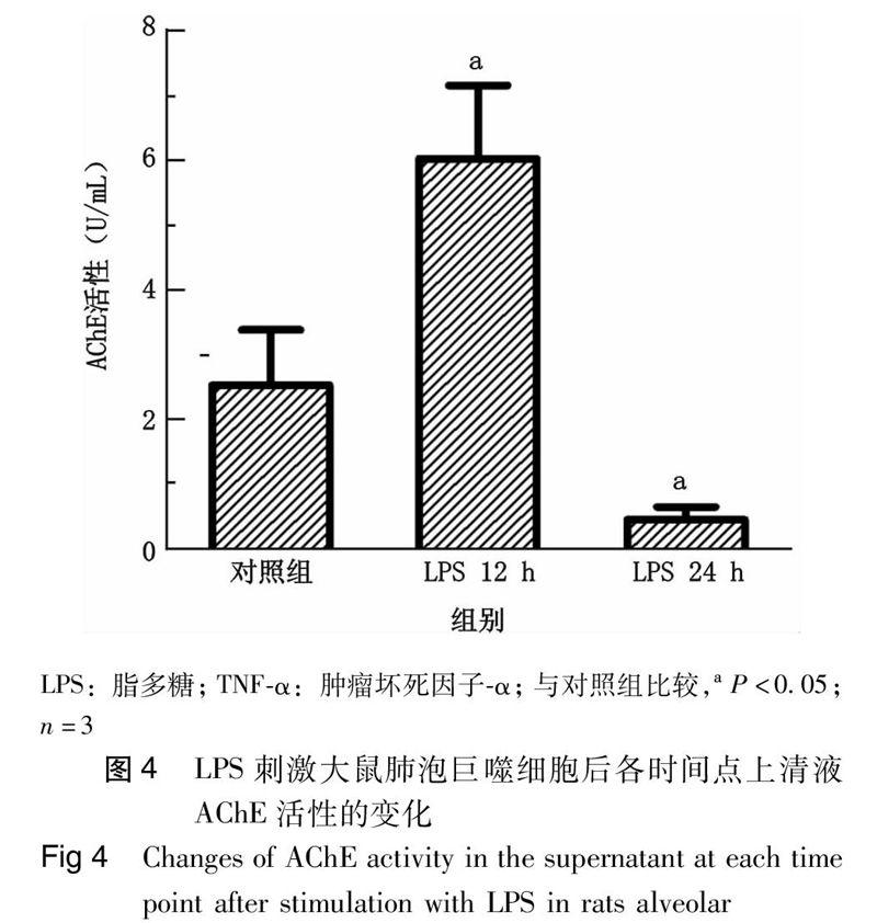
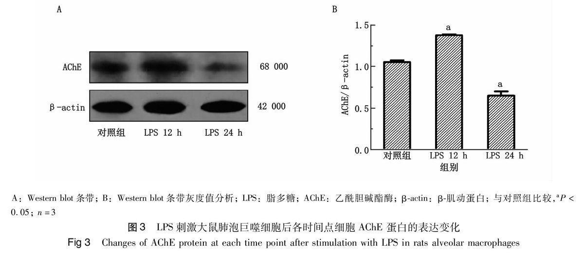
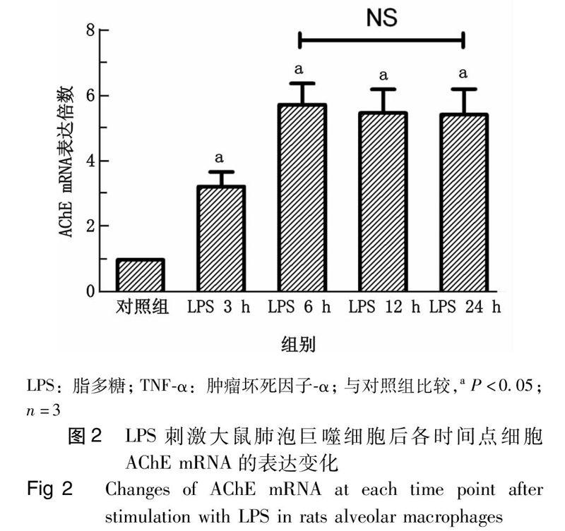
结果 与对照组相比,TNF-α的含量在LPS刺激NR8383细胞后3 h开始显著上升 (P<0.05),12 h达高峰,24 h下降但仍明显高于对照组水平 (P<0.05)。与对照组相比,AChE mRNA于刺激后3 h开始上升,升高(3.19 ± 0.44)倍(P<0.05),6 h后达高水平并维持于此水平,6、12、24 h分别上调[(5.65 ± 0.63)、(5.40 ± 0.71)、(5.35 ± 0.77)[倍(与对照组相比,均P<0.05);AChE蛋白的表达及上清液中AChE的活性在LPS刺激后12 h与各自对照组相比均上升[AChE蛋白:(1.37 ± 0.01) vs.(1.05 ± 0.02),P<0.05;AChE活性:(6.14 ± 1.13) U/mL vs. (2.64 ± 0.85) U/mL,P<0.05],随后呈下降趋势,于LPS作用24 h时均低于各自对照组[AChE蛋白:0.65 ± 0.05,P<0.05,AChE活性:(0.56 ± 0.19 )U/mL,P<0.05]。
结论 LPS刺激大鼠肺泡巨噬细胞后,AChE的表达增加,提示其可能参与肺泡巨噬细胞炎症反应的调控;LPS刺激后24 h,AChE mRNA仍处于高表达水平,AChE蛋白及活性却下降,提示在肺泡巨噬细胞炎症反应中可能存在AChE转录后水平的调控。
【关键词】乙酰胆碱酯酶;脂多糖;肺泡巨噬细胞;炎症反应;脓毒症
Changes of acetylcholinesterase expression in alveolar macrophages stimulated by lipopolysaccharide Liu Fen, Jiang Rong, Li Yong, Zeng Zhenguo, Nie Cheng, Zhao Ning, Huang Caixue, Xia Liang, Qian Kejian. Department of Critical Care Medicine, the First Affiliated Hospital of Nanchang University, Nanchang 330006, China
Corresponding author: Qian Kejian, Email: qkj0607@sohu.com
【Abstract】Objective To observe the changes of acetylcholinesterase (AChE) in rat alveolar macrophages NR8383 stimulated by lipopolysaccharide (LPS) in order to provide a novel experimental evidence for studying the regulation of the cholinergic anti-inflammatory pathway.Methods The NR8383 cells cultured with pyrogen-free in vitro were divided into control group and LPS (1 μg/mL) stimulation group. Culture supernatants and cell pellets were collected by centrifugation at 3 h, 6 h, 12 h and 24 h after stimulation, respectively. The level of tumor necrosis factor-α (TNF-α) in the supernatant was assayed by using enzyme-linked immunosorbent assay (ELISA). The expression of AChE mRNA in cells was detected by using real time quantitative RT-PCR, The level of AChE protein in cells was analyzed by using Western blot, The activity of AChE in the supernatant was measured by using True Choline esterase assay kit (T-CHE). One-way analysis of variance (ANOVA) was used for comparisons between two groups, and LSD-t test was performed for further comparison, and difference was statistically significant at P < 0.05. Results The level of TNF-α began to increased significantly at 3 h after stimulation of NR8383 cells with LPS (P<0.05) compared with control group, and reached its peak at 12 h, then decreased until 24 h but the level was still significantly higher than that in control group (P<0.05); The expression of AChE mRNA began to elevate at 3 h after stimulation compared with control group [ (3.19 ± 0.44) times, P<0.05 ], at 6 h reached the high level and then maintained this level at 12 h and 24 h [ (5.65 ± 0.63), (5.40 ± 0.71), (5.35 ± 0.77) times, compared with control group, all P<0.05 ]; The level of AChE protein in cells and the activity of AChE in the supernatant increased at 12 h after LPS stimulation compared with control group [AChE protein: (1.37 ± 0.01) vs. (1.05 ± 0.02), P<0.05; AChE activity:( 6.14 ± 1.13) U/ml vs. (2.64 ± 0.85) U/ml, P<0.05], followed by a downward trend, and were lower than those in control groups at 24 h after LPS stimulation [AChE protein: (0.65 ± 0.05), P<0.05; AChE activity: (0.56 ± 0.19) U/ml, P<0.05].Conclusions The expression of AChE mRNA increased after stimulation with LPS in rat alveolar macrophages, suggesting that AChE may be involved in regulation of alveolar macrophages inflammatory response; AChE mRNA expression is still high while its protein and activity are declined at 24 h after LPS stimulation, suggesting that there may be some mechanisms existed in post-transcriptional regulation of AChE in alveolar macrophages during inflammatory response.
【Key words】Acetylcholinesterase; Lipopolysaccharide; Alveolar macrophage; Inflammatory response; Sepsis
乙酰胆碱酯酶(acetylcholinesterase, AChE)是一种特异性催化水解神经递质乙酰胆碱 (acetylcholine, ACh)的酶[1]。最近有研究表明炎癥反应时迷走神经末梢通过释放ACh可与巨噬细胞上的α7烟碱型乙酰胆碱受体(α7 nicotinic acetylcholine receptor, α7nAChR)相互作用,减少肿瘤坏死因子-α(tumor necrosis factor-α, TNF-α)等促炎因子的释放,形成一条胆碱能抗炎通路调控炎症反应[2-3]。……