Osteochondral lesions of the talus
2012-11-08AubeeluckAshwinYuGuangRong
Aubeeluck Ashwin,Yu Guang Rong
Department of Orthopedics,Tongji Hospital of Tongji University,Shanghai 200065,China
Introduction
The term“osteochondral lesion”is a general term which encompasses several other terms such as“osteochondritis dissecans”,“transchondral fractures of the talus”,“osteochondral fracture”,or“talar dome fracture”.Monro was the first to describe loose cartilaginous fragments floating in the ankle joint.In 1888,Konig,used the term“osteochondritis dessicans”to describe a spontaneous necrosis of cartilage along with a fragment of its underlying bone,giving rise to loose bodies in the knee joint.Similar lesions in the talus were termed“osteochondritis dessicans”by Max Kappis in 1922.The cause of this lesion remains unclear till date.Furthermore the term“osteochondritis dissecans”implies an inflammatory process,yet most studies report a traumatic etiology.Finally in 1959,in their classic article,Berndt and Harty found that 88%of their patients with osteochondral lesions reported a traumatic origin.They then proposed the term“transchondral fractures of the talus”.By that time these lesions were already known to predominantly occur at two locations of the talar dome:anterolaterally and posteromedially.Berndt and Harty also created anterolateral lesions through a combination of inversion and dorsiflexion of the ankle.Likewise,a posteromedial lesion was created through inversion,plantarflexion and external rotation of the ankle.In 1985,Flick and Gould reported that 98% of anterolateral and 70% of posteromedial lesions,out of a total of 500 lesions,presented a traumatic etiology.Van Buecken,et al.(1989)found that these lesions are present in 6.5% of ankle sprains.These lesions are bilateral in 10% of cases,with one or both side symptomatic[1].
The purpose of this paper is to present the latest concepts about diagnosis,classification and treatment of these lesions.
Diagnosis
Most osteochondral lesions manifest either as acute ankle pain following an inversion injury or as chronic ankle pain.One therefore has to make a distinction between the acute and the chronic situations.
In the acute setting,the clinical picture may be that of an ankle sprain,i.e.a painful,swollen ankle with limited range of motion.If these symptoms persist after 2-3 weeks,an OCL has to be ruled out.
In the chronic situation,intermittent or persistent deep ankle pain,recurrent swelling,weakness,locking,click,stiffness and moderately limited range of motion are often described,with locking being indicative of a loose fragment within the joint.The pain may not necessarily correspond to the location of the lesion and may be exacerbated by exercise or fatigue.A strong link has also been established between OCL and chronic ankle instability[2].
Radiography plays a major role in the diagnosis of OCL,starting with plain anteroposterior,lateral and mortise views.Plain X-rays do not show cartilage defects or undisplaced lesions.Moreover,the lesion may not be visible on the initial films,but appear at a later stage.Therefore,in case initial X-rays prove to be negative,further investigations should be requested,starting with CT scan.The latter reveals the location,size,degree of displacement,and shape of the lesion.CT is essential in the preoperative planning but it lacks the ability to accurately assess the cartilage,bone bruises or undisplaced fragment.Here MRI provides a more accurate evaluation of the subchondral bone,cartilage,and surrounding soft tissue.MRI can also detect early subchondral anomalies.
If occult OCL is suspected and radiography is negative,bone scintigraphy can be used,giving 96%specificity and 94% sensitivity[3].
To summarise,any lesion found on X-rays has to be evaluated with a CT scan,MRI or bone scan being performed in cases where radiography is negative.
Classification
Several systems have been proposed to stage osteochondral lesions of the talus,using X-ray,CT,MRI,arthroscopy.
In 1959 Berndt and Harty proposed a classification system based on radiographic findings.This system is the most frequently used:Stage Ⅰ,subchondral bone compression;Stage Ⅱ,partially detached osteochondral fragment;Stage Ⅲ,completely detached osteochondral fragment without displacement;Stage Ⅳ,loose osteochondral fragment in the joint;Later in 1993 Loomer and coworkers added stage Ⅴ,representing subchondral cysts.

Fig.1 Berndt and Harty classification system图1 本特及哈迪分类系统
A CT-classification was introduced with stagesⅠto Ⅳidentical to Berndt and Harty’s classification but with a stage Ⅴstanding for a radiolucent defect[4].
Hepple,et al[5].reported a classification system based on MRI findings:Stage Ⅰ:articular damage only;Stage ⅡA:articular damage with underlying fracture and surrounding bone edema;Stage ⅡB:similar to ⅡA but without bone edema;Stage Ⅲ:detached but undisplaced osteochondral fragment;Stage Ⅳ:detached and displaced fragment;Stage Ⅴ:formation of subchondral cyst.
The first ones to devise a classification system based on arthroscopic findings were Pritsch,et al[6].This classification was further developed by Cheng,et al[7]:Stage A:smooth,intact but soft cartilage;Stage B:rough articular cartilage surface;Stage C:fissuring or fibrillation present in the cartilage;Stage D:osteochondral flap,or exposed bone;Stage E:detached but undisplaced osteochondral fragment;Stage F:loose osteochondral fragment.
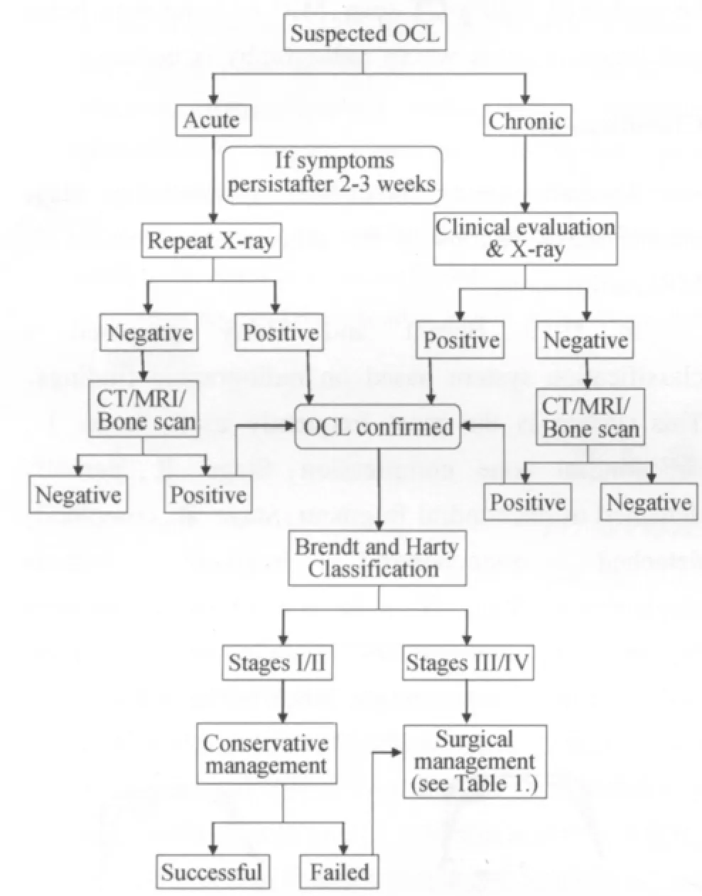
Fig.2 Algorithm for diagnosis and treatment of OCL图2 距骨软骨缺损(OCL)的诊断治疗程式
Although each staging system is useful,none of them suffices to clearly describe the lesions.
Treatment
There are several management strategies for osteochondral lesions of the talus,from conservative treatment,various surgical techniques,to biological repair and cartilage regeneration.Management decisions for some authors are based on the size of the lesion:surgery being mandatory for lesions >1.5 cm.For other authors,the decision is based on the stage of the lesion.
Conservative management
The surgeon has to remember that studies on non operative treatment have shown a relatively low rate of good or very good results in the literature[8,9].Conservative treatment comprises of strict unloading(with or without joint immobilization)of various duration but not exceeding 4 weeks,as well as restriction from strenuous activities,followed by a period of partial weight bear and gradual mobilisation.The main question here is the selection of patients who would achieve acceptable results with non surgical treatment.Spontaneous resolution of the lesion has been reported in the pediatric population,however this is rarely the case in adults[10].In their meta analysis of 14 studies with a total of 201 patients who had undergone non operative treatment for stages I and II osteochondral lesions,Tol and colleagues reported a success rate of 45%[10].Shearer and colleagues report an average success rate of 56% for patients with chronic cystic talar lesions treated non operatively[11].Thus patients with chronic symptoms seem to achieve better results.Shearer and colleagues also noted that a diminution in the size of the lesion after conservative management does not imply a corresponding symptomatic improvement.Since the progression of the lesion is relatively slow,many authors have suggested a trial of conservative management for stages I and II for one year before considering operative management.
Surgical treatment
The optimal treatment strategy for OCL is still subject of debate.The main objective is revascularization of the necrotic lesion.The cartilage being avascular,it has poor reparative ability.In cases where the lesion extends into the subchondral bone,an inflammatory reaction follows and marrow cells are stimulated and eventually the defect is filled with fibrocartilage.This is the rationale behind techniques such as debridement and drilling or microfracturing.The fibrocartilage is suitable in cases where the lesion is small.But in cases where the lesion is large,this replacement tissue does not have the biomechanical properties necessary to make it an acceptable long term treatment.Therefore the latest developments in the treatment of OCL aim at filling the defect with tissue biomechanically closer to hyaline cartilage.This includes autograft transplantation from a donor site,allograft transplantation,or chondrocyte implantation.
Debridement,microfracture and drilling (bone marrow stimulation)
This technique is reserved for symptomatic,<15 mm lesions.It can be done open or arthroscopically.The principles and approaches of the arthroscopic procedure are not within the scope of this article but it is worth to mention the contribution of the posterolateral and posteromedial approaches,which were first described by Van Dijk,making very posterior lesions amenable for arthroscopic treatment[9].With this technique,all unstable cartilage is excised.The underlying devitalised or calcified tissue is curetted and,by microfracturing or drilling,connections are created with deeper healthy tissue.This disrupts intra osseous blood vessels,with formation of a fibrin clot.New blood vessels invest the clot and concomitantly marrow elements stimulate the formation of a fibrocartilaginous scar tissue which fills the defect.Although fibrocartilage lacks the biomechanical abilities of native hyaline cartilage,studies have found it a suitable option when dealing with small lesions<15mm at Berndt and Harty stagesⅠ-Ⅳ.Favourable outcomes have been reported in 85% of patients,with success rates varying from 46%-100%[12,13].
Retrograde drilling
The aim with this technique is to revascularise the subchondral bone and promote the formation of new bone.Its advantage is that the overlying cartilage is not disrupted.It is a viable option when dealing with subchondral cysts with intact overlying cartilage.
Using a drill-targetting device,medial lesions are accessed by arthroscopically drilling through the sinus tarsi while lateral lesions are accessed from anteromedial.Computer-assisted techniques are also used at some centres to enhance the accuracy of the procedure.
Decompressing the cyst brings about the question of structural integrity of the bone.To prevent articular collapse,cancellous bone grafting has been described but in some cases it can be technically difficult to adequately fill the defect with solid bone inserted through the drill path[14].Some authors have introduced surgical-grade calcium sulphate in liquid form introduced into the defect as an alternative to solid bone grafting[15].Pluripotent cells obtained from bone marrow aspirate can also be mixed with the calcium sulphate to promote healing[16].
Range of motion exercises can be started in the immediate post operative period and partial weight bear allowed at 6 weeks.Reported success rate for this technique in previous studies is 88%[16].
Transmalleolar drilling
This technique is for lesions which are difficult to reach because of their location.Its disadvantage is that when drilling through the malleolus and articular cartilage,healthy tibial cartilage is damaged.A success rate of 63% has been reported for this technique.It has to be mentioned that nowadays most lesions are reachable via arthroscopic approaches,which is not in favour of this technique[16].
Internal fixation
Large osteochondral lesions with a significant amount of bone attached are preferably stabilised.This basically concerns acute/subacute cases as well as children.Internal fixation can be achieved with screws,pins fibrin glue,K-wire or bio absorbable screws made of polyglycolic acid (PGA)or polylactic acid (PLLA).Larsen and coworkers[15]reported a small series with PGA/PLLA copolymer bioabsorbable screws,with 6 out of 7 cases demonstrating healing without inflammatory process.In their meta-analysis of treatment using screws,pins,K-wires,Tol et al (2000)reported 73% good to excellent results.
Autologous chondrocyte implantation (ACI)
This is a two-stage procedure which aims at repairing the cartilage defect with hyaline-like cartilage.Initially introduced for treatment of osteochondral defects of the knee,this technique has been adapted for the same situation at the talus.As mentioned by Niemeyer and coworkers[15]in their meta-analysis on ACI,various authors consider this technique as gold standard treatment for large lesions of more than 3-4 cm2.However,controlled studies evaluating the superiority of ACI over other techniques are not available.
The first stage involves harvesting cartilage from a non weight bearing site such as the intercondylar notch,and sending it to the laboratory where chondrocytes are isolated and cultured.The second stage takes place at least 4 weeks after harvest.This step is done open,with a medial or lateral osteotomy.The lesion at the talus is debrided and a periosteal flap is taken from the distal tibia and sutured over the defect,leaving a small opening.The interface is sealed water-tight with fibrin glue before injection of the cultured cells.The opening is then sutured and sealed.The osteotomy is then reduced and fixed with 2 malleolar screws inserted into predrilled holes.Post operatively a short leg back slab is applied and the patient is kept non weight bear until radiographic evidence of osteotomy healing.Koulalis et al.16reported complete cartilage coverage at 6 months.In their systemic analysis,Zengerink,et al[12].Reported a success rate of 76% for this technique.But in their meta-analysis,Niemeyer et al.noted a significant lack of evidence concerning the use of ACI.
Osteochondral transplantation
The aim of this technique is to fill the defect with healthy hyaline cartilage harvested most often from the lesser weight bearing area of the ipsilateral knee.There are 2 procedures that have been developed:mosaicplasty and osteochondral autologous transfer system (OATS).Both procedures consist of harvesting one (OATS)or more (mosaicplasty)cylindrical plugs and transplanting them into the lesion.The graft has to be inserted flush or recessed in order to avoid graft height mismatch,which increases pressure on the graft thus causing graft failure[17].Because of the configuration of the ankle joint,and since the graft has to be delivered perpendicular to the recipient site,osteochondral transplantation is best performed open,with a medial or lateral malleolar osteotomy.This technique can be used for large lesions (>10 mm),provided adequate amount of graft can be harvested.It can also be used as a secondary procedure after a failed primary surgical treatment.Its drawbacks are knee morbidity and the limited amount of cartilage that can be harvested.12% knee morbidity has been found by Zengerink,et al.12(0- 37%),success rates varied from 74%-100% and good results accounted for 87%.

Tab.1 Overview of surgical management of OCL表1 OCL 外科治疗与总览
Possible future forms of treatment
Intra articular injection of platelet-rich plasma(PRP)or hyaluronic acid (HA)are promising prospects for the future.In a recently published paper,Omer[18]and coworkers have evaluated the short term efficacy of platelet-rich plasma and hyaluronate injections as part of conservative management in a series of 32 patients.They found that OCL treated with injections of PRP or HA resulted in an amelioration of function and a decrease in pain for at least 6 months.Some centres are already using HA or PRP injection as an adjunct to surgicaltreatment[19].Parenteral illoprost,a prostacyclin analogue,has been used to treat stage I OCL.It was found to be successful in reducing the bone edema within 3 months[13].Electromagnetic stimulation,ultrasound stimulation,computer-aided navigation,robot assisted surgery as well as tissue engineering are all new avenues for future research.
Conclusion
Patients with OCL can easily be missed in clinical practice.Thus there should be a high index of suspicion especially in forceful inversion injuries of ankle in dorsiflexion or plantarflexion.Berndt and Harty stages Ⅰ and Ⅱ can be given a trial of conservative management.Patients with Berndt and Harty stages Ⅲ or IV or with failed conservative management require surgical treatment.Debridement and bone marrow stimulation is,according to the available information,the surgical technique which currently yields best results for small lesions(<15 mm) while ACI or osteochondral transplantation are recommended by several authors for large lesions.
[1]Thordarson DB.Talar body fractures[J].Orthop Clinics of North America,2001,32(1):1.
[2]Saxena A,Eakin C.Articular talar injuries in athletes:results of microfracture and autogenous bone graft[J].Am J Sports Med,2007,35(10):1680-1687.
[3]Urman M,Amman W,et al.The role of bone scintigraphy in the evaluation of talar dome fractures[J].J Nucl Med,1991,32(12):2241-2244.
[4]Scranton Jr PE,Mc Dermott JE.Treatment of type V osteochondral lesions of the talus with ipsilateral knee osteochondral autografts[J].Foot Ankle Int,2001,22(5):380-384.
[5]Hepple S,Winston I,Glew D:Osteochondral lesions of the talus:A revised classification[J].Foot Ankle Int,1999,20(12):789-793.
[6]Pritsch M,Horoshovski H,Farine I.Arthroscopic treatment of osteochondral lesions of the talus[J].J Bone Joint Surg Am,1986,68(6):862-865.
[7]Cheng MS,Ferkel RD,Applegate GR.Osteochondral lesions of the talus:a radiologic and surgical comparison.Oral presentation presented at the annual meeting of the American Academy of Orthopedic Surgeons.New Orleans,1995 Feb 16-21.[C]∥New Orleans:American Academy of Orthopedic Surgeons,1995.
[8]Shearer C,Loomer R,et al.Non operatively managed stage 5 osteochondral talar lesions[J].Foot Ankle Int,2002,23(7):651-654.
[9]Laffenetre O.Osteochondral lesions of the talus:Current concepts[J].Orthopaedics & Traumatology:Surgery &Research,2010,96(5):554-566.
[10]Tol JL,Struijs PA,et al.Treatment strategies in osteochondral defects of the talar dome:a systematic review[J].Foot Ankle Int,2000,21(12):119-126.
[11]Padhraig F O’Loughlin,Kennedy JG.Current concepts in the diagnosis and treatment of osteochondral lesions of the ankle[J].Am Journal Sports Med,2010,38(2):392-404.
[12]Zengerink M,Peter A,et al.Treatment of osteochondral lesions of the talus:a systematic review[J].Knee Surg Sports Traumatol Arthrosc,2010,18(2):238-246.
[13]Chew Kelvin TL,Tay Eileen,Yue Shuen Wong.Osteochondral lesions of the talus[J].Ann Acad Med Singapore,2008,37(1):63-68.
[14]Kennedy JG,Suero EM,O’Loughlin PF,et al.Clinical tips:retrograde drilling of talar osteochondral defects[J].Foot Ankle Int,2008,29(6):616-619.
[15]Niemeyer P,Salzmann G,Schmal H,et al.Autologous chondrocyte implantation for the treatment of osteochondral defects of the talus:a meta-analysis of available evidence[J].Knee Surg Sports Traumatol Arthrosc,2011,20 (9):1696-1673.
[16]Koulalis D,Schulz W,Heyden M.Autologous chondrocyte transplantation for osteochondritis dissecans of the talus[J].Clin Orthop Relat Res,2002,39(5):186-192.
[17]Latt D,Glisson R,Montijo H,et al.Effect of graft height mismatch on contact pressures with osteochondral grafting of the talus[J].Am J Sports Med,2011,39(12):2662.
[18]Omer M,Carmont C,Laver L,et al.Platelet-rich plasma or hyaluronate in the management of osteochondral lesions of the talus[J].Am J Sports Med,2012,40(3):534-541.
[19]Doral M,Bilge O,Batmaz G,et al.Treatment of osteochondral lesions of the talus with microfracture technique and postoperative hyaluronan injection[J].Knee Surg Sports Traumatol Arthrosc 2012,20(7):1398-1403.