持续高剂量电磁辐射对小鼠外周血免疫细胞数量影响的长期效应
2012-06-20张怡堃黄根山苏振涛
张怡堃,李 慧,黄根山,董 波,苏振涛
(1.第二炮兵总医院血液科,北京 100088;2.总装司令部黄寺门诊部,北京 100021;3.军事医学科学院放射与辐射医学研究所,北京 100850)
持续高剂量电磁辐射对小鼠外周血免疫细胞数量影响的长期效应
张怡堃1,李 慧1,黄根山2,董 波3,苏振涛3
(1.第二炮兵总医院血液科,北京 100088;2.总装司令部黄寺门诊部,北京 100021;3.军事医学科学院放射与辐射医学研究所,北京 100850)
目的:观察慢性持续高剂量电磁辐射照射后小鼠外周血免疫细胞数量及比例变化,探讨慢性持续高剂量电磁辐射对小鼠免疫系统的影响。方法:采用随机、平行对照分组法,将30只雄性Balb/c小鼠平均分为正常对照组、10mW·cm-2辐照组及环磷酰胺给药组,每组10只。辐照组动物予以30min·d-1、每周辐照5d,持续4周;给药组在辐照组开始辐照时给药,每次每只30mg·kg-1,共7次;各组小鼠于辐照结束后30、45、60、75、90、105和120d分别采集尾静脉血,应用流式细胞术检测外周血中CD4+T细胞、CD8+T细胞、CD4+/CD8+比率、CD49+NK细胞数量及比例的变化。结果:经持续高剂量电磁辐射后,小鼠的外周血白细胞数量显著上升,CD4+T细胞数量于辐照后75d开始明显升高,辐照后90d开始下降但仍高于正常组(P<0.05),观察周期结束时,CD4+T细胞数量下降到与正常组相同(120d);CD8+T细胞数量于辐照后75d明显降低,直到观察周期结束时(120d),T细胞的数量恢复至与正常组的T细胞数量相同;CD4+/CD8+比率在持续高剂量电磁辐射诱导下呈上升趋势,并且明显高于正常对照组(P<0.05);CD49+NK细胞数量在辐射后明显低于正常对照组(P<0.05)。结论:慢性、持续性大剂量电磁辐射可导致小鼠免疫系统的紊乱,通过检测外周血中CD4+T细胞、CD8+T细胞、CD4+/CD8+T细胞、CD49+NK细胞数量及比例的变化,可了解辐照后机体的免疫状态。
电磁辐射;免疫细胞;小鼠,近交BALB/C
电磁污染已成为新的环境污染因素,其对人群的健康威胁日益凸显[1-2]。近来国内外报道:在电磁辐射超出卫生标准的作业环境中的人群,发生神经症状、行为能力下降、免疫功能减弱和血液细胞突变的几率增加,提示应当重视该人群的健康追踪研究[3-4]。高剂量电磁辐射对生物的损伤效应的研究取得了一定的进展[5-6]。但是电磁辐射对人体淋巴细胞免疫活性的影响及进而产生的远期效应尚无定论[7-8]。因此,本研究拟建立高剂量电磁辐射的动物研究模型,通过检测辐照后小鼠外周血中免疫细胞数量及比例的变化,初步探讨电磁辐射对免疫系统的损伤以及损伤程度与辐照剂量之间的关系,为深入研究电磁辐射的生物效应提供借鉴。
1 材料与方法
1.1 主要仪器及试剂 辐射源由军事医学科学院放射与辐射医学研究所提供,流式细胞仪 (BD pharmingen公司,美国),MEK-7222K全自动血细胞分析仪 (日本光电工业株式会社,日本),高速冷冻离心机(德国Heraeus贺力氏),台式高速离心机(上海安亭科学仪器厂)。肝素、FITC antimouse CD3ε抗体、APC anti-mouse CD8a抗体、PE anti-mouse CD4抗体和 PE anti-mouse CD49b抗体(BioLegend公司,美国)。
1.2 实验动物及分组 30只雄性Balb/c小鼠,6~8周龄,体质量(20±2)g,由军事医学科学院动 物 实 验 中 心 提 供 [BASK-(军 )(2007-004)],30只小鼠随机分为正常对照组、环磷酰胺(CTX)及辐照组共3组,每组10只。
1.3 照射及给药处理 小鼠供试前在清洁动物房中适应性喂养1周,动物基本状态良好即开始实验。辐照组小鼠接受照射,照射剂量10mW·cm-2,每天照射30min,连续4周;给药组小鼠,每次每只给予CTX 30mg·kg-1,共7次;正常对照组放置于电磁辐射室外未接受照射。
1.4 小鼠外周血象白细胞的检测 将小鼠固定于小鼠固定器,在尾静脉的中下1/3尾静脉,采50μL静脉血置于已抗凝的 (EDTA-K2)的EP管中,混匀,吸出20μL加到2mL稀释液中,加入对应的荧光抗体,在室温下避光孵育15min,加500μL红细胞溶解素,室温避光孵育10min,1 500r·min-1离心5min,弃上清,加1mL PBS洗细胞重悬细胞,1 500r·min-1离心5min,弃上清,加300μL 0.5%多聚甲醛的PBS重悬细胞,混匀后利用流式细胞仪检测白细胞的总数。
1.5 小鼠外周血免疫细胞的检测 取小鼠外周血约500μL置于肝素抗凝的EP管中。依据外周血WBC计数,按照抗体所需标准量分别加入anti-mouse CD4抗体0.625μL、anti-mouse CD8抗体0.625μL,常温避光,孵育30min。按比例加入红细胞裂解液NH4Cl-NaHCO3液,振荡混匀,常温避光,孵育15min,溶血。加PBS终止溶血,离心,1 700r·min-1×5min,弃上清,后再用PBS洗涤细胞2遍,1 700r·min-1×5min,离心,弃上清,用250μL PBS重悬细胞,加入250μL 4%多聚甲醛混匀后利用流式细胞仪进行分析,检测CD4+T细胞、CD8+T细胞的数量,计算CD4+/CD8+比例。
1.6 观察指标 各实验组均于照射后30、45、60、75、90、105和120d于尾静脉的中下1/3处采静脉血,取10μL抗凝血至1.5mL EP管中,加入10μL 1∶50倍稀释大鼠血清10μL,封闭Fc受体,加入1∶300稀释的anti-CD49单抗(1mg·L-1)10μL,避 光 标 记 15min,加 入FACS专用裂解液200μL,充分混匀,避光室温作用5min应用流式细胞仪检测淋巴细胞表面标志CD49+。
1.7 统计学分析 使用Chiss 2005统计学软件进行统计学分析,CD4+T 细胞、CD8+T 细胞、CD4+/CD8+比率等指标以±s表示,多组间样本均数比较采用单因素方差分析。
2 结 果
2.1 电磁辐射对小鼠体温的影响 为排除电磁辐射的热效应而检测其生物效应,于辐照前后检测小鼠肛温的变化,对于肛温变化≤1℃的小鼠进行生物效应的检测。照射组10只小鼠肛温度变化均小于1℃。
2.2 持续高剂量电磁辐射作用下小鼠外周血白细胞总数的变化 慢性持续高剂量的电磁辐射后,与正常对照组及环磷酰胺组比较,辐照组小鼠外周血白细胞总数在检测时间内显著降低(P<0.05)。见表1。
表1 各组小鼠外周血白细胞总数的变化Tab.1 Changes of the number of white blood cells in peripheral blood of the mice in various groups (n=10,±s)
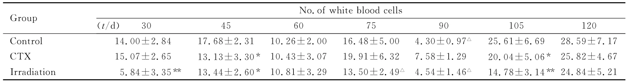
表1 各组小鼠外周血白细胞总数的变化Tab.1 Changes of the number of white blood cells in peripheral blood of the mice in various groups (n=10,±s)
*P<0.05compared with control group;△P<0.05compared with CTX group.
25.61±6.69 28.59±7.17 CTX 15.07±2.65 13.13±3.30* 10.43±3.07 19.91±6.32 7.58±1.29 20.04±5.06* 25.82±4.67 Irradiation 5.84±3.35** 13.44±2.60* 10.81±3.29 13.50±2.49△ 4.54±1.46△ 14.78±3.14**30 45 60 75 90 105 120 Control 14.00±2.84 17.68±2.31 10.26±2.00 16.48±5.00 4.30±0.97△Group No.of white blood cells(t/d)24.84±5.21
2.3 持续高剂量电磁辐射诱导小鼠外周血CD4+T细胞数量的变化 电磁辐射后,与正常对照组比较,照射组小鼠外周血淋巴细胞CD4+T细胞数量均明显升高;与环磷酰组比较,在照射后75d及105dCD4+T细胞数量升高,且差异有统计学意义 (P<0.05)。见表2。
表2 各组小鼠外周血淋巴细胞CD4+T细胞数量的变化Tab.2 Changes of the number of lymphocytes CD4+ T cells in peripheral blood of the mice in various groups (n=10,±s)

表2 各组小鼠外周血淋巴细胞CD4+T细胞数量的变化Tab.2 Changes of the number of lymphocytes CD4+ T cells in peripheral blood of the mice in various groups (n=10,±s)
*P<0.05compared with control group;△P<0.05,△△P<0.01compared with CTX group.
24.20±8.94 CTX 31.82±3.95 34.01±4.99 33.00±3.16 27.28±2.57 24.95±3.49 Irradiation 33.65±5.75 49.92±5.31*△△ 35.85±2.48 36.61±4.49△60 75 90 105 120 Control 32.05±7.15 36.69±6.59 33.16±5.51 35.13±2.62△Group No.of CD4+T cells(t/d)23.77±4.85
2.4 持续高剂量电磁辐射诱导小鼠外周血CD8+T细胞数量的变化 电磁辐射后,与对照组比较,辐照组小鼠淋巴细胞CD8+T细胞数量显著下降;与加药组比较,辐照组小鼠在照射后60、75及105dCD8+T细胞数量明显降低,各组之间比较差异均有统计学意义 (P<0.05)。见表3。
表3 各组小鼠外周血淋巴细胞CD8+T细胞数量的变化Tab.3 Changes of the number of lymphocytes CD8+ T cells in peripheral blood of the mice in various groups (n=10,±s)

表3 各组小鼠外周血淋巴细胞CD8+T细胞数量的变化Tab.3 Changes of the number of lymphocytes CD8+ T cells in peripheral blood of the mice in various groups (n=10,±s)
*P<0.05compared with control group;△P<0.05,△△P<0.01compared with CTX group.
75.80±8.94 CTX 68.18±3.95 66.00±4.99 67.00±3.16 72.72±2.57 75.05±3.49 Irradiation 66.35±5.75 50.08±5.30*△△ 64.15±2.48 63.40±4.49△60 75 90 105 120 Control 67.95±7.15 63.31±6.59 66.84±5.51 64.87±2.62△Group No.of CD8+T cells(t/d)76.23±4.85
2.5 持续高剂量电磁辐射诱导小鼠外周血CD4+/CD8+比值的变化 电磁辐射后,辐照组小鼠外周血淋巴细胞绝对值呈上升趋势,辐照后75d及105d辐照组与对照组相比显著上升 (P<0.05)。见表4。
表4 各组小鼠外周血CD4+/CD8+比值的变化Tab.4 Changes of the ratios of CD4+/CD8+in peripheral blood of the mice in various groups (n=10,±s)

表4 各组小鼠外周血CD4+/CD8+比值的变化Tab.4 Changes of the ratios of CD4+/CD8+in peripheral blood of the mice in various groups (n=10,±s)
*P<0.05compared with control group;△P<0.05compared with CTX group.
+0.06 0.33±0.16 CTX 0.47±0.09 0.52±0.12 0.49±0.07 0.37±0.05 0.33±0.06 Irradiation 0.52±0.13 1.02±0.24*△60 75 90 105 120 Control 0.49±0.16 0.59±0.17 0.50±0.13 0.54±Group CD4+/CD8(t/d)0.56±0.06 0.58±0.12 0.32±0.08
2.6 持续高剂量电磁辐射诱导小鼠外周血CD49+NK细胞数量的变化 电磁辐射后与正常对照组比较,辐照组小鼠淋巴细胞CD49+NK细胞数量明显降低 (P<0.05),表明小鼠免疫系统出现紊乱。见表5。
表5 各组小鼠外周血CD49+T细胞数量的变化Tab.5 Changes of the number of CD49+ T cells in peripheral blood of the mice in various groups (n=10,±s)
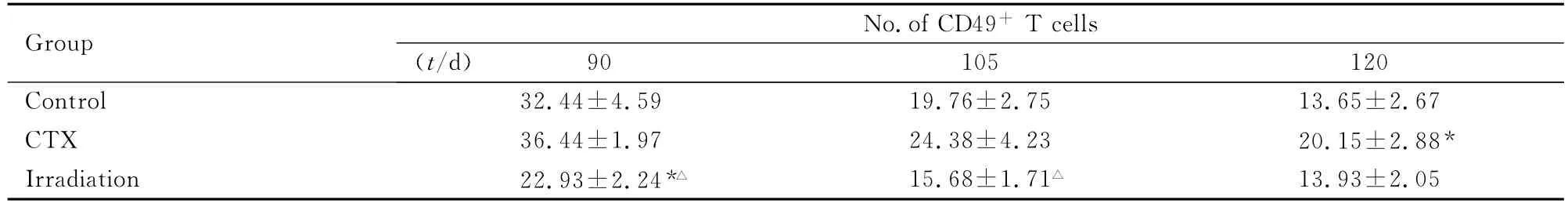
表5 各组小鼠外周血CD49+T细胞数量的变化Tab.5 Changes of the number of CD49+ T cells in peripheral blood of the mice in various groups (n=10,±s)
*P<0.05compared with control group;△P<0.05compared with CTX group.
Group No.of CD49+T cells(t/d)90 105 120 Control 32.44±4.59 19.76±2.75 13.65±2.67 CTX 36.44±1.97 24.38±4.23 20.15±2.88*Irradiation 22.93±2.24*△ 15.68±1.71△13.93±2.05
3 讨 论
本研究采用随机、平行以及独立的动物实验方法,系统建立了慢性高剂量电磁照射小鼠模型,检测10mW·cm-2大功率辐照对小鼠外周血免疫细胞的影响。为排除热效应的干扰,采用间断暴露[3]的方法,最终测定小鼠肛温在照射前后基本没有变化,保证实验研究的非热效应;同时,在实验过程中采用共同饲养、辐照,检测时分别采集动物样品,排除感染、疾病等生物学效应对血象的影响,保证实验结果具有良好的稳定性及重复性。
慢性高剂量密度电磁辐射对小鼠外周血免疫细胞的影响主要表现在CD4+T、CD8+T细胞以及CD4+/CD8+比值的变化。本研究结果显示:电磁辐射后小鼠外周血淋巴细胞CD4+T细胞数量高于正常对照组,与对照组相比较,照射后75和105d淋巴细胞CD4+T细胞数量增高,差异有统计学意义;电磁辐射后,辐照组小鼠淋巴细胞CD8+T细胞数量与对照组相比显著下降,在照射后60、75及105dCD8+T细胞数量明显低于给药组;电磁辐射后辐照组外周血淋巴细胞CD4+/CD8+绝对值呈上升趋势,且明显高于正常对照组,在照射后75及105d照射组高于正常对照组。本研究结果表明:慢性高剂量电磁辐射可导致小鼠外周血淋巴细胞数降低,外周血淋巴细胞CD4+/CD8+比值有所增大,在检测周期结束时小鼠外周血淋巴细胞数有恢复的趋势,提示去除高剂量辐射后,小鼠机体的免疫细胞数量可逐渐恢复。
综上所述,慢性大功率电磁辐射对生物体的免疫系统具有一定程度的损伤效应[9],但该方面研究目前还处于基础研究阶段,需要与辐射后小鼠的组织病理损伤、造血干细胞的变化情况相结合,从而进行更深入、系统的研究。
[1]Jean HUSS,Luxembourg,Socialist Group.The potential dangers of electromagnetic fields and their effect on the environment [A]. Parliamentary Assembly Assemblee parlementaire,Report of the Committee on the Environment,Agriculture and Local and Regional Affairs [C].Sweden,2011.
[2]Levitt BB,Lai H.Biological effects from exposure to electromagnetic radiation emitted by cell tower base stations and other antenna arrays[J].Environ Rev,2010(18):369-395.
[3]Macri MA,Di Luzio S.Biological effects of electromagnetic fields[J].Immunopath Pharmacol,2002,15 (2):95-105.
[4]Goldsworthy A. The biological effects of weak electromagnetic fields[R].London Imperial College,2007.
[5]Hardell L,Sage C.Biological effects from electromagnetic field exposure and public exposure standards [J].Biomed Pharmacother,2008,62 (2):104-109.
[6]Pirogova E,Vojisavljevic V,Cosic I.Biological effects of electromagnetic radiation,Recent Advances in Biomedical Engineering [M].Austria:In-Tech Vienna,2009.
[7]SimkóM, Mattsson MO. Extremely low frequency electromagnetic fields as effectors of cellular responses in vitro:possible immune cell activation [J].Cell Biochem,2004,93 (1):83-92.
[8]Marino C,Cristalli G, Galloni P,et al. Effects of microwaves(900MHz)on the cochlear receptor:exposure systems and preliminary results[J].Radiat Environ Biophys,2000,39 (2):131-136.
[9]Nageswari KS,Sarma KR,Rajvanshi VS,et al.Effects of chronicmicrowave radiation on T cell-mediated immunity in the rabbit[J].Int J Biometeorol,1991,35 (2):92-97.
Long-term effects of lasting and high-dose electromagnetic radiation on peripheral blood immune cells in mice
ZHANG Yi-kun1,LI Hui2,HUANG Gen-shan2,DONG Bo3,SU Zhen-tao3
(1.Department of Hematology,General Hospital of PLA Second Artillery Forces,Beijing 100088,China;2.Huangsi Out-patient Department,General Equipment Department Headquarters,Beijing 100021,China;3.Institute of Radiation Medicine,Academy of Military Medical Sciences,Beijing 100850,China)
Objective To study the long-term effects of lasting and high-dose electromagnetic radiation on number and percent of peripheral immune cells in mice and explore the influence of the lasting and high-dose electromagnetic radiation in murine immune system.Methods Thirty BALB/c mice were randomly divided into control group,CTX group and irradiation group.The mice in irradiation group were exposed to electromagnetic radiation at the power density of 10mW·cm-2,30min a day,for 5consecutive days every week,lasting for 4weeks.The mice in CTX group were fed with CTX with the concentration of 30mg·kg-1,for seven times.The number and percents of CD4+T cells,CD8+T cells,CD4+/CD8+,CD49+NK cells in peripheral blood cells were detected 30,45,60,75,90,105and 120dafter electromagnetic radiation.Results After high-dose electromagnetic radiation,the number of WBC was increased obviously(P<0.05).The number of CD4+T cells was increased on the 75th day(P<0.05)and began to decrease on the 90th day,while the number was much more than that in control group(0mW·cm-2).Until the 120th day,the number of T cells was as the same as the control.After the long-term effect of lasting and high-dose electromagnetic radiation,the number of CD8+T cells was decreased,and from the 90th day to the 120th day the number of T cells became normal(P<0.05).The ratio of CD4+/CD8+and the number of CD49+NK cells were lower than those in control group after irradiation (P<0.05).Conclusion Chronic,persistent large doses of electromagnetic radiation can lead to immune system disorders,through detecting the changes in the number of peripheral blood CD4+T cells,CD8+T cells,CD49+NK cells and the proportion of CD4+/CD8+,the immune status of mice after irradiation can be found.
electromagnetic radiation;immune cells;mice,inbred BALBC
R358.4
A
1671-587Ⅹ(2012)05-0856-05
2012-03-07
国家高技术研究发展计划项目资助课题 (2009AA8030421E)
张怡堃 (1972-),女,吉林省长春市人,副主任医师,医学博士,主要从事血液肿瘤的基础与临床研究。
张怡堃 (Tel:010-66355109,E-mail:zhangyikun1989@yahoo.com.cn)