Effects of miR-203 targeting ΔNp63 on proliferation,invasion,apoptosis and stem-cell characteristics of squamous cell carcinoma cells
2021-11-13ChanGaoAiHeGuizhongXiongNiXieMinLi
Chan Gao,Ai'e He,Guizhong Xiong,Ni Xie,Min Li
Abstract Objective:To investigate the effect of miR-203 targeting ΔNp63 on proliferation,invasion,apoptosis and stemcell characteristics of oral squamous cell carcinoma (OSCC)cells.Methods:TCGA database was used to analyze the expression of miR-203 and ΔNp63 in head and neck squamous cell carcinoma and adjacent normal tissues.RT-qPCR was used to detect the expression of miR-203 and ΔNp63 mRNA in OSCC cells (Tca8113,CAL-27,and SCC-9) and normal human oral keratinocytes(NHOK).The CAL-27 cells were transfected with miR-NC,miR-203 mimic,pcDNA,or pcDNAΔNp63,and co-transfected with miR-203 mimic and pcDNAΔNp63.The expressions of ΔNp63,Bax,Bcl-2,Ki67,MMP-2,E-cadherin,N-cadherin,Lin28B,SOX2 and OCT4 were detected by Western blotting.Luciferase reporter assay was used to analyze the targeting relationship between miR-203 and ΔNp63.CCK8,Transwell,and flow cytometry were performed respectively to detect cell viability,invasion,apoptosis and the number of ALDH positive cells.A nude mouse xenograft tumor was constructed to detect the effect of miR-203 on tumors in vivo.Results:The expression of miR-203 was down-regulated while the expression of ΔNp63 was up-regulated in head and neck squamous cell carcinoma and OSCC cells (P<0.05).miR-203 targeted and negatively regulated the expression of ΔNp63.The expressions of ΔNp63,Ki67,MMP-2,N-cadherin,Bcl-2,Lin28B,SOX2 and OCT4,the viability of CAL-27 cells,the number of invasive cells and ALDH-positive cells were decreased significantly,while the apoptosis rate and the protein expression levels of Bax and E-cadherin were increased in miR-203 group(P<0.01).The results of ΔNp63 group were opposite.miR-203 mimic reversed the promotive effects of overexpressed ΔNp63 on CAL-27 cell viability,invasion and stem cell-like properties (P<0.05).miR-203 inhibited tumor growth in nude mice translanted with OSCC cell line and downregulated ΔNp63 expression.Conclusion:miR-203 plays a tumor-suppressive role in OSCC by inhibiting the expression of ΔNp63.
Keywords oral squamous cell carcinoma;miR-203;ΔNp63;invasion;OCT4
Introduction
Oral squamous cell carcinoma (OSCC) is the most common malignant tumor in oral and maxillofacial region [1].The morbidity and mortality of OSCC rank first among oral and maxillofacial malignant tumors.OSCC is prone to cervical lymph node metastasis with rapid progression,which threats human health severely [2].Although great progress has been made in medical technology,the prognosis of patients with OSCC is still not satisfactory.Therefore,there is an urgent need to better understand the molecular mechanism of OSCC tumor growth and metastasis.microRNA (miR) is an endogenous small non-coding RNA that regulates the expression of target genes after transcription.Studies have shown that the expression of miR-203 is significantly down-regulated in head and neck squamous cell carcinoma [3],hematopoietic malignant tumor[4]and colon cancer[5],and negatively correlated with the proliferation of cancer cells.TP63 is a homologue of the TP53 family of transcription factors,which plays a key role in the development of epithelial cells.According to the different transcriptional initiation,it can be divided into two subtypes,ΔNp63 and TAp63 [6].ΔNp63 is generally overexpressed in epithelial tumors including prostate cancer,breast cancer and head and neck cancer [7-9].In esophageal squamous cell carcinoma,miR-203 inhibits tumor growth and metastasis by targeting ΔNp63[10].The expression and targeting relationship between miR-203 and ΔNp63 in OSCC have not been reported.The main purpose of this study is to explore whether miR-203 affects the biological behavior of OSCC cells by regulating ΔNp63.
Materials and methods
Main reagents
Fetal bovine serum (FBS),DMEM medium,trypsin,Lipofectamine 2000,CCK8 kit,Trizol kit and TUNEL apoptosis kit were purchased from Thermo Fisher.Reverse transcription kit and SYBR Green PCR Master Mix kit were purchased from Japan Takara company.Annexin V-FITC apoptosis detection kit was from BD Pharmingen.Primary antibodies rabbit polyclonal anti-β-actin,rabbit monoclonal anti-Ki67,rabbit polyclonal anti-MMP-2,rabbit monoclonal anti-Bax,rabbit polyclonal anti-Bcl-2,rabbit monoclonal Lin28B,rabbit monoclonal anti-SOX2,and rabbit monoclonal anti-OCT4 were from Abcam.Rabbit monoclonal anti-E-cadherin antibody,rabbit monoclonal anti-N-cadherin antibody,and HRP-coupled goat antirabbit IgG were purchased from Cell Signaling Technology.
Data collection
The gene expression profiles of miR-203 and ΔNp63 in head and neck squamous cell carcinoma and adjacent normal tissues were obtained by The Cancer Genome Atlas(TCGA)(https://cancergenome.nih.gov/).
Cell culture
OSCC cell line (Tca8113,CAL-27,SCC-9) and normal human oral keratinocytes (NHOK) were cultured in an atmosphere containing 5% CO2and 95% O2at 37 ℃with RPMI 1640 medium which supplemented with 10%fetal bovine serum(FBS)and 1%penicillin/streptomycin(100 U/mL,Thermo Fisher Scientific).
RT-qPCR
The total RNAs of OSCC cell lines,NHOK cell and transfected CAL-27 cell were extracted by TRIzol kit and synthesized into cDNA by reverse transcription kit,and then amplified by PCR instrument.Quantitative analysis of cDNA was carried out according to the instructions of the fluorescence quantitative kit.U6 was used as the internal reference for miR-203 and GAPDH as the internal reference for ΔNp63.The relative expression levels of miR-203 and ΔNp63 were calculated by 2-ΔΔCtmethod.The reaction procedure was as follows:95℃,30 s;95℃,3 s;60℃,30 s,40 cycles.All the primers used in the experiment were designed by Primer 5 and synthesized by Shanghai Sangon Biotech.The primers are as follows:miR-203 forward primer:5’-AACCTTGCTCGTGAAATGTTTAG-3’,reverse primer:5’-TATGCTTGTTCTCGTCTCTGTGTC-3’;U6 forward primer:5’-TGCGGGTGCTCGCTTCGGCAGC-3’,reverse primer:5’-CCAGTGCAGGGTCCGAGGT-3’;ΔNp63 forward primer:5’-GGGTGAGCGTGTTATTGATGCT-3’,reverse primer:5’-GAGTGGAATGACTTCAACTTT-3’;GAPDH forward primer:5’-GCTGAGTATGTCGTGGAGTC-3’;reverse primer:5’-AGTTGGTGGTGCAGGATGC-3’.
Western blotting
Total protein obtained from OSCC cell lines,NHOK cells and transfected CAL-27 cells were separated by electrophoresis on SDS-PAGE and electrotransferred onto polyvinylidene difluoride (PVDF)-plus membranes.After blocking with skim milk for 1 h,the membranes were incubated overnight at 4 ℃with the following primary antibodies:ΔNp63 (1:1,000),Bax(1:500),Bcl-2 (1:1,000),Ki67 (1:1,000),MMP-2 (1:1,000),E-cadherin (1:1,000),N-cadherin (1:1,000),Lin28B (1:1,000),SOX2 (1:1,000),OCT4 (1:1,000)and β-actin antibody (1:2,000).Then,the membranes were washed three times in TBST and incubated with secondary antibodies conjugated with HRP (1:2,000)at 37 ℃for 1 h.Immunopositive bands were visualized by enhanced chemiluminescence (ECL) and analyzed using ImagePro plus 6.0 software.
Cell transfection
miR negative control(NC),miR-203 mimic,NC plasmid pcDNA,overexpression plasmid pcDNA-ΔNp63 and miR-203 mimic+ΔNp63 were transfected into CAL-27 cells by Lipofectamine 2000 following the manufactures’instructions.The control group cells were treated with Lipofectamine 2000 only.After 24 h of post-transfection,cells were collected for the indicated experiments.
Luciferase reporter assay
The potential binding sites between miR-203 and ΔNp63 3’UTR were analyzed by online software Targetscan.CAL-27 cells growing in logarithmic phase were collected and inoculated in 24-well plates and incubated for 24 h.A total of 1 g luciferase reporter gene constructor in firefly PGL3-ΔNp63-wild type(ΔNp63-wt) or PGL3-ΔNp63-mutant (ΔNp63 mut)and miR-NC or miR-203 mimic and PRL-CMV algal luciferase reporter plasmids were used to transfect cells.Forty-eight hours after transfection,CAL-27 cells were lysed for 15 min,and the relative activity of luciferase was measured by double luciferase detection system.The ratio of firefly luciferase activity to renal luciferase activity was used to represent the relative activity of luciferase.
Cell viability assay
Cell viability was assessed using CCK8 assay.The transfected CAL-27 cells were seeded onto 96-well plates,followed by incubation at 37 ℃for 24 h,48 h,and 72 h.CCK8 (10 μL) solution was added to the culture medium and incubated for another 1 h.The optical density (OD) value at 450 nm was recorded with a microplate reader(Bio-Rad).
Cell invasion assay
Cell invasion assay was performed using a transwell chamber.The transfected CAL-27 cells were placed in the upper chamber of the Transwell scaffold with coated matrix glue (1 μg/mL),and the lower chamber contained a medium with 10% FBS.After incubation for 24 h,the cells retained in the upper chamber were wiped with cotton swabs,and the cells infiltrated into the membrane were fixed.The stained cells were stained with 0.1% crystal violet and counted under the microscope.
Cell apoptosis analysis
Cell apoptosis analysis was performed using flow cytometry with an Annexin V/PI apoptotic detection kit.Briefly,transfected CAL-27 cells were washed in PBS and re-suspended in binding buffer containing 10 μL of Annexin V and 5 μL PI according to the manufacturer’s instructions.The apoptotic cells were analyzed using a flow cytometry (BD Biosciences) equipped with FlowJo software(Tree Star).
ALDH positive cells sorting by flow cytometry
CAL-27 cells were inoculated into a 6-well plate with a density of 1×10⁶cells per well and cultured overnight.After 48 h of transfection,the cells were re-suspended in 1 mL ALDEFLUOR buffer,5 μL BODIPYaminoacetaldehyde (BAAA) was added into cells and briefly mixed;300 μL cell suspension was transferred to a centrifuge tube containing 5 μL diethylaminoazobenzene (DEAB) and mixed evenly with a pipette;then two centrifuge tubes were put into a cell incubator and incubated for 40 min.The cells were washed twice with 2 mL ALDEFLUOR buffer and re-suspended in 500 μL ALDEFLUOR buffer supplemented with DAPI to stain dead cells.The sample was analyzed by flow cytometry.
Subcutaneous transplantation of xenograft in nude mice
Sixteen 18-20 g SPF 4-week-old BALB/c nude mice were purchased from Beijing Vital River Laboratory Animal Technology Co.,Ltd.,with the license number SCXK (Beijing) 2016-0011.The nude mice were raised in a sterile environment at 22 ℃,humidity of 50%to 60%,light/night time of 12/12 h,free drinking water and food,and the experiment was carried out after a week of adaptive feeding.2×106CAL-27 cells transfected with miR-NC or miR-203 mimic were subcutaneously injected into the lateral side of nude mice (n=8 in each group).After feeding for 30 days,the tumor volume (V) was monitored every 3 days from the 6th day,V=0.5×length×width2.After 30 days,the mice were euthanized by neck dislocation,and the tumor was removed by operation.The tumor sample was photographed,weighed and paraffin embedded section for follow-up experiment.
TUNEL staining
The paraffin sections of tumor tissue preserved were dewaxed and then incubated with TUNEL reaction mixture containing TdT and biotinylated dUTP at 37 ℃for 1 h.After washing,DAB chromogenic solution was added to develop color.The number of TUNEL positive cells was counted under light microscope.
Immunohistochemical experiment
The paraffin sections of tumor tissue were dewaxed and repaired with sodium citrate-hydrochloric acid buffer solution.The slices were incubated overnight at 4 ℃with primary antibodies Ki67 (1:200) and SOX2(1:200).The goat anti-rabbit IgG secondary antibody coupled with HRP (1:500) was added at 37 ℃for 1 h,and horseradish peroxidase labeled streptavidin was added for 40 min at 37 ℃.After washing,the nucleus was re-stained with hematoxylin.After sealing,the Ki67 and SOX2 positive cells were observed by light microscope.
Statistical method
The data were analyzed by SPSS 16.0.The measurement data were expressed as mean±standard deviation(SD).Student’sttest and one-way ANOVA analysis were used for comparison between groups,P<0.05.
Results
Expressions of miR-203 and ΔNp63 in OSCC cells
The expressions of miR-203 and ΔNp63 in OSCC cells were analyzed by TCGA data online analysis tool.Compared with normal tissues,the expression of miR-203 in head and neck squamous cell carcinoma was decreased,while the expression of ΔNp63 was increased (P<0.05).Compared with NHOK cells,the expression of miR-203 in OSCC cells was significantly down-regulated,while the expression of ΔNp63 mRNA and protein was significantly up-regulated(P<0.05)(Figure 1).
Targeting inhibition of ΔNp63 expression by miR-203
Compared with the control group,the expression of miR-203 in miR-203 group was significantly up-regulated (P<0.01,Figure 2A),and the expression of ΔNp63 was significantly down-regulated (P<0.01,Figure 2B).Targetscan website analysis predicted that there were miR-203 binding sites on ΔNp63 3’UTR(Figure 2C).Compared with miR-NC+ΔNp63-wt group and miR-203+ΔNp63-mut group,the fluorescence activity of miR-203+ΔNp63-wt group was significantly decreased(P<0.01,Figure 2D).
miR-203 targeting ΔNp63 inhibits the prolifera⁃tion and invasion of OSCC cells
Compared with the control group,the expression levels of ΔNp63,Ki67,MMP-2 and N-cadherin protein,the cell viability,and the number of invaded cells were significantly decreased,while the E-cadherin protein expression level was significantly increased in miR-203 group (P<0.05).The expressions of ΔNp63,Ki67,MMP-2 and N-cadherin protein,the cell viability,and the number of invaded cells were significantly increased,and the E-cadherin protein expression was obviously down-regulated in cells transfected with pcDNA-ΔNp63(P<0.05).miR-203 mimic reversed the effects of ΔNp63 overexpression on the cell viability,cell invasion and the protein levels of ΔNp63,Ki67,MMP-2,N-cadherin and E-cadherin(P<0.05)(Figure 3).
miR-203 targeting ΔNp63 promotes apoptosis of OSCC cells
Compared with the control group,the apoptosis rate and the expression of Bax protein were elevated,while the expression of Bcl-2 protein was significantly reduced in cells transfected with miR-203 mimic(P<0.05).In ΔNp63 group,the apoptosis rate of cells and the expression of Bax protein were significantly decreased,while the expression of Bcl-2 protein was notably increased (P<0.01).Compared with ΔNp63 group,the apoptosis rate of cells and the Bax protein level in miR-203+ΔNp63 group were evidently increased,while the expression of Bcl-2 protein was significantly down-regulated (P<0.01) (Figure 4).
miR-203 targeting ΔNp63 inhibits the expression of cancer stem-cell markers in OSCC cells
Compared with the control group,the number of ALDH positive cells and the expression of Lin28B,SOX2 and OCT4 protein were decreased significantly in miR-203 group,while the number of ALDH positive cells and the expression of Lin28B,SOX2 and OCT4 protein were increased significantly in ΔNp63 group (P<0.05).miR-203 mimic significantly reversed the effects of ΔNp63 on cancer stem-cell markers expression(P<0.05)(Figure 5).
miR-203 inhibits the growth of xenograft in nude mice
Compared with miR-NC group,the tumor weight and volume,the positive rates of Ki67 and SOX2,and the expressions of ΔNp63 and MMP-2 protein in miR-203 group were significantly decreased,while the expressions of miR-203 and TUNEL-positive cells in tumor tissues were significantly increased (P<0.05)(Figure 6).
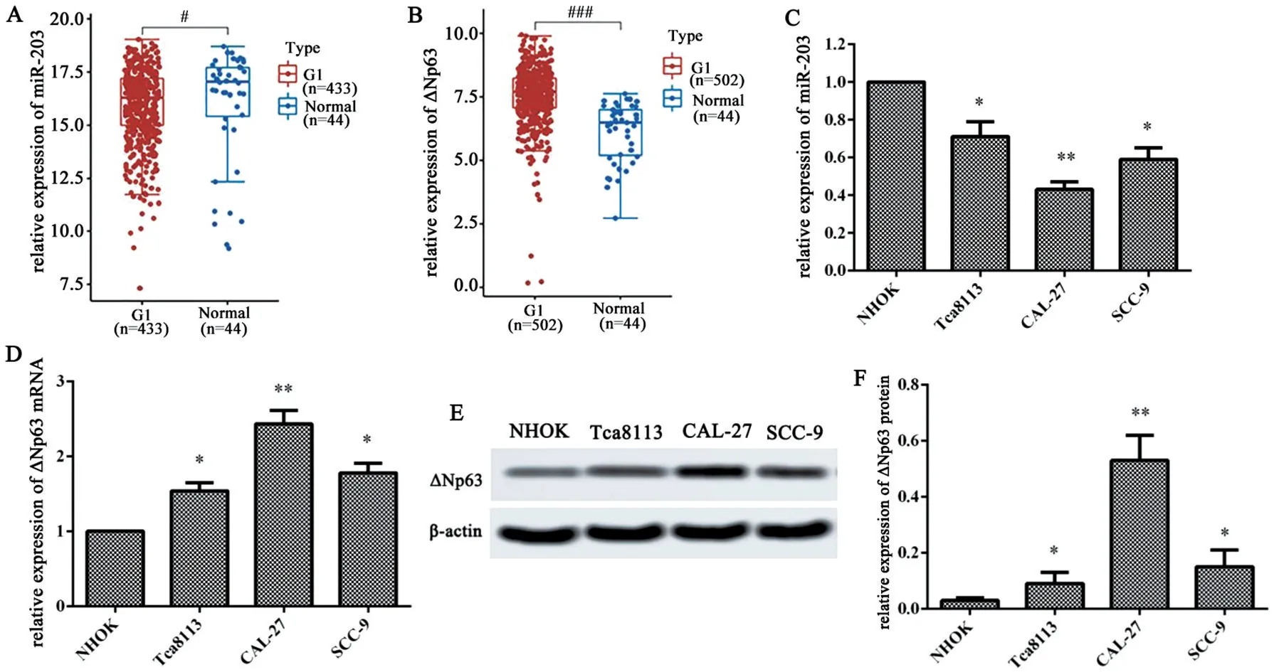
Figure 1 Expressions of miR-203 and ΔNp63 in OSCC cells.A-B:Analysis of miR-203 and ΔNp63 expression in head and neck squamous cell carcinoma by TCGA database;C-D:The miR-203 and ΔNp63 mRNA expressions were detected by RT-qPCR;E-F:The ΔNp63 protein expression was determined by Western blotting.#P<0.05 or###P<0.001 vs. Normal tissues;*P<0.05 or**P<0.01 vs.NHOK cells.
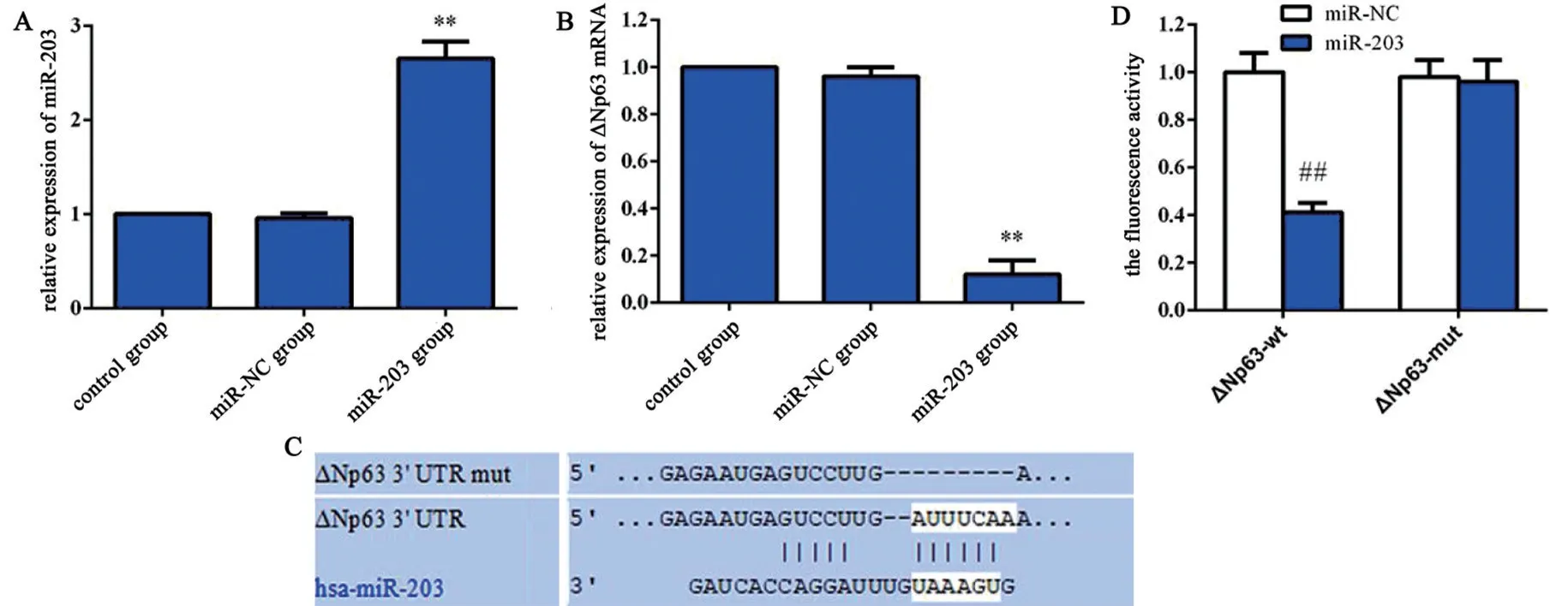
Figure 2 Targeting inhibition of ΔNp63 expression by miR-203.A:miR-203 relative expression;B:ΔNp63 relative expression;C:Analysis of binding sites between miR-203 and Δ Np63 3’UTR by Targetscan website;D:Luciferase reporter gene confirmed that miR-203 binded directly to ΔNp63 3’UTR.**P<0.01 vs.Control group;##P<0.01 vs.miR-203+ΔNp63-mut group.
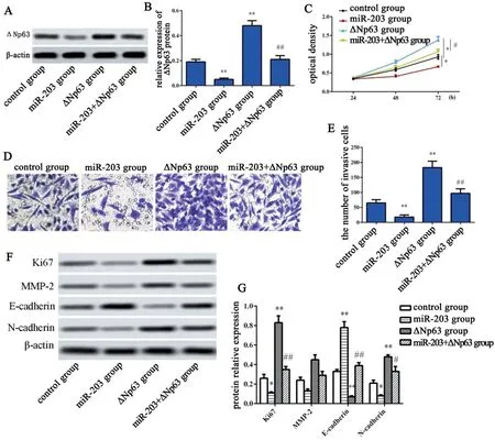
Figure 3 miR-203 targeting ΔNp63 inhibits the proliferation and invasion of OSCC cells.A-B:Expression of ΔNp63 protein in each group;C:Cell activity was detected by CCK8;D-E:The number of invasive cells was detected by Transwell(×200);F-G:Proliferation and invasion protein expressions were detected by Western blotting.*P<0.05 or**P<0.01 vs.Control group;#P<0.05 or##P<0.01 vs.ΔNp63 group.
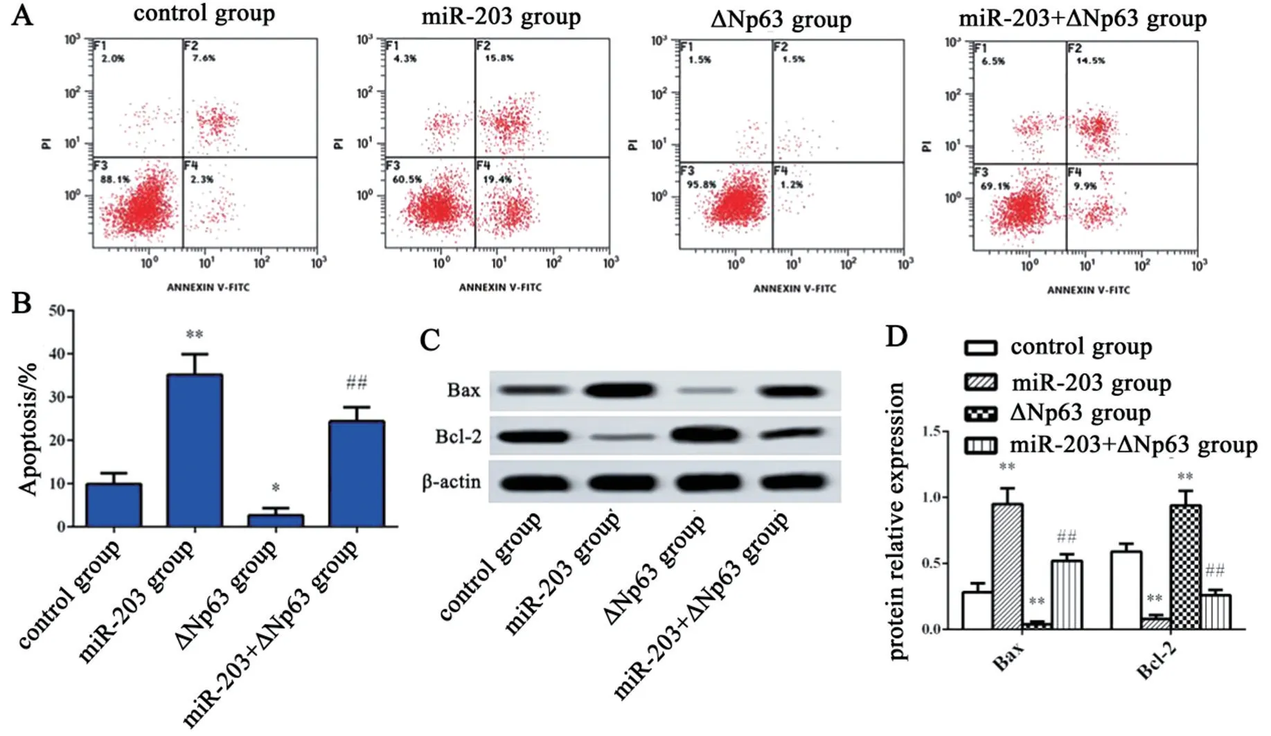
Figure 4 miR-203 targeting ΔNp63 promotes apoptosis of OSCC cells.A-B:Flow cytometry was used to detect the apoptosis rate;C-D:The Bax and Bcl-2 protein expression was assessed by Western blotting.*P<0.05 or**P<0.01 vs. Control group;##P<0.01 vs.ΔNp63 group.

Figure 5 miR-203 targeting ΔNp63 inhibits the expression of cancer stem-cell markers in OSCC cells.A:Flow cytometry sorting of ALDH positive cells;B-C:Detection of Lin28B,SOX2 and OCT4 protein expression by Western blotting.**P<0.01 vs. Control group;#P<0.05 or##P<0.01 vs.ΔNp63 group.

Figure 6 Effect of miR-203 on the growth of xenograft in nude mice.A:xenograft in nude mice(n=5);B:Tumor weight;C:Tumor volume;D:The expression of miR-203;E:Detection of ΔNp63 and MMP-2 protein expression by Western blotting;F:Apoptosis(×400) was detected by TUNEL,and the expression of Ki67 and SOX2 (×400) was detected by immunohistochemistry.*P<0.05 or**P<0.01 vs.miR-NC group.
Discussion
It is well known that miRNAs can regulate a variety of cellular pathways by affecting the expression of a variety of target genes,and the change of miRNAs expression may be related to human carcinogenesis [11-12].Therefore,understanding the specific microRNA involved in tumor development will provide valuable insights for the diagnosis and treatment of tumors.It is known that miR-203 targeting regulation ΔNp63 controls the proliferation and differentiation of keratinocytes[13],which are evaluated in some types of epithelial tumorigenesis and metastasis.Studies have shown that the expression of miR-203 is down-regulated in cervical cancer [14],gastric cancer [15]and non-small cell lung cancer [16],and is related to the poor prognosis of patients.Study results from Liu et al.[17]showed that miR-203 regulates the proliferation and apoptosis of ovarian cancer cells by targeting SOCS3.ΔNp63 was overexpressed in cervical cancer,head and neck cancer,lung cancer and breast cancer[18-19].Therefore,ΔNp63 and miR-203 seem to play both carcinogenic and tumor inhibitory roles in the development of cancer.The dry characteristics of cancer cells are related to the invasiveness of tumors,which enable tumor cells to acquire unlimited self-renewal potential,thus promoting subsequent tumor growth and metastasis [20].Recent studies have shown that the interaction between ΔNp63 and miR-138-5p regulates the growth,metastasis of oral squamous cell carcinoma [21].Studies have shown that ΔNp63 promotes the stem cell-like phenotype of OSCC cells by regulating SOX2,CD44 and KLF4 [21-22].Therefore,ΔNp63 plays a key role in maintaining the selfrenewal potential of tumor cells.Previous studies have shown that miR-203 acts as an inhibitor of tumor stem cells in a variety of cancers,for example,miR-203 reduces the stem cell characteristics of human colon cancer cells by inhibiting the expression of GATA6 [23].In addition,miR-203 inhibits the drylike characteristics of renal cancer cells by regulating dry-related genes,such as SOX2,CD44 and KLF4[24].
In this study,we searched for the expressions of miR-203 and ΔNp63 in TCGA head and neck cancer data set,and found that miR-203 was down-regulated and ΔNp63 expression was up-regulated in head and neck cancer.Results of qRT-PCR and WB confirmed that miR-203 was low expressed in OSCC cell line,while ΔNp63 was highly expressed in OSCC cell line.The target gene of miR-203 was predicted by bioinformatics analysis.Double luciferase reporter gene assay confirmed the targeting relationship between miR-203 and ΔNp63.Further experiments showed that miR-203 exerted its anti-tumor effect in OSCC by inhibiting ΔNp63.Both miR-203 and ΔNp63 were overexpressed in CAL-27 cells.It was found that miR-203 mimic reversed the effects of ΔNp63 overexpression on promotion of cell proliferation and invasion,and suppression on cell apoptosis.Therefore,miR-203 inhibits the proliferation and invasion of OSCC cells by targeting ΔNp63.The main cause of tumor-related death is local recurrence and metastasis.This study showed that miR-203 inhibited the expression of cancer stem-cell markers such as ALDH,Lin28B,SOX2 and OCT4 in OSCC cells by targeting ΔNp63,thus inhibiting tumor metastasis and growth.
The experiment of xenograftin vivofurther confirmed the inhibitory effect of miR-203 on tumor growth,which is consistent with previous reports.miR-203 plays an inhibitory role in colon cancer [25]and ovarian cancer[26].The results of this studyin vi-voare consistent with thosein vitro.Overexpression of miR-203 could inhibit tumor growth,promote apoptosis of cancer cells in tumor tissue,and downregulate the expression of MMP,Ki67 and SOX2.
In conclusion,miR-203 expression is down-regulated in OSCC,and overexpression of miR-203 exerts an anti-tumor effect in OSCC by targeting ΔNp63,indicating that miR-203 may be an effective target for the treatment of OSCC.
AcknowledgmentsThis study was funded by Knowledge Innovation Project of Hubei Province in 2018(Natural Science Foundation of Hubei Province) (No.2018CFC834)
