A new automatic cell smear and laser release system for near-infrared light responsive release of nucleated red blood cells
2021-10-10GUOZhongyangYOUQiannanGEMingfengWANGGuoweiMEIQianDONGWenfei
GUO Zhong-yang,YOU Qian-nan,GE Ming-feng,WANG Guo-wei,MEI Qian ,DONG Wen-fei
(1. School of Biomedical Engineering (Suzhou), Division of Life Sciences and Medicine,
University of Science and Technology of China, Hefei 230026, China;2. Suzhou Institute of Biomedical Engineering and Technology,Chinese Academy of Science, Suzhou 215163, China)
* Corresponding author,E-mail: qmei@sibet.ac.cn
Abstract: In order to realize the separation and release of nucleated red blood cells from peripheral blood and develop a safe and effective non-invasive technique to separate nucleated red blood cells for prenatal diagnosis of fetal diseases, an automatic cell smear preparation system based on hydrogel material was established, and a laser focusing and microscopic imaging system for recognizing and releasing nucleated red blood cells was constructed. Firstly, the mechanical structure of cell smear preparation machine was designed, the upper computer control software was designed based on single chip microcomputer, and a hydrogel membrane substrate smear was prepared by optimizing the slide-pushing angle and speed. MXene, a twodimensional material, was introduced into temperature-sensitive hydrogel gelatin, and the near-infrared light response was realized on the surface of hydrogel membrane by using the near-infrared photothermal conversion characteristics of MXene. Then, the whole cell smear experiment was carried out on the surface of the hydrogel substrate membrane. A monolayer cell smear was prepared by optimizing the parameters of blood slide. Finally, the optical path of laser focusing and microscopic imaging was established. After the nucleated red blood cells were recognized and located, the light from an 808 nm laser source passed through a collimator lens and a convergent lens and was focused on the surface of the cell smear, which released cells under photothermal effect. A monolayer cell smear was processed and prepared, and then a photothermal effect was produced under the near-infrared light of 808 nm. After the control of the laser focusing system, a fixed cell-releasing area with a spot diameter of 300 μm was finally obtained. In this paper, the automatic slidepushing technology was applied to the preparation of a monolayer cell smear based on hydrogel membrane,
Key words: cell smear; hydrogel; prenatal diagnostics; near-infrared light response; cell release
1 Introduction
Prenatal diagnosis is essential for the early diagnosis and screening of birth defects such as Down syndrome, neural tube defects and single-gene diseases[1]. The existing prenatal diagnoses can be divided into invasive diagnoses and non-invasive diagnoses. Invasive diagnoses include amniocentesis,umbilical cord blood sampling, and chorionic puncture, etc.[2]. They are performed by invasive methods, and are often accompanied by miscarriage, amniotic fluid overflow, infection and other risks[3].Therefore, establishing a non-invasive prenatal diagnosis method is the focus and mainstream trend of the current prenatal diagnosis technology development[4]. Fetal Nucleated Red Blood Cells (NRBCs)contain the entire fetal genome and the early gestational expression (expressed in peripheral blood at the 6th week and maximized between 12thand 14thweek). They are highly distinguishable in maternal blood cell population and are hard to be confused with maternal cells. With obvious cellular morphological characteristics, they are easy to identify and are the preferred fetal cells for prenatal diagnosis[5].However, fetal NRBCs are very few in maternal peripheral blood. How to identify and separate NRBCs from peripheral blood is a major challenge in prenatal diagnosis[6].
The existing methods for separation and enrichment of peripheral blood cells include density gradient centrifugation, magnetic activated cell sorting, fluorescence activated cell sorting, microfluidic chip technology, etc[7-9]. Among these methods, the separation and detection are mainly realized by the specific binding of antibody and cells. The procedures have a complex operation process, long detection cycle and high cost. Simplifying the process of cell separation and enrichment is an urgent problem to be solved for non-invasive prenatal diagnosis.
Using the slide-pushing technique to make a good cell smear for blood cell morphology examination is critical in clinical application[10]. The traditional man-made cell smear cannot be standardized due to the uncontrollability of manual operation,which leads to cell overlap and rupture. The automatic cell smear preparation machine can standardize the preparation of monolayer cell smears and identify the NRBCs by optical imaging based on their special morphology. In order to achieve the collection of NRBCs for downstream application,the thermal response release system reported so far can release the captured cells at physiological temperature, and help the released cells maintain enough activity. This method can release cells in a large area, however, it is hard to release cells in a fixed area. Correspondingly, light-stimulated response system is considered as an ideal controllable release system due to its operability[11]. Compared with ultraviolet light, near-infrared light produces less damage to cells and has stronger penetration, so it is more suitable for the construction of light-responsive release system. By introducing a light response system into the thermal response release system, more efficient fixed-point release can be achieved. This paper proposes the use of the filmforming and photothermal properties of hydrogel[12],in which MXene two-dimensional material is introduced into thermo-sensitive hydrogel gelatin, the near-infrared photothermal effect of MXene can be combined with the thermal response of hydrogel substrate. Thus an automatic monolayer cell smear preparation machine is prepared, and a laser convergence and microscopic imaging system are established to solve the traditional problem of cell capture, separation and release depending on the specific binding of antigen and antibody. By optimizing the structure of each module and material properties,the recognition and fixed-point release of NRBCs provide a new approach for noninvasive prenatal testing.
2 Design of cell smear preparation machine system and recognition &release system
2.1 Overall design of control system
The scheme of automatic pushing and laser convergence system used to recognize and release the NRBCs is shown in Figure 1. The automatic slide pushing system consists of a mechanical motion subsystem, an adaptive smear system, a microstructural slide-loading platform and an electronic control subsystem. The movement of the XY stage is controlled by the mechanical motion subsystem,which controls the pushing speed and angle. The adaptive smear structure has two DOFs, which can adaptively fine-tune the condition of the smear and the surface in contact with it. By combining the two-dimensional motion of the motion subsystem, the three-dimensional motion of the smear and slide can be controlled. The microstructural slide-loading platform can carry the standard microscope slides(25 mm × 75 mm) for the preparation of hydrogelbased smears and cell smears. The electronic control subsystem is used to realize the communication and interactive operation among the modules of the whole smear system.
Firstly, a hydrogel membrane is prepared on a slide by using the automatic slide-pushing system.The membrane is taken as substrate, and then covered with a monolayer blood cell by spreading the blood sample. In this way, a cell smear is obtained. Secondly, the blood cell membrane is stained by Wright-Giemsa solution to examine cell morphology. As a clinical hematological detection method,this method has the advantages of good staining effect, obvious nucleo-cytoplasm contrast, easy operation and rapid staining[13]. After the staining of the cell smear, the NRBCs can be identified and located under optical microscope according to unique cell morphology. Then the cell smear is placed in a laser convergence and microscopic imaging system.According to the near-infrared response properties of hydrogel substrate, an 808 nm laser is selected for fixed-point irradiation to realize the photothermal conversion and release NRBC in the spot area.
2.2 Design of an automatic smear system
The automatic smear system includes four parts: a mechanical motion subsystem, an adaptive smear system, a microstructural slide-loading platform and an electronic control subsystem. The design principle of mechanical motion subsystem is shown in Figure 2. The system is divided into two modules: speed regulation and angle adjustment.The speed regulation module, which consists of an X stage, a carrier base and a closed-loop stepper motor, can adjust the speed within the range of 0−150 mm/s. The angle adjustment module is composed of a Y stage, a smear structure base, a closedloop stepper motor and an R-axis decelerating stepper motor. It can tune the angle within the range of 20°−50° when spreading the blood sample.
The adaptive smear system consists of smear clips and two sets of hinges that are perpendicular to each other in the pushing direction. With two DOFs,the system can adaptively fine-tune the smear while keeping it fit to the contact surface. The microstructural slide carrier is designed with a slide-loading groove and a waste liquid collection groove.
A 200 μm height difference between the slideloading groove and the slide surface is designed to ensure a hydrogel substrate membrane with uniform thickness during pushing. The waste liquid collection groove is used to collect excess hydrogel when pushing the slide to the end, so as to avoid the backflow-caused damage and contamination. The electronic control subsystem consists of an upper computer control system, a motion control card, a motor driver, a photoelectric limit switch and a power supply. The upper computer control software is programmed based on a single chip microcomputer. The serial port is connected with the motion control card to realize the interactive operation and control of pushing speed module and angle module.
2.3 Design of cell recognition and release system
According to the properties of hydrogel substrate membrane and the design principle of cell smear, the cell recognition and release system shown in Figure 3 has been designed in order to achieve the recognition and fixed-point release of NRBCs. It is composed of a laser-focusing subsystem, a bright field illumination subsystem and a microscopic imaging subsystem. According to the near-infrared response characteristics of hydrogel substrate membrane, an 808 nm laser source is combined with a collimating lens and a convergent lens(C1) into a laser convergence subsystem to irradiate a fixed area of the cell smear, which, in turn, produces the photothermal effect and releases the cells.In order to observe the cell release, a bright field illumination subsystem and a microscopic imaging subsystem are constructed on the basis of the laser convergence subsystem. The bright field illumination subsystem is composed of the bright field source and Kohler lens group. Through the application of dichromatic mirror, the bright field source and the 808 nm laser source can share an optical path. Subsequently, the microscopic imaging subsystem module composed of a microscopic objective, a reflecting mirror, a convergent lens (C2) and an imaging detector is used to observe the cell-releasing effect.
The laser convergence system is built to produce the near-infrared response, which enables the cell release at a fixed point. The laser light is focused to form a tiny light spot. A smaller light spot means a smaller spot area projected on the cell smear and higher efficiency and precision of cell release. To control the size of the light spot, the spot diameter should be reduced as much as possible so as to narrow the range of cell release at a fixed point. According to Gauss formula[14]:

whereω1is the spot diameter after focusing;Fis the focal length of the convergent lensC1,F=F1+F2,whereF1is the distance from the convergent lens to the dichromatic mirror andF2is the reflection distance from the dichromatic mirror to the cell smear;andω0is the waist radius of the laser Gaussian beam. The spot size and light intensity distribution on the equiphase planes on both sides of the convergent lensC1 are the same, soω0is the spot radius after collimation. By substituting the laser wavelengthλ(808 nm) and the collimated spot radiusω0(850 μm) into Equation (1), the spot diameter after laser convergence can be obtained as follows:

The bright field light is radiated to the cell smear surface through the Kohler lens group, and then is reflected to the detector by the imaging system composed of a microscope objective, a mirror and a convergent lens. By moving the stage, the whole cell smear can be imaged and the NRBCs can be identified and located. The laser light irradiates the identified target area through the collimating lens and convergent lens, and triggers the near-infrared response and photothermal conversion followed by fixed-point cell release. Finally, the cellreleasing performance is characterized by a microscopic imaging system.
3 Experiment and results
3.1 Preparation and optimization of hydrogel pusher
The experimental hydrogel was two-dimensional MXene composite gelatin (C102H151O39N31,gelatin) prepared by our group. The gelatin and MXene were stirred and mixed evenly at a mass ratio of 200:1 in a water bath at 38 ℃ to obtain the hydrogel. The prepared hydrogel was kept at 38 ℃to ensure the melting state through magnetic stirring, pushed and spread on the slide surface by a home-made automatic pushing machine, and then naturally cooled to form a layer of hydrogel membrane with smooth surface and uniform thickness.The prototype machine designed and fabricated based on a single-chip microcomputer is shown in Figure 4. The slide-carrier is located in a horizontal position. The pusher-loading platform has an angle to the slide carrier, but with a base parallel to the slide carrier. The preparation of hydrogel membrane was optimized by adjusting the pusher’s speed and angle. At first, a certain amount of hydrogel solution was draw with a pipette, and dropped evenly on the front end of the slide. Then the pusher was started and descended until its bottom touched the slide horizontally. The pusher was pushed forward with an angle to the slide so that a hydrogel membrane was formed on the surface of the slide.
In the experimental process, orthogonal experiments were carried out at different pushing speeds(20 mm/s, 30 mm/s and 40 mm/s), pushing angles(25°, 30° and 35°) and hydrogel amounts (200 μL,250 μL and 300 μL) to optimize the preparation parameters of hydrogel membrane. The results showed that a faster pushing speed and a larger pushing angle could cause the damage to the hydrogel membrane more easily. The hydrogel amount is mainly determined whether the hydrogel membrane could completely cover the slide. After repeated experiments, the preparation parameters of hydrogel membrane were optimized at a pushing speed of 30 mm/s, a pushing angle of 30° and a hydrogel amount of 250 μL.
The prepared hydrogel membrane was observed and characterized by Scanning Electron Microscope (SEM) and Atomic Force Microscope(AFM). As shown in Figure 5 (a, b), the hydrogel membrane had a uniform thickness of about 200 μm and an average surface roughness ofRa= 1.31 nm,providing a good surface smoothness for subsequent whole blood experiment. An ultraviolet absorption test was carried out on the slide spread with a composite gelatin. As can be seen from the test results in Figure 5(c), the prepared composite gelatin has an obvious absorption peak at about 800 nm, which indicates an achievable near-infrared response and photothermal conversion. Based on the above optimized conditions, a hydrogel film was prepared.Its photothermal properties were tested by 808 nm laser irradiation, and the characterization results are shown in Fig. 5(d), including the photothermal response curves of gelatin, composite gelatin and stained composite gelatin under the irradiation of 808 nm laser (laser power: 150 mW/mm2). Under the laser irradiation, the temperature of gelatin was almost unchanged, and no photothermal effect was produced. However, within the irradiated area of composite gelatin, the temperature increased and quickly rose to 37 ℃ within 120 s and even up to 47 ℃ within 360 s (this temperature could be applied to the subsequent cell release). To verify whether the Wright-Giemsa staining method would affect the photothermal properties of stained composite gelatin, a laser irradiation experiment was performed under the same conditions. The stained composite gelatin exposed to 150 mW/mm2laser irradiation could be heated to 37 ℃ within 20 s and up to 62.9 ℃ within 320 s. When the laser power was reduced to 100 mW/mm2, the photothermal conversion effect on the stained composite gelatin was similar as that on the unstained composite gelatin.
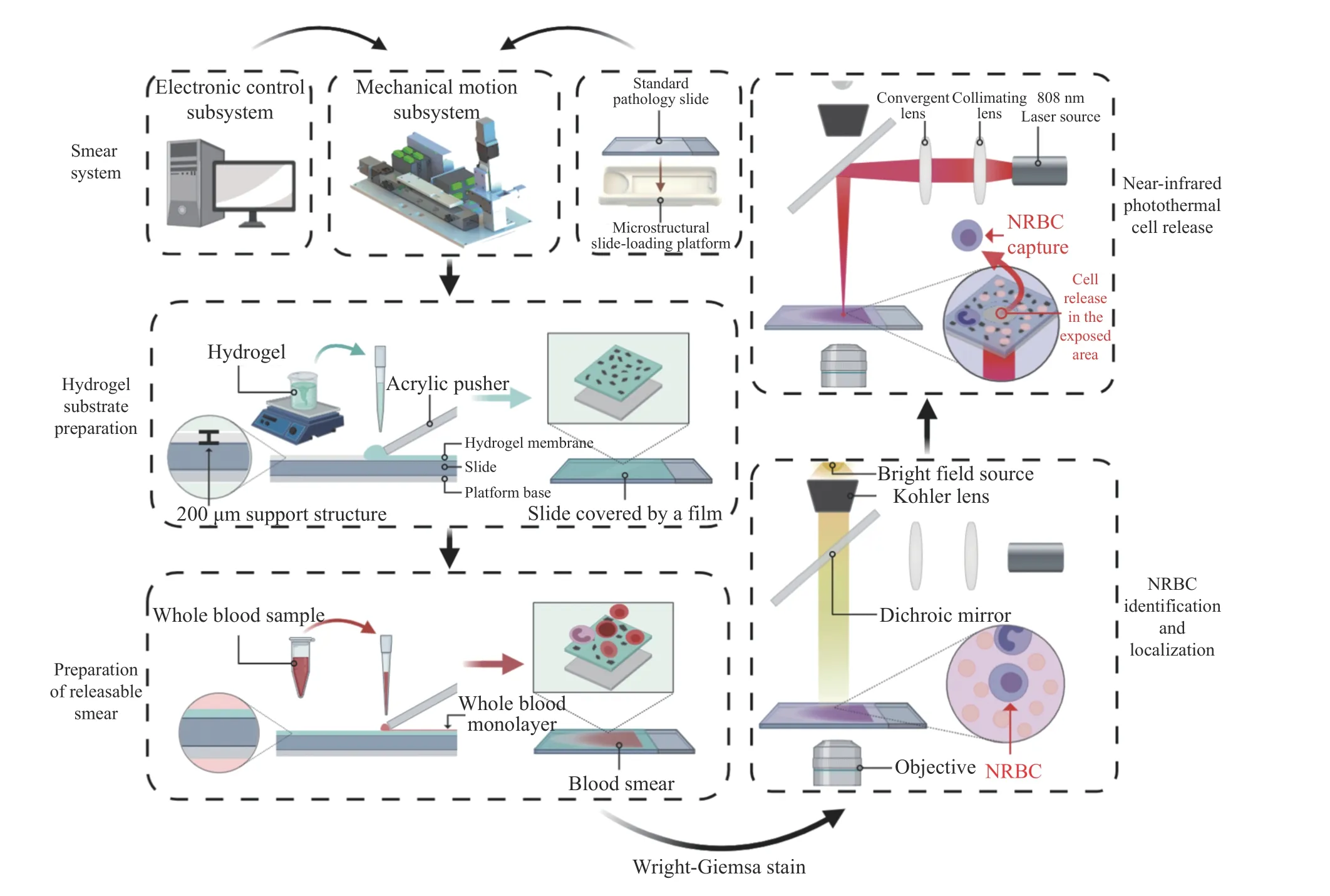
Fig. 1 Schematic diagram of cell smear preparation and laser response system图 1 细胞涂片制备及激光响应系统示意图
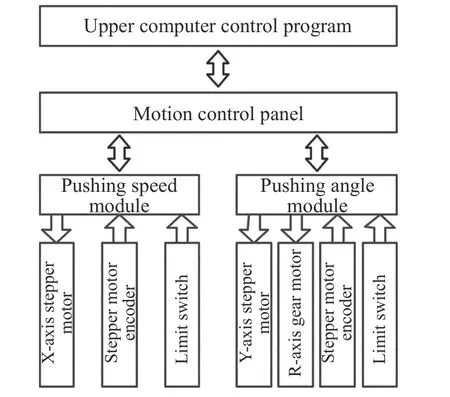
Fig. 2 Schematic diagram of mechanical system of cell smear图 2 细胞涂片机械系统原理示意图
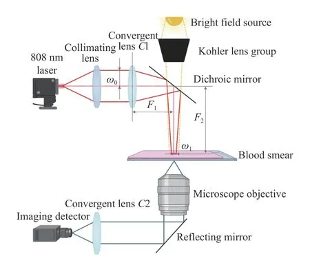
Fig. 3 Schematic diagram of laser focusing and microscopic imaging system图 3 激光会聚与显微成像系统示意图
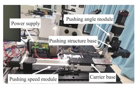
Fig. 4 Prototype of preparation machine of automatic cell smear图 4 自动细胞涂片制备机样机
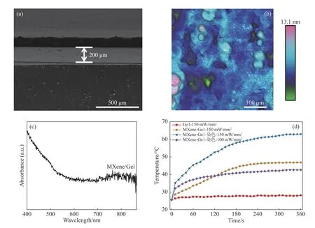
Fig. 5 (a) Characterization by SEM; (b) surface roughness characterization by AFM; (c) ultraviolet absorption spectrum;(d) photo-thermal curves图 5 (a)SEM表征;(b)AFM表面粗糙度表征;(c)紫外吸收图谱;(d)光热曲线
3.2 Preparation and optimization of a cell smear on hydrogel membrane
An automatic cell smear preparation machine was designed to carry out blood slide-pushing experiment on the surface of hydrogel membrane. The blood samples were from the peripheral blood with a hematocrit (HCT) ranging from 0.39 to 0.45 examined in a clinic. The reference value of HCT for normal females is 0.35−0.45. However, during pregnancy, the HCT value will vary within 0.33−0.46 as the mother undergoes a series of physiological changes with the growth and development of her fetus[15].
4 groups of peripheral blood samples with the HCT values of 0.39−0.45 were pushed on the surface of hydrogel substrate. According to the parameters mentioned above, the pushing speed, the pushing angle and the whole-blood volume were investigated and optimized in the process of cell smear preparation. After optimization, the pushing effect was shown in Figure 6 (Color online) (smear speed: 100 mm/s, pushing angle: 40°, blood volume:5 μL). The Fig. 6 compares the effects of a stained cell smear on slide substrate, a cell smear on hydrogel membrane substrate and a standard cell smear prepared by a fully automatic blood analyzer Mindray SC-120 which is currently used in clinical tests. The front, middle and tail ends of the three cell smears were analyzed by microscopic imaging system, repectively. Five sampling points were randomly selected from each part within the 0.3 mm2FOV (field of view) of the imaging system to obtain the cell distribution images of the smears. The cells were then counted using ImageJ software. The cell distributions on the smears with 4 groups of blood samples are shown in Fig. 7. As can be seen from the figure, the cells aggregate and overlap on the smear with slide substrate, while the cells are in a single layer format adhere to hydrogel membrane substrate and are distributed uniformly. By comparing the front and tail parts of the cell smears, it can be seen that the cell smear with hydrogel membrane substrate shares the same monolayer cell distribution with the standard smear. In addition, the average of cell density is 19.3% higher than that of the standard smear, thus improving the efficiency of the NRBC observation and detection based on cell smear.
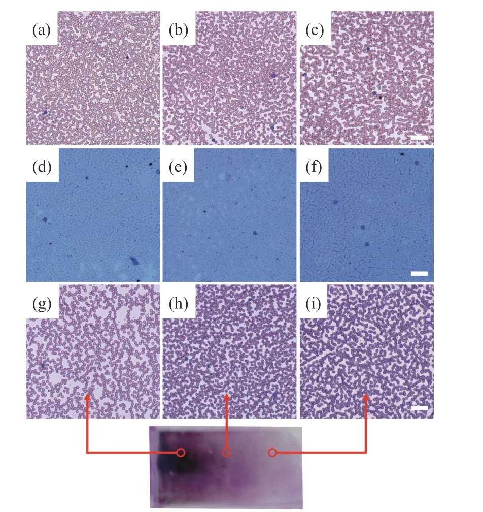
Fig. 6 Comparison of self-made cell smear and Mindray SC-120 standard cell smear: (a-c) slide; (d-f) hydrogel membrane; (g-i) Mindray SC-120图 6 自制细胞涂片与迈瑞SC-120标准细胞涂片效果比对分析:(a-c)玻片;(d-f)水凝胶膜;(g-i)迈瑞SC-120
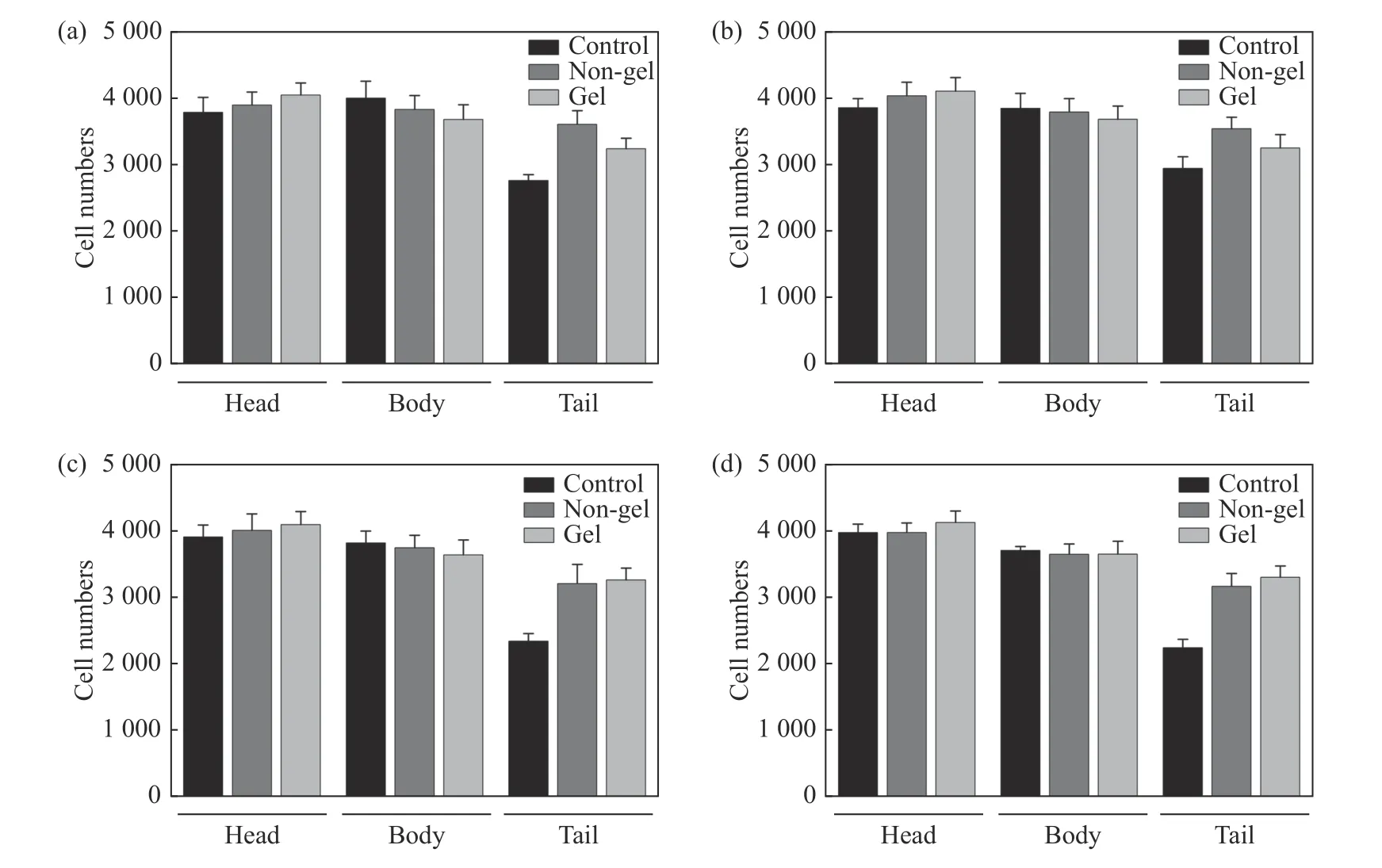
Fig. 7 Cell distribution statistics图 7 细胞分布统计
The performance parameters of the prototype machine proposed in this paper were compared with those of the commercial Mindray SC-120 automatic pushing machine. The comparison results are summarized in Table 1. Compared with Mindray SC-120, the proposed prototype has the following advantages: (1) The pusher-loading platform is modularized so that the various substrates (glass/hydrogel/releasable) can be made by changing the platform; (2) the scraping operation in traditional scraping-pushing process is not necessary, so the blood consumption is reduced down to only 4 μL; (3) the pusher can be a standard medical type, rather than a customized product, so the maintenance cost is reduced.
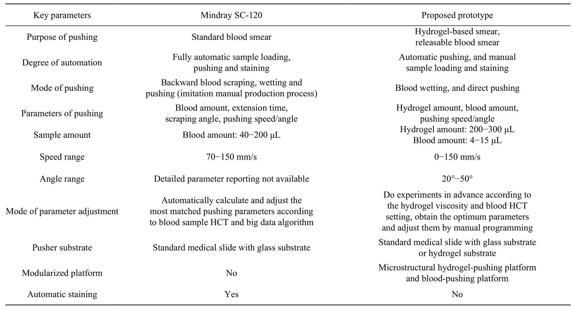
Tab. 1 Performance comparisons between the proposed prototype and Mindray SC-120 automatic pushing-staining machine表 1 本样机与迈瑞SC-120全自动推片染色机的性能参数对比
3.3 NRBC release under near-infrared response
3.3.1 Recognition and release of NRBCs
The NRBC recognition and release based on cell smear preparation technique were achieved by using the photothermal response properties of hydrogel. The prepared cell smear was placed in the microscopic imaging system. Then the NRBC was identified and determined according to its morphological features, such as round nucleus, a nucleuscytoplasm ratio of less than 1/2, no cytoplasmic granules, and nucleus amesiality[16]. As shown in Fig. 8(a), the NRBCs in the cell layer on the smear were identified and located, and then irradiated for 90 s by an 808 nm laser source (laser power:100 mW/mm2) fixed at a distance where a 1.7 mm spot was formed. Finally, they were rinsed with deionized water and dried, and observed under a microscope.
As can be seen from the characterization results (Fig. 8(b)), almost all the cells fall off with the photothermal melting of the hydrogel membrane within the fixed area of hydrogel membrane irradiated by laser. Therefore, the fixed-point release of cells can be achieved by introducing photothermally responsive hydrogel.
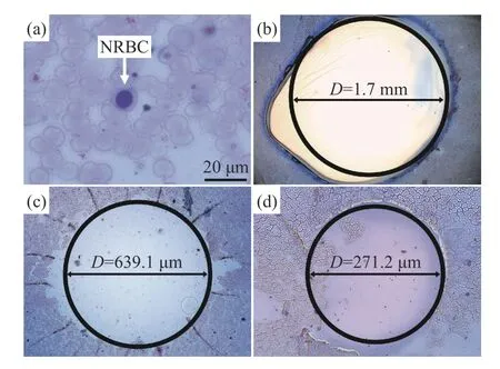
Fig. 8 (a) Recognition and localization of NRBC; (b-d)comparison of cell release areas before and after laser convergence with different spot diameters.(b) D = 1 700 μm; (c) D = 600 μm; (d) D = 300 μm图 8 (a)NRBC识别定位结果;(b-d)激光会聚前后细胞释放区域比较:(b)光斑直径D = 1 700 μm;(c)光斑直径D = 600 μm;(d)光斑直径D = 300 μm
3.3.2 Optimization of laser convergence system
The fixed-point release of NRBCs was achieved through direct irradiation of 808-nm laser.However, the spot diameter directly projected onto the cell smear surface was 1.7 mm and required further adjustment relative to the cell size. Therefore, a laser convergence system was built by connecting the laser with a collimating lens and a convergent lens to reduce the spot diameter on the smear surface. This system could not only ensure the function of cell release, but also improve the accuracy of NRBC release. By substitutingω0= 300 μm andω0= 150 μm into Equation (2) respectively, the focal lengthsF= 100 cm andF=50 cm can be obtained. SinceF2is a fixed distance (F2= 20 cm), the focus diameter can be adjusted by adjustingF1=80 cm andF1= 30 cm, whereF1is the distance between the convergent lensC1 and the dichromatic mirror. The photothermal releases generated by the converged spots of about 600 μm and 300 μm are shown in Figure 8(c) and Figure 8(d) respectively. The micro-characterization results show that the proposed laser convergence system can scale down the cell-releasing area to about 271.2 μm (in diameter), while achieving the same photothermal conversion effect.
4 Conclusion
In this paper, a monolayer cell smear based on hydrogel membrane was prepared by automatic pushing technique. The prepared hydrogel membrane had the property of near-infrared light response and showed photothermal conversion under 808 nm laser irradiation. The processing parameters of hydrogel membrane substrate and cell smear were optimized. The whole blood cells were spread into a single layer on the surface of the hydrogel membrane substrate with a uniform thickness of 200 μm. A laser convergence system and a microscopic imaging system were constructed using an 808 nm laser source and applied to cell smears. By designing and constructing the light path for laser convergence and microscopic imaging, the light source was focused on a fixed area of the cell smear to realize the cell recognition and release. The results showed that when the pushing speed was 100 mm/s and the pushing angle was 40°, the prepared cell smear had uniform cell distribution and an average cell density 19.3% higher than a standard smear. While keeping laser power at the cell-releasing level, this technique can reduce the spot diameter to about 300 μm, so as to realize the cell release and enrichment. This paper provides a new technical approach that can be combined with automatic microscanning imaging and microneedle extraction in subsequent studies to efficiently and accurately extract the NRBCs for noninvasive prenatal diagnoses.
——中文对照版——
1 引 言
产前诊断对于唐氏综合征、神经管缺陷、单基因疾病等出生缺陷的早期诊断和筛查至关重要[1],目前产前诊断主要分为有创性诊断和无创性诊断。有创性诊断包含羊膜枪穿刺、脐血取样以及绒毛膜穿刺等[2],是通过侵入式方法进行的,在诊断过程中往往伴随着流产或羊水溢出、感染等风险[3]。因此,开发以非侵入式进行无创性产前诊断是当前产前诊断技术发展的研究重点和主流趋势[4]。胎儿有核红细胞(NRBCs)包含了胎儿全部基因组,具有孕早期表达(6周在外周血中表达,12−14周表达量达到最高),在母血细胞群中可鉴別性高,不会出现与母体细胞混淆的情况,具备明显的细胞形态学特征,易识别,是应用于产前诊断的首选胎儿细胞[5]。然而,其在母体外周血中数量极其稀少,如何从外周血中对有核红细胞进行识别分离是产前诊断中的一大挑战[6]。
目前已有的对外周血细胞进行分离富集的方法主要有密度梯度离心富集、磁激活细胞分选法、荧光激活细胞分选法以及微流控芯片等[7-9],这些方法多以抗体与细胞特异性结合为核心进行分离检测,操作流程复杂、检测周期较长且成本较高。……
杂志排行
中国光学的其它文章
- Surface plasmon resonance characteristics of a graphene nano-disk based on three-dimensional boundary element method
- Design of optical wedge demodulation system for fiber Fabry-Perot sensor
- Photon-assisted Fano resonance tunneling periodic double-well potential characteristics
- Formation mechanism of the continuous spectral profile of lightning plasma
- 中国遥感卫星辐射校正场敦煌戈壁场区光环境变化研究
- 油气井下光纤光栅温度压力传感器